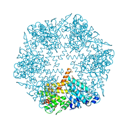
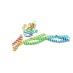
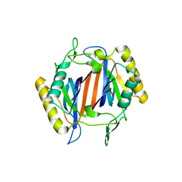
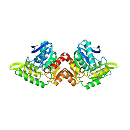
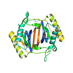
3KDO
 
 | | Crystal structure of Type III Rubisco SP6 mutant complexed with 2-CABP | | 分子名称: | 2-CARBOXYARABINITOL-1,5-DIPHOSPHATE, MAGNESIUM ION, Ribulose bisphosphate carboxylase | | 著者 | Nishitani, Y, Fujihashi, M, Doi, T, Yoshida, S, Atomi, H, Imanaka, T, Miki, K. | | 登録日 | 2009-10-23 | | 公開日 | 2010-10-06 | | 最終更新日 | 2023-11-22 | | 実験手法 | X-RAY DIFFRACTION (2.36 Å) | | 主引用文献 | Structure-based catalytic optimization of a type III Rubisco from a hyperthermophile
J.Biol.Chem., 285, 2010
|
|
3KDS
 | | apo-FtsH crystal structure | | 分子名称: | Cell division protein FtsH, N-{(2R)-2-[2-(hydroxyamino)-2-oxoethyl]-4-methylpentanoyl}-3-naphthalen-2-yl-L-alanyl-L-alaninamide, ZINC ION | | 著者 | Bieniossek, C, Niederhauser, B, Baumann, U. | | 登録日 | 2009-10-23 | | 公開日 | 2009-12-01 | | 最終更新日 | 2023-11-01 | | 実験手法 | X-RAY DIFFRACTION (2.601 Å) | | 主引用文献 | The crystal structure of apo-FtsH reveals domain movements necessary for substrate unfolding and translocation
Proc.Natl.Acad.Sci.USA, 106, 2009
|
|
3KGK
 | | Crystal structure of ArsD | | 分子名称: | Arsenical resistance operon trans-acting repressor arsD | | 著者 | Ye, J, Rosen, B.P. | | 登録日 | 2009-10-29 | | 公開日 | 2010-02-16 | | 最終更新日 | 2024-02-07 | | 実験手法 | X-RAY DIFFRACTION (1.4 Å) | | 主引用文献 | The 1.4 A crystal structure of the ArsD arsenic metallochaperone provides insights into its interaction with the ArsA ATPase.
Biochemistry, 49, 2010
|
|
4M9P
 | |
3KJT
 | |
3KBT
 | |
2YZ1
 | |
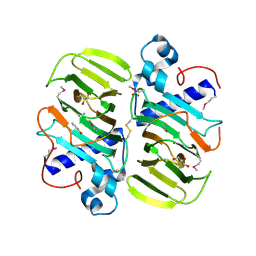
3KDN
 | | Crystal structure of Type III Rubisco SP4 mutant complexed with 2-CABP | | 分子名称: | 2-CARBOXYARABINITOL-1,5-DIPHOSPHATE, MAGNESIUM ION, Ribulose bisphosphate carboxylase | | 著者 | Nishitani, Y, Fujihashi, M, Doi, T, Yoshida, S, Atomi, H, Imanaka, T, Miki, K. | | 登録日 | 2009-10-23 | | 公開日 | 2010-10-06 | | 最終更新日 | 2023-11-22 | | 実験手法 | X-RAY DIFFRACTION (2.09 Å) | | 主引用文献 | Structure-based catalytic optimization of a type III Rubisco from a hyperthermophile
J.Biol.Chem., 285, 2010
|
|
3KBU
 | |
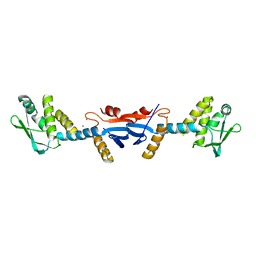
3KCG
 | | Crystal structure of the antithrombin-factor IXa-pentasaccharide complex | | 分子名称: | (4S)-2-METHYL-2,4-PENTANEDIOL, 3,4-di-O-methyl-2,6-di-O-sulfo-alpha-D-glucopyranose-(1-4)-2,3-di-O-methyl-beta-D-glucopyranuronic acid-(1-4)-2,3,6-tri-O-sulfo-alpha-D-glucopyranose-(1-4)-3-O-methyl-2-O-sulfo-alpha-L-idopyranuronic acid-(1-4)-methyl 2,3,6-tri-O-sulfo-alpha-D-glucopyranoside, Antithrombin-III, ... | | 著者 | Huntington, J.A, Johnson, D.J.D. | | 登録日 | 2009-10-21 | | 公開日 | 2010-02-02 | | 最終更新日 | 2023-11-01 | | 実験手法 | X-RAY DIFFRACTION (1.7 Å) | | 主引用文献 | Molecular basis of factor IXa recognition by heparin-activated antithrombin revealed by a 1.7-A structure of the ternary complex.
Proc.Natl.Acad.Sci.USA, 107, 2010
|
|
3KDK
 | |
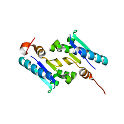
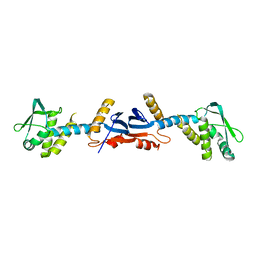
3KJL
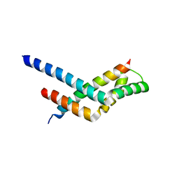 | | Sgf11:Sus1 complex | | 分子名称: | Protein SUS1, SAGA-associated factor 11 | | 著者 | Stewart, M, Ellisdon, A.M. | | 登録日 | 2009-11-03 | | 公開日 | 2009-12-08 | | 最終更新日 | 2023-09-06 | | 実験手法 | X-RAY DIFFRACTION (2.7 Å) | | 主引用文献 | Structural basis for the interaction between yeast Spt-Ada-Gcn5 acetyltransferase (SAGA) complex components Sgf11 and Sus1.
J.Biol.Chem., 285, 2010
|
|
3KMN
 | |
3KNP
 | |
3KDG
 | |
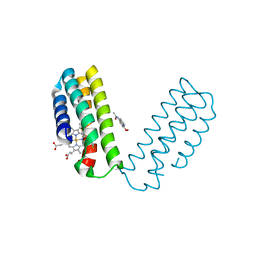
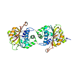
3KCP
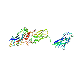 | | Crystal structure of interacting Clostridium thermocellum multimodular components | | 分子名称: | CALCIUM ION, CHLORIDE ION, Cellulosomal-scaffolding protein A, ... | | 著者 | Adams, J.J, Currie, M.A, Bayer, E.A, Jia, Z, Smith, S.P. | | 登録日 | 2009-10-21 | | 公開日 | 2010-02-09 | | 最終更新日 | 2024-02-21 | | 実験手法 | X-RAY DIFFRACTION (1.94 Å) | | 主引用文献 | Insights into Higher-Order Organization of the Cellulosome Revealed by a Dissect-and-Build Approach: Crystal Structure of Interacting Clostridium thermocellum Multimodular Components
J.Mol.Biol., 396, 2010
|
|
3KWY
 | |
3KXP
 | | Crystal Structure of E-2-(Acetamidomethylene)succinate Hydrolase | | 分子名称: | Alpha-(N-acetylaminomethylene)succinic acid hydrolase, CHLORIDE ION | | 著者 | McCulloch, K.M, Mukherjee, T, Begley, T.P, Ealick, S.E. | | 登録日 | 2009-12-03 | | 公開日 | 2010-02-09 | | 最終更新日 | 2017-11-01 | | 実験手法 | X-RAY DIFFRACTION (2.26 Å) | | 主引用文献 | Structure determination and characterization of the vitamin B(6) degradative enzyme (E)-2-(acetamidomethylene)succinate hydrolase.
Biochemistry, 49, 2010
|
|
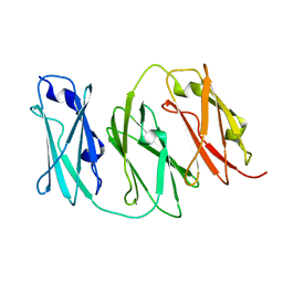
3L11
 | | Crystal Structure of the Ring Domain of RNF168 | | 分子名称: | E3 ubiquitin-protein ligase RNF168, MALONATE ION, ZINC ION | | 著者 | Neculai, D, Yermekbayeva, L, Crombet, L, Weigelt, J, Bountra, C, Edwards, A.M, Arrowsmith, C.H, Bochkarev, A, Dhe-Paganon, S, Structural Genomics Consortium (SGC) | | 登録日 | 2009-12-10 | | 公開日 | 2010-01-19 | | 最終更新日 | 2023-09-06 | | 実験手法 | X-RAY DIFFRACTION (2.12 Å) | | 主引用文献 | Molecular insights into the function of RING finger (RNF)-containing proteins hRNF8 and hRNF168 in Ubc13/Mms2-dependent ubiquitylation.
J.Biol.Chem., 287, 2012
|
|
3L1M
 | |
3KMH
 | |
3KN6
 | |
3KOB
 | |
3KP2
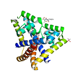 | |
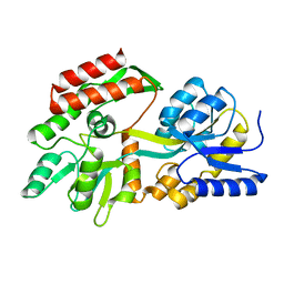
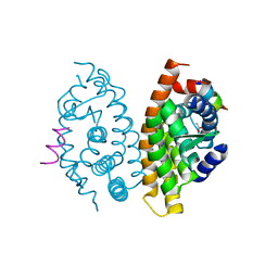
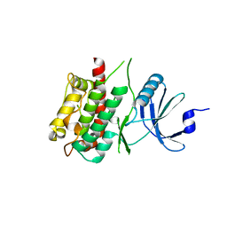
3KPY
 | | Crystal Structure of hPNMT in Complex AdoHcy and 6-Chlorooxindole | | 分子名称: | 6-chloro-1,3-dihydro-2H-indol-2-one, Phenylethanolamine N-methyltransferase, S-ADENOSYL-L-HOMOCYSTEINE | | 著者 | Drinkwater, N, Martin, J.L. | | 登録日 | 2009-11-17 | | 公開日 | 2010-09-29 | | 最終更新日 | 2023-09-06 | | 実験手法 | X-RAY DIFFRACTION (2.4 Å) | | 主引用文献 | Fragment-based screening by X-ray crystallography, MS and isothermal titration calorimetry to identify PNMT (phenylethanolamine N-methyltransferase) inhibitors.
Biochem.J., 431, 2010
|
|


