1W24
 
 | | Crystal Structure Of human Vps29 | | 分子名称: | VACUOLAR PROTEIN SORTING PROTEIN 29 | | 著者 | Wang, D, Guo, M, Teng, M, Niu, L. | | 登録日 | 2004-06-26 | | 公開日 | 2005-03-23 | | 最終更新日 | 2024-05-08 | | 実験手法 | X-RAY DIFFRACTION (2.1 Å) | | 主引用文献 | Crystal Structure of Human Vacuolar Protein Sorting Protein 29 Reveals a Phosphodiesterase/Nuclease-Like Fold and Two Protein-Protein Interaction Sites.
J.Biol.Chem., 280, 2005
|
|
3WLV
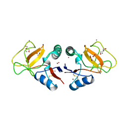
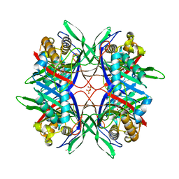 | | Thermostable urate oxidase from Bacillus sp. TB-90 | | 分子名称: | 8-AZAXANTHINE, CHLORIDE ION, POTASSIUM ION, ... | | 著者 | Hibi, T, Hayashi, Y, Itoh, T. | | 登録日 | 2013-11-14 | | 公開日 | 2014-06-18 | | 最終更新日 | 2024-05-29 | | 実験手法 | X-RAY DIFFRACTION (1.747 Å) | | 主引用文献 | Intersubunit salt bridges with a sulfate anion control subunit dissociation and thermal stabilization of Bacillus sp. TB-90 urate oxidase.
Biochemistry, 53, 2014
|
|
1VQ2
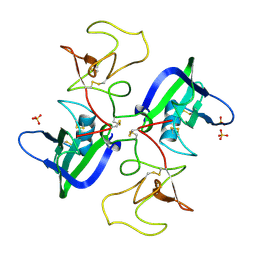
 | | CRYSTAL STRUCTURE OF T4-BACTERIOPHAGE DEOXYCYTIDYLATE DEAMINASE, MUTANT R115E | | 分子名称: | 3,4-DIHYDRO-2'-DEOXYURIDINE-5'-MONOPHOSPHATE, DEOXYCYTIDYLATE DEAMINASE, ZINC ION | | 著者 | Almog, R, Maley, F, Maley, G.F, Maccoll, R, Van Roey, P. | | 登録日 | 2004-12-15 | | 公開日 | 2004-12-21 | | 最終更新日 | 2023-12-27 | | 実験手法 | X-RAY DIFFRACTION (2.2 Å) | | 主引用文献 | Three-Dimensional Structure of the R115E Mutant of T4-Bacteriophage 2'-Deoxycytidylate Deaminase
Biochemistry, 43, 2004
|
|
2QJ4
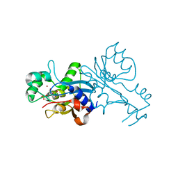
 | | A Mechanistic Basis for Converting a Receptor Tyrosine Kinase Agonist to an Antagonist | | 分子名称: | Hepatocyte growth factor, SULFATE ION | | 著者 | Tolbert, W.D, Daugherty, J, Gao, C.-F, Xe, Q, Miranti, C, Gherardi, E, Vande Woude, G, Xu, H.E. | | 登録日 | 2007-07-06 | | 公開日 | 2007-09-18 | | 最終更新日 | 2023-08-30 | | 実験手法 | X-RAY DIFFRACTION (2.5 Å) | | 主引用文献 | A mechanistic basis for converting a receptor tyrosine kinase agonist to an antagonist
Proc.Natl.Acad.Sci.Usa, 104, 2007
|
|
3TW4
 | | Crystal Structure of Human Septin 7 GTPase Domain | | 分子名称: | GUANOSINE-5'-DIPHOSPHATE, Septin-7 | | 著者 | Serrao, V.H.B, Alessandro, F, Pereira, H.M, Thiemann, O.T, Garratt, R.C. | | 登録日 | 2011-09-21 | | 公開日 | 2011-11-23 | | 最終更新日 | 2023-09-13 | | 実験手法 | X-RAY DIFFRACTION (3.35 Å) | | 主引用文献 | Promiscuous interactions of human septins: The GTP binding domain of SEPT7 forms filaments within the crystal.
Febs Lett., 585, 2011
|
|
4BHX
 | | Crystal structure of the SCAN domain from human paternally expressed gene 3 protein | | 分子名称: | 1,2-ETHANEDIOL, DI(HYDROXYETHYL)ETHER, PATERNALLY-EXPRESSED GENE 3 PROTEIN, ... | | 著者 | Rimsa, V, Eadsforth, T.C, Hunter, W.N. | | 登録日 | 2013-04-09 | | 公開日 | 2013-04-17 | | 最終更新日 | 2023-12-20 | | 実験手法 | X-RAY DIFFRACTION (1.95 Å) | | 主引用文献 | Structure of the Scan Domain of Human Paternally Expressed Gene 3 Protein.
Plos One, 8, 2013
|
|
1M02
 | | NMR Structure of PW2 Bound to SDS Micelles: A Tryptophan-rich Anticocidial Peptide Selected from Phage Display Libraries | | 分子名称: | HIS-PRO-LEU-LYS-GLN-TYR-TRP-TRP-ARG-PRO-SER-ILE | | 著者 | Tinoco, L.W, da Silva Jr, A, Leite, A, Valente, A.P, Almeida, F.C. | | 登録日 | 2002-06-11 | | 公開日 | 2002-08-14 | | 最終更新日 | 2024-05-22 | | 実験手法 | SOLUTION NMR | | 主引用文献 | NMR structure of PW2 bound to SDS micelles. A tryptophan-rich anticoccidial peptide selected from phage display libraries
J.Biol.Chem., 277, 2002
|
|
1AMK
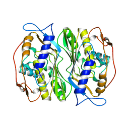
 | | LEISHMANIA MEXICANA TRIOSE PHOSPHATE ISOMERASE | | 分子名称: | 2-PHOSPHOGLYCOLIC ACID, TRIOSE PHOSPHATE ISOMERASE | | 著者 | Williams, J.C, Wierenga, R. | | 登録日 | 1997-06-17 | | 公開日 | 1997-12-17 | | 最終更新日 | 2024-05-22 | | 実験手法 | X-RAY DIFFRACTION (1.83 Å) | | 主引用文献 | Structural and mutagenesis studies of leishmania triosephosphate isomerase: a point mutation can convert a mesophilic enzyme into a superstable enzyme without losing catalytic power.
Protein Eng., 12, 1999
|
|
3EIH
 | | Crystal structure of S.cerevisiae Vps4 in the presence of ATPgammaS | | 分子名称: | 1,2-ETHANEDIOL, MAGNESIUM ION, PHOSPHOTHIOPHOSPHORIC ACID-ADENYLATE ESTER, ... | | 著者 | Gonciarz, M.D, Whitby, F.G, Eckert, D.M, Kieffer, C, Heroux, A, Sundquist, W.I, Hill, C.P. | | 登録日 | 2008-09-15 | | 公開日 | 2008-09-30 | | 最終更新日 | 2024-02-21 | | 実験手法 | X-RAY DIFFRACTION (3.25 Å) | | 主引用文献 | Biochemical and structural studies of yeast vps4 oligomerization.
J.Mol.Biol., 384, 2008
|
|
1NCN
 | |
1T70
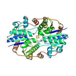
 | | Crystal structure of a novel phosphatase from Deinococcus radiodurans | | 分子名称: | Phosphatase | | 著者 | Shin, D.H, Wang, W, Kim, R, Yokota, H, Kim, S.H, Berkeley Structural Genomics Center (BSGC) | | 登録日 | 2004-05-07 | | 公開日 | 2004-12-07 | | 最終更新日 | 2024-02-14 | | 実験手法 | X-RAY DIFFRACTION (2.3 Å) | | 主引用文献 | Structural and enzymatic characterization of DR1281: A calcineurin-like phosphoesterase from Deinococcus radiodurans.
Proteins, 70, 2008
|
|
8U1L
 | |
5EZT
 | | Peracetylated Bovine Carbonic Anhydrase II | | 分子名称: | Carbonic anhydrase 2, ZINC ION | | 著者 | Whitesides, G.M, Kang, K, Choi, J.-M, Fox, J.M. | | 登録日 | 2015-11-26 | | 公開日 | 2016-07-20 | | 最終更新日 | 2016-07-27 | | 実験手法 | X-RAY DIFFRACTION (1.54 Å) | | 主引用文献 | Acetylation of Surface Lysine Groups of a Protein Alters the Organization and Composition of Its Crystal Contacts.
J.Phys.Chem.B, 120, 2016
|
|
1BKO
 | |
1BRS
 | |
1BUO
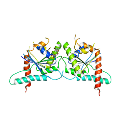
 | | BTB DOMAIN FROM PLZF | | 分子名称: | PROTEIN (PROMYELOCYTIC LEUKEMIA ZINC FINGER PROTEIN PLZF) | | 著者 | Ahmad, K.F, Engel, C.K, Prive, G.G. | | 登録日 | 1998-09-04 | | 公開日 | 1998-10-14 | | 最終更新日 | 2024-02-07 | | 実験手法 | X-RAY DIFFRACTION (1.9 Å) | | 主引用文献 | Crystal structure of the BTB domain from PLZF.
Proc.Natl.Acad.Sci.USA, 95, 1998
|
|
1NIP
 | | CRYSTALLOGRAPHIC STRUCTURE OF THE NITROGENASE IRON PROTEIN FROM AZOTOBACTER VINELANDII | | 分子名称: | ADENOSINE-5'-DIPHOSPHATE, IRON/SULFUR CLUSTER, MAGNESIUM ION, ... | | 著者 | Komiya, H, Georgiadis, M.M, Chakrabarti, P, Woo, D, Kornuc, J.J, Rees, D.C. | | 登録日 | 1992-09-29 | | 公開日 | 1993-10-31 | | 最終更新日 | 2024-02-14 | | 実験手法 | X-RAY DIFFRACTION (2.9 Å) | | 主引用文献 | Crystallographic structure of the nitrogenase iron protein from Azotobacter vinelandii.
Science, 257, 1992
|
|
1NH9
 | | Crystal Structure of a DNA Binding Protein Mja10b from the hyperthermophile Methanococcus jannaschii | | 分子名称: | DNA-binding protein Alba | | 著者 | Wang, G, Bartlam, M, Guo, R, Yang, H, Xue, H, Liu, Y, Huang, L, Rao, Z. | | 登録日 | 2002-12-19 | | 公開日 | 2003-12-23 | | 最終更新日 | 2023-10-25 | | 実験手法 | X-RAY DIFFRACTION (2 Å) | | 主引用文献 | Crystal structure of a DNA binding protein from the hyperthermophilic euryarchaeon Methanococcus jannaschii
Protein Sci., 12, 2003
|
|
1BKP
 | |
1BYF
 | | STRUCTURE OF TC14; A C-TYPE LECTIN FROM THE TUNICATE POLYANDROCARPA MISAKIENSIS | | 分子名称: | ACETATE ION, CALCIUM ION, GLYCEROL, ... | | 著者 | Poget, S.F, Legge, G.B, Bycroft, M, Williams, R.L. | | 登録日 | 1998-10-14 | | 公開日 | 1999-07-23 | | 最終更新日 | 2023-12-27 | | 実験手法 | X-RAY DIFFRACTION (2 Å) | | 主引用文献 | The structure of a tunicate C-type lectin from Polyandrocarpa misakiensis complexed with D -galactose.
J.Mol.Biol., 290, 1999
|
|
3FPC
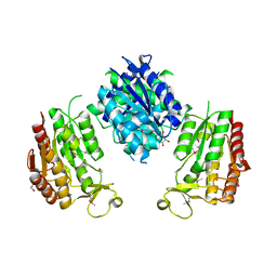
 | | Chimera of alcohol dehydrogenase by exchange of the cofactor binding domain res 153-294 of T. brockii ADH by E. histolytica ADH | | 分子名称: | 1,2-ETHANEDIOL, CACODYLATE ION, IMIDAZOLE, ... | | 著者 | Felix, F, Goihberg, E, Shimon, L, Burstein, Y. | | 登録日 | 2009-01-05 | | 公開日 | 2010-01-19 | | 最終更新日 | 2023-11-01 | | 実験手法 | X-RAY DIFFRACTION (1.4 Å) | | 主引用文献 | Biochemical and structural properties of chimeras constructed by exchange of cofactor-binding domains in alcohol dehydrogenases from thermophilic and mesophilic microorganisms
Biochemistry, 49, 2010
|
|
3WRW
 | |
1P6A
 | | STRUCTURAL BASIS FOR VARIATION IN ADENOVIRUS AFFINITY FOR THE CELLULAR RECEPTOR CAR (S489Y MUTANT) | | 分子名称: | Coxsackievirus and adenovirus receptor, Fiber protein | | 著者 | Howitt, J, Bewley, M.C, Graziano, V, Flanagan, J.M, Freimuth, P. | | 登録日 | 2003-04-29 | | 公開日 | 2004-05-11 | | 最終更新日 | 2018-08-22 | | 実験手法 | X-RAY DIFFRACTION (2.9 Å) | | 主引用文献 | Structural basis for variation in adenovirus affinity for the cellular coxsackievirus and adenovirus receptor.
J.Biol.Chem., 278, 2003
|
|
1OT8
 | | Structure of the Ankyrin Domain of the Drosophila Notch Receptor | | 分子名称: | MAGNESIUM ION, Neurogenic locus Notch protein | | 著者 | Zweifel, M.E, Leahy, D.J, Hughson, F.M, Barrick, D. | | 登録日 | 2003-03-21 | | 公開日 | 2003-10-28 | | 最終更新日 | 2024-02-14 | | 実験手法 | X-RAY DIFFRACTION (2 Å) | | 主引用文献 | Structure and stability of the ankyrin domain of the Drosophila Notch receptor
Protein Sci., 12, 2003
|
|
1UEL
 | | Solution structure of ubiquitin-like domain of hHR23B complexed with ubiquitin-interacting motif of proteasome subunit S5a | | 分子名称: | 26S proteasome non-ATPase regulatory subunit 4, UV excision repair protein RAD23 homolog B | | 著者 | Fujiwara, K, Tenno, T, Jee, J.G, Sugasawa, K, Ohki, I, Kojima, C, Tochio, H, Hiroaki, H, Hanaoka, H, Shirakawa, M, RIKEN Structural Genomics/Proteomics Initiative (RSGI) | | 登録日 | 2003-05-19 | | 公開日 | 2004-02-10 | | 最終更新日 | 2023-12-27 | | 実験手法 | SOLUTION NMR | | 主引用文献 | Structure of the Ubiquitin-interacting Motif of S5a Bound to the Ubiquitin-like Domain of HR23B
J.Biol.Chem., 279, 2004
|
|


