3SM4
 
 | |
3SLP
 | |
3SMB
 | |
3SME
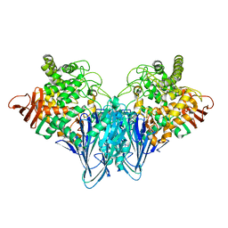
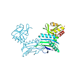
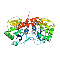
 | | Structure of PTP1B inactivated by H2O2/bicarbonate | | 分子名称: | MAGNESIUM ION, Tyrosine-protein phosphatase non-receptor type 1 | | 著者 | Tanner, J.J, Singh, H. | | 登録日 | 2011-06-27 | | 公開日 | 2011-10-19 | | 最終更新日 | 2023-09-13 | | 実験手法 | X-RAY DIFFRACTION (1.7 Å) | | 主引用文献 | The Biological Buffer Bicarbonate/CO(2) Potentiates H(2)O(2)-Mediated Inactivation of Protein Tyrosine Phosphatases.
J.Am.Chem.Soc., 133, 2011
|
|
3S4B
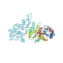
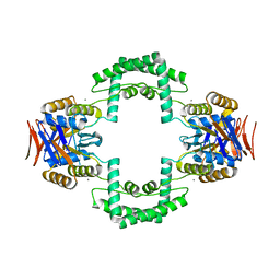
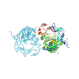
 | | Cellobiose phosphorylase from Cellulomonas uda in complex with glucose | | 分子名称: | Cellobiose phosphorylase, alpha-D-glucopyranose | | 著者 | Van Hoorebeke, A, Stout, J, Soetaert, W, Van Beeumen, J, Desmet, T, Savvides, S. | | 登録日 | 2011-05-19 | | 公開日 | 2012-06-27 | | 最終更新日 | 2024-02-28 | | 実験手法 | X-RAY DIFFRACTION (2.4 Å) | | 主引用文献 | Cellobiose phosphorylase: reconstructing the structural itinerary along the catalytic pathway
To be Published
|
|
8S8A
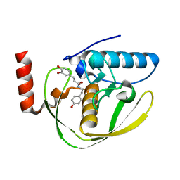
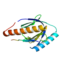
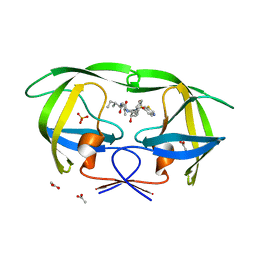
 | | Human pyridoxal phosphatase in complex with 7,8-dihydroxyflavone without phosphate | | 分子名称: | 7,8-bis(oxidanyl)-2-phenyl-chromen-4-one, CHLORIDE ION, Chronophin, ... | | 著者 | Brenner, M, Gohla, A, Schindelin, H. | | 登録日 | 2024-03-06 | | 公開日 | 2024-06-12 | | 最終更新日 | 2024-06-26 | | 実験手法 | X-RAY DIFFRACTION (1.5 Å) | | 主引用文献 | 7,8-Dihydroxyflavone is a direct inhibitor of human and murine pyridoxal phosphatase.
Elife, 13, 2024
|
|
3S5O
 | |
2EW6
 | | Structure of Helicobacter Pylori peptide deformylase in complex with inhibitor | | 分子名称: | (2E)-3-(3,4-DIHYDROXYPHENYL)-N-[2-(4-HYDROXYPHENYL)ETHYL]ACRYLAMIDE, COBALT (II) ION, peptide deformylase | | 著者 | Cai, J. | | 登録日 | 2005-11-02 | | 公開日 | 2006-10-24 | | 最終更新日 | 2023-10-25 | | 実験手法 | X-RAY DIFFRACTION (2.2 Å) | | 主引用文献 | Peptide deformylase is a potential target for anti-Helicobacter pylori drugs: reverse docking, enzymatic assay, and X-ray crystallography validation
Protein Sci., 15, 2006
|
|
3SNO
 | |
3S5U
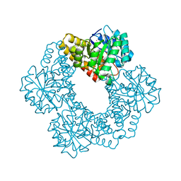
 | | Crystal structure of CRISPR associated protein | | 分子名称: | CALCIUM ION, Putative uncharacterized protein | | 著者 | Ke, A, Nam, K.H. | | 登録日 | 2011-05-23 | | 公開日 | 2011-06-22 | | 最終更新日 | 2024-02-28 | | 実験手法 | X-RAY DIFFRACTION (2.7 Å) | | 主引用文献 | Crystal structure of clustered regularly interspaced short palindromic repeats (CRISPR)-associated Csn2 protein revealed Ca2+-dependent double-stranded DNA binding activity.
J. Biol. Chem., 286, 2011
|
|
3SO6
 | |
3S4P
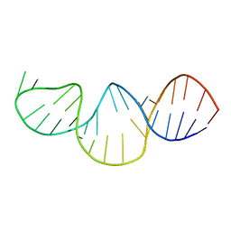
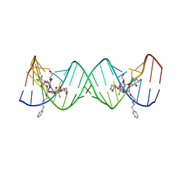 | | Crystal structure of the bacterial ribosomal decoding site complexed with an amphiphilic paromomycin O2''-ether analogue | | 分子名称: | (1R,2R,3S,4R,6S)-4,6-diamino-2-{[3-O-(2,6-diamino-2,6-dideoxy-beta-L-idopyranosyl)-2-O-{2-[(2-phenylethyl)amino]ethyl}-beta-D-ribofuranosyl]oxy}-3-hydroxycyclohexyl 2-amino-2-deoxy-alpha-D-glucopyranoside, RNA (5'-R(P*GP*CP*GP*UP*CP*AP*CP*AP*CP*CP*GP*GP*UP*GP*AP*AP*GP*UP*CP*GP*C)-3') | | 著者 | Szychowski, J, Kondo, J, Zahr, O, Auclair, K, Westhof, E, Hanessian, S, Keillor, J.W. | | 登録日 | 2011-05-20 | | 公開日 | 2011-09-21 | | 最終更新日 | 2023-11-01 | | 実験手法 | X-RAY DIFFRACTION (2.56 Å) | | 主引用文献 | Inhibition of aminoglycoside-deactivating enzymes APH(3')-IIIa and AAC(6')-Ii by amphiphilic paromomycin O2''-ether analogues
Chemmedchem, 6, 2011
|
|
3S5R
 | |
3SD2
 | |
3S6E
 | |
3SDI
 | | Structure of yeast 20S open-gate proteasome with Compound 20 | | 分子名称: | 2-(N-MORPHOLINO)-ETHANESULFONIC ACID, MAGNESIUM ION, N~4~-(2,2-dimethylpropyl)-N~1~-{(2S)-1-[(4-methylbenzyl)amino]-1-oxo-4-phenylbutan-2-yl}-N~2~-[(5-methyl-1,2-oxazol-3-yl)carbonyl]-L-aspartamide, ... | | 著者 | Sintchak, M.D. | | 登録日 | 2011-06-09 | | 公開日 | 2012-06-13 | | 最終更新日 | 2023-09-13 | | 実験手法 | X-RAY DIFFRACTION (2.65 Å) | | 主引用文献 | Optimization of a series of dipeptides with a P3 threonine residue as non-covalent inhibitors of the chymotrypsin-like activity of the human 20S proteasome.
Bioorg.Med.Chem.Lett., 20, 2010
|
|
3SF8
 | |
3SFG
 | |
3SA3
 | | Crystal structure of wild-type HIV-1 protease in complex with AG23 | | 分子名称: | ACETATE ION, N~2~-acetyl-N-[(2S,3R)-4-{(1,3-benzothiazol-6-ylsulfonyl)[(2S)-2-methylbutyl]amino}-3-hydroxy-1-phenylbutan-2-yl]-L-isoleucinamide, PHOSPHATE ION, ... | | 著者 | Schiffer, C.A, Nalam, M.N.L. | | 登録日 | 2011-06-02 | | 公開日 | 2012-06-06 | | 最終更新日 | 2023-09-13 | | 実験手法 | X-RAY DIFFRACTION (1.65 Å) | | 主引用文献 | Protease Inhibitors that protrude out from substrate envelope are more susceptible to developing drug resistance
To be Published
|
|
2F88
 | |
3SAB
 | |
8G6B
 | | Crystal structure of PfAMA1-RON2L chimera | | 分子名称: | Apical membrane antigen 1, rhoptry neck protein 2 chimera, SULFATE ION | | 著者 | Boulanger, M.J, Ramaswamy, R. | | 登録日 | 2023-02-14 | | 公開日 | 2023-09-13 | | 最終更新日 | 2024-02-14 | | 実験手法 | X-RAY DIFFRACTION (1.55 Å) | | 主引用文献 | Structure guided mimicry of an essential P. falciparum receptor-ligand complex enhances cross neutralizing antibodies.
Nat Commun, 14, 2023
|
|
3SG4
 | | Crystal Structure of GCaMP3-D380Y, LP(linker 2) | | 分子名称: | CALCIUM ION, Myosin light chain kinase, Green fluorescent protein, ... | | 著者 | Schreiter, E.R, Akerboom, J, Looger, L.L. | | 登録日 | 2011-06-14 | | 公開日 | 2012-06-20 | | 最終更新日 | 2023-12-06 | | 実験手法 | X-RAY DIFFRACTION (2.4 Å) | | 主引用文献 | Optimization of a GCaMP calcium indicator for neural activity imaging.
J.Neurosci., 32, 2012
|
|
7VQ1
 | | Structure of Apo-hsTRPM2 channel | | 分子名称: | Transient receptor potential cation channel subfamily M member 2 | | 著者 | Yu, X.F, Xie, Y, Zhang, X.K, Ma, C, Guo, J.T, Yang, F, Yang, W. | | 登録日 | 2021-10-18 | | 公開日 | 2021-12-22 | | 最終更新日 | 2024-06-19 | | 実験手法 | ELECTRON MICROSCOPY (3.76 Å) | | 主引用文献 | Structural and functional basis of the selectivity filter as a gate in human TRPM2 channel.
Cell Rep, 37, 2021
|
|
3SMM
 | | Crystal structure of human 14-3-3 sigma C38N/N166H in complex with task-3 peptide and stabilizer Fusicoccin J aglycone | | 分子名称: | 14-3-3 protein sigma, CHLORIDE ION, Fusicoccin J aglycone, ... | | 著者 | Anders, C, Schumacher, B, Ottmann, C. | | 登録日 | 2011-06-28 | | 公開日 | 2012-07-11 | | 最終更新日 | 2023-09-13 | | 実験手法 | X-RAY DIFFRACTION (2 Å) | | 主引用文献 | A semisynthetic fusicoccane stabilizes a protein-protein interaction and enhances the expression of K+ channels at the cell surface.
Chem. Biol., 20, 2013
|
|


