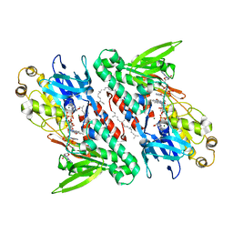
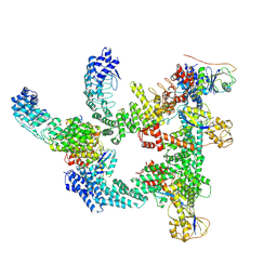
6UUB
 
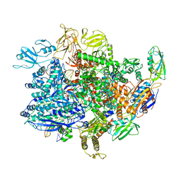 | | E. coli sigma-S transcription initiation complex with a mismatching UTP ("Fresh" crystal soaked with UTP for 2 hours) | | 分子名称: | DNA-directed RNA polymerase subunit alpha, DNA-directed RNA polymerase subunit beta, DNA-directed RNA polymerase subunit beta', ... | | 著者 | Zuo, Y, De, S, Steitz, T.A. | | 登録日 | 2019-10-30 | | 公開日 | 2020-08-26 | | 最終更新日 | 2023-10-11 | | 実験手法 | X-RAY DIFFRACTION (3.955 Å) | | 主引用文献 | Structural Insights into Transcription Initiation from De Novo RNA Synthesis to Transitioning into Elongation.
Iscience, 23, 2020
|
|
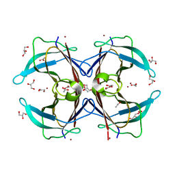
3RB7
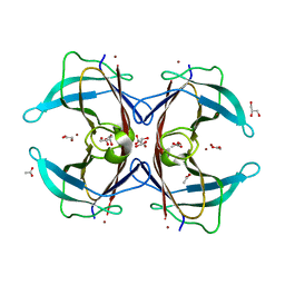
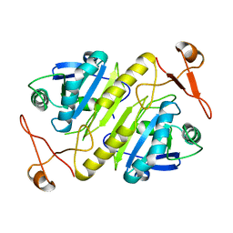
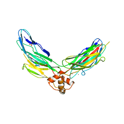 | | Crystal structure of CBD12 from CALX1.2 | | 分子名称: | CALCIUM ION, Na/Ca exchange protein, SULFATE ION | | 著者 | Wu, M, Zheng, L. | | 登録日 | 2011-03-28 | | 公開日 | 2011-11-02 | | 最終更新日 | 2024-02-21 | | 実験手法 | X-RAY DIFFRACTION (2.9 Å) | | 主引用文献 | Structural Basis of the Ca(2+) Inhibitory Mechanism of Drosophila Na(+)/Ca(2+) Exchanger CALX and Its Modification by Alternative Splicing.
Structure, 19, 2011
|
|
6VCK
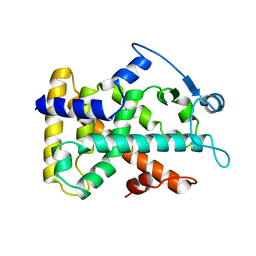
 | | Crystal structure of E.coli RppH-DapF in complex with GDP, Mg2+ and F- | | 分子名称: | CHLORIDE ION, Diaminopimelate epimerase, FLUORIDE ION, ... | | 著者 | Gao, A, Vasilyev, N, Kaushik, A, Duan, W, Serganov, A. | | 登録日 | 2019-12-21 | | 公開日 | 2020-02-05 | | 最終更新日 | 2023-10-11 | | 実験手法 | X-RAY DIFFRACTION (2.69 Å) | | 主引用文献 | Principles of RNA and nucleotide discrimination by the RNA processing enzyme RppH.
Nucleic Acids Res., 48, 2020
|
|
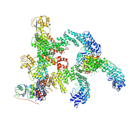
3IYD
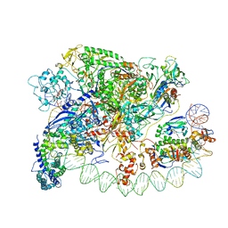 | | Three-dimensional EM structure of an intact activator-dependent transcription initiation complex | | 分子名称: | ADENOSINE-3',5'-CYCLIC-MONOPHOSPHATE, Catabolite gene activator, DNA (98-MER), ... | | 著者 | Hudson, B.P, Quispe, J, Lara, S, Kim, Y, Berman, H, Arnold, E, Ebright, R.H, Lawson, C.L. | | 登録日 | 2009-08-01 | | 公開日 | 2009-11-10 | | 最終更新日 | 2024-02-21 | | 実験手法 | ELECTRON MICROSCOPY (19.799999 Å) | | 主引用文献 | Three-dimensional EM structure of an intact activator-dependent transcription initiation complex
Proc.Natl.Acad.Sci.USA, 106, 2009
|
|
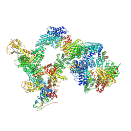
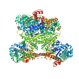
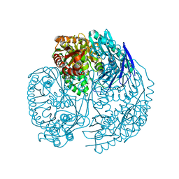
6WM4
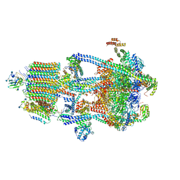 | | Human V-ATPase in state 3 with SidK and ADP | | 分子名称: | 2-acetamido-2-deoxy-beta-D-glucopyranose, ADENOSINE-5'-DIPHOSPHATE, Renin receptor, ... | | 著者 | Wang, L, Wu, H, Fu, T.M. | | 登録日 | 2020-04-20 | | 公開日 | 2020-11-11 | | 最終更新日 | 2024-11-06 | | 実験手法 | ELECTRON MICROSCOPY (3.6 Å) | | 主引用文献 | Structures of a Complete Human V-ATPase Reveal Mechanisms of Its Assembly.
Mol.Cell, 80, 2020
|
|
3GPS
 | | Crystal structure of the F87M/L110M mutant of human transthyretin at pH 5.5 | | 分子名称: | ACETATE ION, GLYCEROL, Transthyretin, ... | | 著者 | Palmieri, L.C, Freire, J.B.B, Foguel, D, Lima, L.M.T.R. | | 登録日 | 2009-03-23 | | 公開日 | 2010-04-07 | | 最終更新日 | 2023-09-06 | | 実験手法 | X-RAY DIFFRACTION (1.78 Å) | | 主引用文献 | Novel Zn2+-binding sites in human transthyretin: implications for amyloidogenesis and retinol-binding protein recognition.
J.Biol.Chem., 285, 2010
|
|
9J79
 | |
7V0K
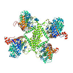
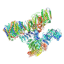 | | Consensus refinement of human erythrocyte ankyrin-1 complex (Composite map) | | 分子名称: | 2-acetamido-2-deoxy-beta-D-glucopyranose, Ammonium transporter Rh type A, Ankyrin-1, ... | | 著者 | Vallese, F, Kim, K, Yen, L.Y, Johnston, J.D, Noble, A.J, Cali, T, Clarke, O.B. | | 登録日 | 2022-05-10 | | 公開日 | 2022-07-20 | | 最終更新日 | 2024-10-16 | | 実験手法 | ELECTRON MICROSCOPY (2.4 Å) | | 主引用文献 | Architecture of the human erythrocyte ankyrin-1 complex.
Nat.Struct.Mol.Biol., 29, 2022
|
|
3ZJC
 | |
4G73
 | | Crystal structure of NDH with NADH and Quinone | | 分子名称: | 1,4-DIHYDRONICOTINAMIDE ADENINE DINUCLEOTIDE, 2,3-DIMETHOXY-5-METHYL-6-(3,11,15,19-TETRAMETHYL-EICOSA-2,6,10,14,18-PENTAENYL)-[1,4]BENZOQUINONE, FLAVIN-ADENINE DINUCLEOTIDE, ... | | 著者 | Li, W, Feng, Y, Ge, J, Yang, M. | | 登録日 | 2012-07-19 | | 公開日 | 2012-10-24 | | 最終更新日 | 2023-11-08 | | 実験手法 | X-RAY DIFFRACTION (2.522 Å) | | 主引用文献 | Structural insight into the type-II mitochondrial NADH dehydrogenases.
Nature, 491, 2012
|
|
9JCE
 | | local refinement of FEM1B bound with TOM20 | | 分子名称: | Mitochondrial import receptor subunit TOM20 homolog, Poly-UNK, Protein fem-1 homolog B | | 著者 | Zhao, S, Xu, C. | | 登録日 | 2024-08-29 | | 公開日 | 2025-04-09 | | 最終更新日 | 2025-05-07 | | 実験手法 | ELECTRON MICROSCOPY (3.59 Å) | | 主引用文献 | TOM20-driven E3 ligase recruitment regulates mitochondrial dynamics through PLD6.
Nat.Chem.Biol., 2025
|
|
9J7B
 | | local refinement of FEM1B bound with TOM20(tetramer) | | 分子名称: | Mitochondrial import receptor subunit TOM20 homolog, Poly-UNK, Protein fem-1 homolog B | | 著者 | Zhao, S, Xu, C. | | 登録日 | 2024-08-18 | | 公開日 | 2025-04-09 | | 最終更新日 | 2025-05-07 | | 実験手法 | ELECTRON MICROSCOPY (4.12 Å) | | 主引用文献 | TOM20-driven E3 ligase recruitment regulates mitochondrial dynamics through PLD6.
Nat.Chem.Biol., 2025
|
|
9J7A
 | | local refinement of FEM1B bound with TOM20 (dimer) | | 分子名称: | Mitochondrial import receptor subunit TOM20 homolog, Poly-UNK, Protein fem-1 homolog B | | 著者 | Zhao, S, Xu, C. | | 登録日 | 2024-08-18 | | 公開日 | 2025-04-09 | | 最終更新日 | 2025-05-07 | | 実験手法 | ELECTRON MICROSCOPY (4.13 Å) | | 主引用文献 | TOM20-driven E3 ligase recruitment regulates mitochondrial dynamics through PLD6.
Nat.Chem.Biol., 2025
|
|
9J77
 | | Cryo-EM structure of CRL2-FEM1B (dimer 1) | | 分子名称: | Cullin-2, E3 ubiquitin-protein ligase RBX1, N-terminally processed, ... | | 著者 | Zhao, S, Xu, C. | | 登録日 | 2024-08-18 | | 公開日 | 2025-04-09 | | 最終更新日 | 2025-05-07 | | 実験手法 | ELECTRON MICROSCOPY (3.56 Å) | | 主引用文献 | TOM20-driven E3 ligase recruitment regulates mitochondrial dynamics through PLD6.
Nat.Chem.Biol., 2025
|
|
9J78
 | | Cryo-EM structure of CRL2-FEM1B (dimer 2) | | 分子名称: | Cullin-2, E3 ubiquitin-protein ligase RBX1, N-terminally processed, ... | | 著者 | Zhao, S, Xu, C. | | 登録日 | 2024-08-18 | | 公開日 | 2025-04-09 | | 最終更新日 | 2025-05-07 | | 実験手法 | ELECTRON MICROSCOPY (3.88 Å) | | 主引用文献 | TOM20-driven E3 ligase recruitment regulates mitochondrial dynamics through PLD6.
Nat.Chem.Biol., 2025
|
|
1Z4U
 | | hepatitis C virus NS5B RNA-dependent RNA polymerase complex with inhibitor PHA-00799585 | | 分子名称: | (2Z)-2-[(1-ADAMANTYLCARBONYL)AMINO]-3-[4-(2-BROMOPHENOXY)PHENYL]PROP-2-ENOIC ACID, CHLORIDE ION, GLYCEROL, ... | | 著者 | Pfefferkorn, J.A, Greene, M, Nugent, R, Gross, R.J, Mitchell, M.A, Finzel, B.C, Harris, M.S, Wells, P.A, Shelly, J.A, Anstadt, R. | | 登録日 | 2005-03-16 | | 公開日 | 2005-06-07 | | 最終更新日 | 2024-04-03 | | 実験手法 | X-RAY DIFFRACTION (2.8 Å) | | 主引用文献 | Inhibitors of HCV NS5B polymerase. Part 2: Evaluation of the northern region of (2Z)-2-benzoylamino-3-(4-phenoxy-phenyl)-acrylic acid
Bioorg.Med.Chem.Lett., 15, 2005
|
|
3RSZ
 | |
3GRG
 | | Crystal structure of the F87M/L110M mutant of human transthyretin at pH 7.5 | | 分子名称: | ACETATE ION, GLYCEROL, Transthyretin, ... | | 著者 | Palmieri, L.C, Freire, J.B.B, Foguel, D, Lima, L.M.T.R. | | 登録日 | 2009-03-25 | | 公開日 | 2010-04-07 | | 最終更新日 | 2023-09-06 | | 実験手法 | X-RAY DIFFRACTION (1.9 Å) | | 主引用文献 | Novel Zn2+-binding sites in human transthyretin: implications for amyloidogenesis and retinol-binding protein recognition.
J.Biol.Chem., 285, 2010
|
|
2F3X
 | |
2VR8
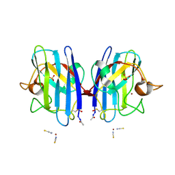 | | Crystal Structure of G85R ALS mutant of Human Cu,Zn Superoxide Dismutase (CuZnSOD) at 1.36 A resolution | | 分子名称: | COPPER (II) ION, SULFATE ION, SUPEROXIDE DISMUTASE [CU-ZN], ... | | 著者 | Antonyuk, S, Cao, X, Seetharaman, S.V, Whitson, L.J, Taylor, A.B, Holloway, S.P, Strange, R.W, Doucette, P.A, Tiwari, A, Hayward, L.J, Padua, S, Cohlberg, J.A, Selverstone Valentine, J, Hasnain, S.S, Hart, P.J. | | 登録日 | 2008-03-28 | | 公開日 | 2008-04-08 | | 最終更新日 | 2024-11-20 | | 実験手法 | X-RAY DIFFRACTION (1.36 Å) | | 主引用文献 | Structures of the G85R Variant of Sod1 in Familial Amyotrophic Lateral Sclerosis.
J.Biol.Chem., 283, 2008
|
|
1XHP
 | |
1PRX
 | | HORF6 A NOVEL HUMAN PEROXIDASE ENZYME | | 分子名称: | HORF6 | | 著者 | Choi, H.-J, Kang, S.W, Yang, C.-H, Rhee, S.G, Ryu, S.-E. | | 登録日 | 1998-04-03 | | 公開日 | 1998-06-17 | | 最終更新日 | 2024-10-30 | | 実験手法 | X-RAY DIFFRACTION (2 Å) | | 主引用文献 | Crystal structure of a novel human peroxidase enzyme at 2.0 A resolution.
Nat.Struct.Biol., 5, 1998
|
|
2GWX
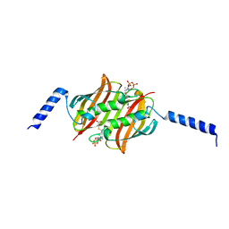
 | | MOLECULAR RECOGNITION OF FATTY ACIDS BY PEROXISOME PROLIFERATOR-ACTIVATED RECEPTORS | | 分子名称: | PROTEIN (PPAR-DELTA) | | 著者 | Xu, H.E, Lambert, M.H, Montana, V.G, Park, D.J, Blanchard, S, Brown, P, Sternbach, D, Lehmann, J, Bruce, G.W, Willson, T.M, Kliewer, S.A, Milburn, M.V. | | 登録日 | 1999-03-11 | | 公開日 | 2000-03-11 | | 最終更新日 | 2023-12-27 | | 実験手法 | X-RAY DIFFRACTION (2.3 Å) | | 主引用文献 | Molecular recognition of fatty acids by peroxisome proliferator-activated receptors.
Mol.Cell, 3, 1999
|
|
3U1K
 | | Crystal structure of human PNPase | | 分子名称: | CITRIC ACID, Polyribonucleotide nucleotidyltransferase 1, mitochondrial | | 著者 | Lin, C.L, Yuan, H.S. | | 登録日 | 2011-09-30 | | 公開日 | 2012-02-01 | | 最終更新日 | 2023-11-01 | | 実験手法 | X-RAY DIFFRACTION (2.13 Å) | | 主引用文献 | Crystal structure of human polynucleotide phosphorylase: insights into its domain function in RNA binding and degradation
Nucleic Acids Res., 40, 2012
|
|
3RT1
 | |


