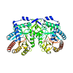
3WY2
 
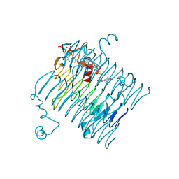
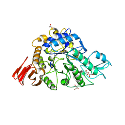 | | Crystal structure of alpha-glucosidase in complex with glucose | | 分子名称: | Alpha-glucosidase, GLYCEROL, MAGNESIUM ION, ... | | 著者 | Shen, X, Gai, Z, Kato, K, Yao, M. | | 登録日 | 2014-08-18 | | 公開日 | 2015-06-10 | | 最終更新日 | 2020-07-29 | | 実験手法 | X-RAY DIFFRACTION (1.471 Å) | | 主引用文献 | Structural analysis of the alpha-glucosidase HaG provides new insights into substrate specificity and catalytic mechanism
Acta Crystallogr. D Biol. Crystallogr., 71, 2015
|
|
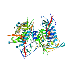
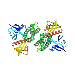
4LUE
 | | Crystal Structure of HCK in complex with 7-[trans-4-(4-methylpiperazin-1-yl)cyclohexyl]-5-(4-phenoxyphenyl)-7H-pyrrolo[2,3-d]pyrimidin-4-amine (resulting from displacement of SKF86002) | | 分子名称: | 7-[trans-4-(4-methylpiperazin-1-yl)cyclohexyl]-5-(4-phenoxyphenyl)-7H-pyrrolo[2,3-d]pyrimidin-4-amine, CALCIUM ION, CHLORIDE ION, ... | | 著者 | Parker, L.J, Tanaka, A, Handa, N, Honda, K, Tomabechi, Y, Shirouzu, M, Yokoyama, S. | | 登録日 | 2013-07-25 | | 公開日 | 2014-02-12 | | 最終更新日 | 2023-12-06 | | 実験手法 | X-RAY DIFFRACTION (3.04 Å) | | 主引用文献 | Kinase crystal identification and ATP-competitive inhibitor screening using the fluorescent ligand SKF86002.
Acta Crystallogr.,Sect.D, 70, 2014
|
|
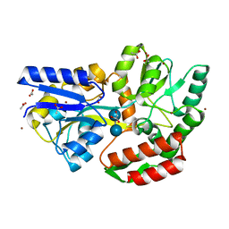
3TGS
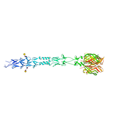
 | | Crystal structure of HIV-1 clade C strain C1086 gp120 core in complex with NBD-556 | | 分子名称: | 2-acetamido-2-deoxy-beta-D-glucopyranose, HIV-1 clade C1086 gp120 core, N-(4-chlorophenyl)-N'-(2,2,6,6-tetramethylpiperidin-4-yl)ethanediamide | | 著者 | Kwon, Y.D, Kwong, P.D. | | 登録日 | 2011-08-17 | | 公開日 | 2012-04-04 | | 最終更新日 | 2020-07-29 | | 実験手法 | X-RAY DIFFRACTION (2.7 Å) | | 主引用文献 | Unliganded HIV-1 gp120 core structures assume the CD4-bound conformation with regulation by quaternary interactions and variable loops.
Proc.Natl.Acad.Sci.USA, 109, 2012
|
|
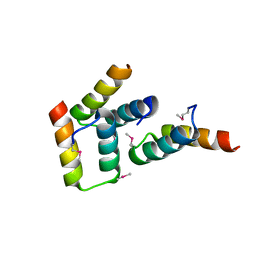
1CBV
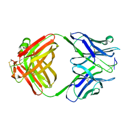 | | AN AUTOANTIBODY TO SINGLE-STRANDED DNA: COMPARISON OF THE THREE-DIMENSIONAL STRUCTURES OF THE UNLIGANDED FAB AND A DEOXYNUCLEOTIDE-FAB COMPLEX | | 分子名称: | DNA (5'-D(*TP*TP*T)-3'), PROTEIN (FAB (BV04-01) AUTOANTIBODY-HEAVY CHAIN), PROTEIN (FAB (BV04-01) AUTOANTIBODY-LIGHT CHAIN) | | 著者 | Herron, J.N, He, X.M, Ballard, D.W, Blier, P.R, Pace, P.E, Bothwell, A.L.M, Voss Junior, E.W, Edmundson, A.B. | | 登録日 | 1993-03-16 | | 公開日 | 1994-01-31 | | 最終更新日 | 2017-11-29 | | 実験手法 | X-RAY DIFFRACTION (2.66 Å) | | 主引用文献 | An autoantibody to single-stranded DNA: comparison of the three-dimensional structures of the unliganded Fab and a deoxynucleotide-Fab complex.
Proteins, 11, 1991
|
|
4D11
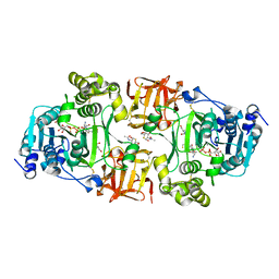 | | GalNAc-T2 crystal soaked with UDP-5SGalNAc, mEA2 peptide and manganese (Lower resolution dataset) | | 分子名称: | 2-acetamido-2-deoxy-5-thio-alpha-D-galactopyranose, MANGANESE (II) ION, PEPTIDE, ... | | 著者 | Lira-Navarrete, E, Iglesias-Fernandez, J, Zandberg, W.F, Companon, I, Kong, Y, Corzana, F, Pinto, B.M, Clausen, H, Peregrina, J.M, Vocadlo, D, Rovira, C, Hurtado-Guerrero, R. | | 登録日 | 2014-05-01 | | 公開日 | 2014-05-28 | | 最終更新日 | 2023-12-20 | | 実験手法 | X-RAY DIFFRACTION (2.85 Å) | | 主引用文献 | Substrate-Guided Front-Face Reaction Revealed by Combined Structural Snapshots and Metadynamics for the Polypeptide N- Acetylgalactosaminyltransferase 2.
Angew.Chem.Int.Ed.Engl., 53, 2014
|
|
1L3M
 | | The Solution Structure of [d(CGC)r(amamam)d(TTTGCG)]2 | | 分子名称: | 5'-D(*CP*GP*C)-R(P*(A39)P*(A39)P*(A39))-D(P*TP*TP*TP*GP*CP*G)-3' | | 著者 | Tsao, Y.P, Wang, L.Y, Hsu, S.T, Jain, M.L, Chou, S.H, Huang, W.C, Cheng, J.W. | | 登録日 | 2002-02-28 | | 公開日 | 2002-04-03 | | 最終更新日 | 2024-05-01 | | 実験手法 | SOLUTION NMR | | 主引用文献 | The solution structure of [d(CGC)r(amamam)d(TTTGCG)]2.
J.Biomol.NMR, 21, 2001
|
|
1T0W
 | | 25 NMR structures of Truncated Hevein of 32 aa (Hevein-32) complex with N,N,N-triacetylglucosamina | | 分子名称: | 2-acetamido-2-deoxy-beta-D-glucopyranose-(1-4)-2-acetamido-2-deoxy-beta-D-glucopyranose-(1-4)-2-acetamido-2-deoxy-beta-D-glucopyranose, Hevein | | 著者 | Aboitiz, N, Vila-Perello, M, Groves, P, Asensio, J.L, Andreu, D, Canada, F.J, Jimenez-Barbero, J. | | 登録日 | 2004-04-13 | | 公開日 | 2004-09-28 | | 最終更新日 | 2020-07-29 | | 実験手法 | SOLUTION NMR | | 主引用文献 | NMR and modeling studies of protein-carbohydrate interactions: synthesis, three-dimensional structure, and recognition properties of a minimum hevein domain with binding affinity for chitooligosaccharides
Chembiochem, 5, 2004
|
|
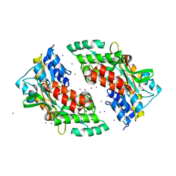
1GYN
 | | Class II fructose 1,6-bisphosphate aldolase with Cadmium (not Zinc) in the active site | | 分子名称: | CADMIUM ION, FRUCTOSE-BISPHOSPHATE ALDOLASE II | | 著者 | Hall, D.R, Kemp, L.E, Leonard, G.A, Berry, A, Hunter, W.N. | | 登録日 | 2002-04-27 | | 公開日 | 2003-02-26 | | 最終更新日 | 2023-12-13 | | 実験手法 | X-RAY DIFFRACTION (2 Å) | | 主引用文献 | The Organization of Divalent Cations in the Active Site of Cadmium Escherichia Coli Fructose 1,6-Bisphosphate Aldolase
Acta Crystallogr.,Sect.D, 59, 2003
|
|
3R0X
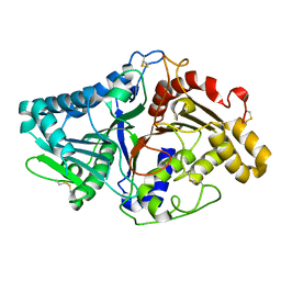
 | | Crystal structure of Selenomethionine incorporated apo D-serine deaminase from Salmonella tyhimurium | | 分子名称: | 1,2-ETHANEDIOL, D-serine dehydratase, SODIUM ION, ... | | 著者 | Bharath, S.R, Shveta, B, Savithri, H.S, Murthy, M.R.N. | | 登録日 | 2011-03-09 | | 公開日 | 2011-06-29 | | 最終更新日 | 2011-07-13 | | 実験手法 | X-RAY DIFFRACTION (1.93 Å) | | 主引用文献 | Crystal structures of open and closed forms of D-serine deaminase from Salmonella typhimurium - implications on substrate specificity and catalysis
Febs J., 2011
|
|
1TBF
 | | Catalytic Domain Of Human Phosphodiesterase 5A in Complex with Sildenafil | | 分子名称: | 5-{2-ETHOXY-5-[(4-METHYLPIPERAZIN-1-YL)SULFONYL]PHENYL}-1-METHYL-3-PROPYL-1H,6H,7H-PYRAZOLO[4,3-D]PYRIMIDIN-7-ONE, GLYCEROL, MAGNESIUM ION, ... | | 著者 | Zhang, K.Y.J, Card, G.L, Suzuki, Y, Artis, D.R, Fong, D, Gillette, S, Hsieh, D, Neiman, J, West, B.L, Zhang, C, Milburn, M.V, Kim, S.-H, Schlessinger, J, Bollag, G. | | 登録日 | 2004-05-20 | | 公開日 | 2004-08-03 | | 最終更新日 | 2023-08-23 | | 実験手法 | X-RAY DIFFRACTION (1.3 Å) | | 主引用文献 | A Glutamine Switch Mechanism for Nucleotide Selectivity by Phosphodiesterases
Mol.Cell, 15, 2004
|
|
4JGF
 | |
1QVY
 | | Crystal structure of RhoGDI K(199,200)R double mutant | | 分子名称: | Rho GDP-dissociation inhibitor 1, SULFATE ION | | 著者 | Czepas, J, Devedjiev, Y, Krowarsh, D, Derewenda, U, Derewenda, Z.S. | | 登録日 | 2003-08-29 | | 公開日 | 2004-02-10 | | 最終更新日 | 2023-08-16 | | 実験手法 | X-RAY DIFFRACTION (1.6 Å) | | 主引用文献 | The impact of Lys-->Arg surface mutations on the crystallization of the globular domain of RhoGDI.
Acta Crystallogr.,Sect.D, 60, 2004
|
|
1NOP
 | | Crystal structure of human tyrosyl-DNA phosphodiesterase (Tdp1) in complex with vanadate, DNA and a human topoisomerase I-derived peptide | | 分子名称: | 5'-D(*AP*GP*AP*GP*TP*T)-3', VANADATE ION, topoisomerase I-derived peptide, ... | | 著者 | Davies, D.R, Interthal, H, Champoux, J.J, Hol, W.G.J. | | 登録日 | 2003-01-16 | | 公開日 | 2003-03-11 | | 最終更新日 | 2024-02-14 | | 実験手法 | X-RAY DIFFRACTION (2.3 Å) | | 主引用文献 | Crystal structure of a transition state mimic for Tdp1 assembled from vanadate, DNA, and a topoisomerase I-derived peptide
Chem.Biol., 10, 2003
|
|
3S6Z
 | |
1V0S
 | |
1NR4
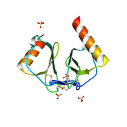 | | High resolution crystal structures of thymus and activation-regulated chemokine | | 分子名称: | SULFATE ION, Thymus and activation-regulated chemokine | | 著者 | Asojo, O.A, Boulegue, C, Hoover, D.M, Lu, W, Lubkowski, J. | | 登録日 | 2003-01-23 | | 公開日 | 2003-08-05 | | 最終更新日 | 2024-04-03 | | 実験手法 | X-RAY DIFFRACTION (1.72 Å) | | 主引用文献 | Structures of thymus and activation-regulated chemokine (TARC).
Acta Crystallogr.,Sect.D, 59, 2003
|
|
1V0W
 | |
3FSB
 | | Crystal structure of QdtC, the dTDP-3-amino-3,6-dideoxy-D-glucose N-acetyl transferase from Thermoanaerobacterium thermosaccharolyticum in complex with CoA and dTDP-3-amino-quinovose | | 分子名称: | COENZYME A, QdtC, [(3R,4S,5S,6R)-4-amino-3,5-dihydroxy-6-methyloxan-2-yl][hydroxy-[[(2R,3S,5R)-3-hydroxy-5-(5-methyl-2,4-dioxopyrimidin-1-yl)oxolan-2-yl]methoxy]phosphoryl] hydrogen phosphate | | 著者 | Holden, H.M, Thoden, J.B. | | 登録日 | 2009-01-09 | | 公開日 | 2009-02-17 | | 最終更新日 | 2023-09-06 | | 実験手法 | X-RAY DIFFRACTION (1.95 Å) | | 主引用文献 | Structural and Functional Studies of QdtC: an N-Acetyltransferase Required for the Biosynthesis of dTDP-3-Acetamido-3,6-Dideoxy-alpha-D-Glucose.
Biochemistry, 48, 2009
|
|
1OAB
 | | CRYSTAL STRUCTURE OF THE TYROSINE REGULATED 3-DEOXY-D-ARABINO-HEPTULOSONATE-7-PHOSPHATE SYNTHASE FROM SACCHAROMYCES CEREVISIAE IN COMPLEX WITH PHOSPHOENOLPYRUVATE AND MANGANESE(II) | | 分子名称: | MANGANESE (II) ION, PHOSPHOENOLPYRUVATE, TYROSINE-REGULATED 3-DEOXY-D-ARABINO-HEPTULOSONATE-7-PHOSPHATE SYNTHASE | | 著者 | Koenig, V, Pfeil, A, Heinrich, G, Braus, G.H, Schneider, T.R. | | 登録日 | 2003-01-06 | | 公開日 | 2004-01-23 | | 最終更新日 | 2023-12-13 | | 実験手法 | X-RAY DIFFRACTION (1.9 Å) | | 主引用文献 | Substrate and Metal Complexes of 3-Deoxy-D-Arabino-Heptulosonate-7-Phosphate Synthase from Saccharomyces Cerevisiae Provide New Insights Into the Catalytic Mechanism
J.Mol.Biol., 337, 2004
|
|
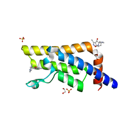
1H4R
 | | Crystal Structure of the FERM domain of Merlin, the Neurofibromatosis 2 Tumor Suppressor Protein. | | 分子名称: | MERLIN, SULFATE ION | | 著者 | Cooper, D.R, Kang, B.S, Sheffield, P, Devedjiev, Y, Derewenda, Z.S. | | 登録日 | 2001-05-14 | | 公開日 | 2002-01-16 | | 最終更新日 | 2023-12-13 | | 実験手法 | X-RAY DIFFRACTION (1.8 Å) | | 主引用文献 | The Structure of the Ferm Domain of Merlin, the Neurofibromatosis Type 2 Gene Product.
Acta Crystallogr.,Sect.D, 58, 2002
|
|
3SEU
 | | Zn-mediated Polymer of Maltose-binding Protein A216H/K220H by Synthetic Symmetrization (Form III) | | 分子名称: | ACETATE ION, GLYCEROL, Maltose-binding periplasmic protein, ... | | 著者 | Zhao, M, Soriaga, A.B, Laganowsky, A, Sawaya, M.R, Cascio, D, Yeates, T.O. | | 登録日 | 2011-06-11 | | 公開日 | 2011-09-21 | | 最終更新日 | 2024-02-28 | | 実験手法 | X-RAY DIFFRACTION (1.85 Å) | | 主引用文献 | An approach to crystallizing proteins by metal-mediated synthetic symmetrization.
Protein Sci., 20, 2011
|
|
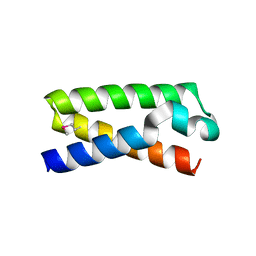
3FYR
 | | Crystal structure of the sporulation histidine kinase inhibitor Sda from Bacillus subtilis | | 分子名称: | Sporulation inhibitor sda | | 著者 | Jacques, D.A, Streamer, M, King, G.F, Guss, J.M, Trewhella, J, Langley, D.B. | | 登録日 | 2009-01-23 | | 公開日 | 2009-06-23 | | 最終更新日 | 2017-11-01 | | 実験手法 | X-RAY DIFFRACTION (1.97 Å) | | 主引用文献 | Structure of the sporulation histidine kinase inhibitor Sda from Bacillus subtilis and insights into its solution state
Acta Crystallogr.,Sect.D, 65, 2009
|
|
3SEX
 | | Ni-mediated Dimer of Maltose-binding Protein A216H/K220H by Synthetic Symmetrization (Form II) | | 分子名称: | IODIDE ION, Maltose-binding periplasmic protein, NICKEL (II) ION, ... | | 著者 | Zhao, M, Soriaga, A.B, Laganowsky, A, Sawaya, M.R, Cascio, D, Yeates, T.O. | | 登録日 | 2011-06-11 | | 公開日 | 2011-09-21 | | 最終更新日 | 2024-02-28 | | 実験手法 | X-RAY DIFFRACTION (1.95 Å) | | 主引用文献 | An approach to crystallizing proteins by metal-mediated synthetic symmetrization.
Protein Sci., 20, 2011
|
|
3T48
 | |
5QXI
 | | PanDDA analysis group deposition -- Crystal Structure of ATAD2 in complex with PC587 | | 分子名称: | (4R,4aR,7aS,9R)-3,10-dimethyl-5,6,7,7a,8,9-hexahydro-4H-4a,9-epiminopyrrolo[3',4':5,6]cyclohepta[1,2-d][1,2]oxazol-4-ol, (4R,4aS,7aS,9S)-3,10-dimethyl-5,6,7,7a,8,9-hexahydro-4H-4a,9-epiminopyrrolo[3',4':5,6]cyclohepta[1,2-d][1,2]oxazol-4-ol, 1,2-ETHANEDIOL, ... | | 著者 | Snee, M, Talon, R, Fowley, D, Collins, P, Nelson, A, Arrowsmith, C.H, Bountra, C, Edwards, A, Von-Delft, F. | | 登録日 | 2020-02-11 | | 公開日 | 2020-04-08 | | 最終更新日 | 2024-03-06 | | 実験手法 | X-RAY DIFFRACTION (1.64 Å) | | 主引用文献 | PanDDA analysis group deposition - Bromodomain of human ATAD2 fragment screening
To Be Published
|
|


