4MIX
 
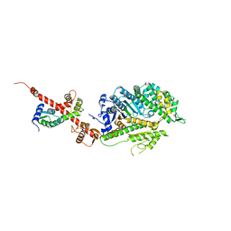
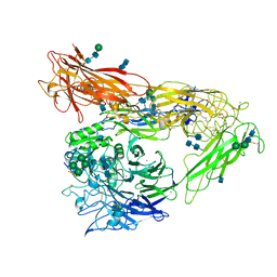
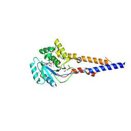 | | PaToxG Glycosyltransferase | | 分子名称: | CALCIUM ION, Putative insecticidal toxin, URIDINE-DIPHOSPHATE-N-ACETYLGLUCOSAMINE | | 著者 | Bogdanovic, X, Wirth, C, Hunte, C. | | 登録日 | 2013-09-02 | | 公開日 | 2013-10-16 | | 最終更新日 | 2017-11-15 | | 実験手法 | X-RAY DIFFRACTION (1.8 Å) | | 主引用文献 | A bacterial toxin catalyzing tyrosine glycosylation of Rho and deamidation of Gq and Gi proteins.
Nat.Struct.Mol.Biol., 20, 2013
|
|
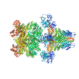
3J99
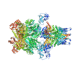 | | Structure of 20S supercomplex determined by single particle cryoelectron microscopy (State IIIb) | | 分子名称: | Alpha-soluble NSF attachment protein, Synaptosomal-associated protein 25, Syntaxin-1A, ... | | 著者 | Zhao, M, Wu, S, Cheng, Y, Brunger, A.T. | | 登録日 | 2014-12-05 | | 公開日 | 2015-01-28 | | 最終更新日 | 2024-02-21 | | 実験手法 | ELECTRON MICROSCOPY (8.2 Å) | | 主引用文献 | Mechanistic insights into the recycling machine of the SNARE complex.
Nature, 518, 2015
|
|
3J92
 | | Structure and assembly pathway of the ribosome quality control complex | | 分子名称: | 28S rRNA, 5.8S rRNA, 5S rRNA, ... | | 著者 | Shao, S, Brown, A, Santhanam, B, Hegde, R.S. | | 登録日 | 2014-12-02 | | 公開日 | 2015-01-21 | | 最終更新日 | 2018-07-18 | | 実験手法 | ELECTRON MICROSCOPY (3.6 Å) | | 主引用文献 | Structure and Assembly Pathway of the Ribosome Quality Control Complex.
Mol.Cell, 57, 2015
|
|
6ZQF
 | | Cryo-EM structure of the 90S pre-ribosome from Saccharomyces cerevisiae, state Dis-B (Poly-Ala) | | 分子名称: | 18S rRNA, 40S ribosomal protein S1-A, 40S ribosomal protein S11-A, ... | | 著者 | Cheng, J, Lau, B, Venuta, G.L, Berninghausen, O, Hurt, E, Beckmann, R. | | 登録日 | 2020-07-09 | | 公開日 | 2020-09-23 | | 最終更新日 | 2024-05-01 | | 実験手法 | ELECTRON MICROSCOPY (4.9 Å) | | 主引用文献 | 90 S pre-ribosome transformation into the primordial 40 S subunit.
Science, 369, 2020
|
|
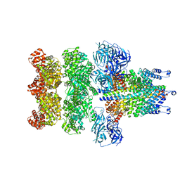
3J96
 | | Structure of 20S supercomplex determined by single particle cryoelectron microscopy (State I) | | 分子名称: | Alpha-soluble NSF attachment protein, Synaptosomal-associated protein 25, Syntaxin-1A, ... | | 著者 | Zhao, M, Wu, S, Cheng, Y, Brunger, A.T. | | 登録日 | 2014-12-05 | | 公開日 | 2015-01-28 | | 最終更新日 | 2024-02-21 | | 実験手法 | ELECTRON MICROSCOPY (7.6 Å) | | 主引用文献 | Mechanistic insights into the recycling machine of the SNARE complex.
Nature, 518, 2015
|
|
6RA9
 | | Novel structural features and post-translational modifications in eukaryotic elongation factor 1A2 from Oryctolagus cuniculus | | 分子名称: | Elongation factor 1-alpha 2, GLYCEROL, GUANOSINE-5'-DIPHOSPHATE, ... | | 著者 | Carriles, A.A, Hermoso, J, Gago, F. | | 登録日 | 2019-04-05 | | 公開日 | 2020-06-03 | | 最終更新日 | 2024-01-24 | | 実験手法 | X-RAY DIFFRACTION (2.7 Å) | | 主引用文献 | Structural Cues for Understanding eEF1A2 Moonlighting.
Chembiochem, 22, 2021
|
|
3J98
 | | Structure of 20S supercomplex determined by single particle cryoelectron microscopy (State IIIa) | | 分子名称: | Alpha-soluble NSF attachment protein, Synaptosomal-associated protein 25, Syntaxin-1A, ... | | 著者 | Zhao, M, Wu, S, Cheng, Y, Brunger, A.T. | | 登録日 | 2014-12-05 | | 公開日 | 2015-01-28 | | 最終更新日 | 2024-02-21 | | 実験手法 | ELECTRON MICROSCOPY (8.4 Å) | | 主引用文献 | Mechanistic insights into the recycling machine of the SNARE complex.
Nature, 518, 2015
|
|
4L79
 | | Crystal Structure of nucleotide-free Myosin 1b residues 1-728 with bound Calmodulin | | 分子名称: | Calmodulin, MAGNESIUM ION, Unconventional myosin-Ib | | 著者 | Shuman, H, Zwolak, A, Dominguez, R, Ostap, E.M. | | 登録日 | 2013-06-13 | | 公開日 | 2014-01-29 | | 最終更新日 | 2023-09-20 | | 実験手法 | X-RAY DIFFRACTION (2.3 Å) | | 主引用文献 | A vertebrate myosin-I structure reveals unique insights into myosin mechanochemical tuning.
Proc.Natl.Acad.Sci.USA, 111, 2014
|
|
4MMY
 | | Integrin AlphaVBeta3 ectodomain bound to the tenth domain of Fibronectin with the IAKGDWND motif | | 分子名称: | 2-acetamido-2-deoxy-beta-D-glucopyranose, 2-acetamido-2-deoxy-beta-D-glucopyranose-(1-4)-2-acetamido-2-deoxy-beta-D-glucopyranose, Fibronectin, ... | | 著者 | van Agthoven, J, Xiong, J, Arnaout, M.A. | | 登録日 | 2013-09-09 | | 公開日 | 2014-03-26 | | 最終更新日 | 2020-07-29 | | 実験手法 | X-RAY DIFFRACTION (3.183 Å) | | 主引用文献 | Structural basis for pure antagonism of integrin alpha V beta 3 by a high-affinity form of fibronectin.
Nat.Struct.Mol.Biol., 21, 2014
|
|
4MY6
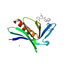 | | EnaH-EVH1 in complex with peptidomimetic low-molecular weight inhibitor Ac-[2-Cl-F]-[ProM-2]-[ProM-1]-OH | | 分子名称: | (3aR,5aS,8S,10aS)-1-[(3S,6R,8aS)-1'-[(2S)-2-acetamido-3-(2-chlorophenyl)propanoyl]-5-oxidanylidene-spiro[1,2,3,8a-tetrahydroindolizine-6,2'-pyrrolidine]-3-yl]carbonyl-10-oxidanylidene-2,3,3a,5a,8,10a-hexahydrodipyrrolo[3,2-b:3',1'-f]azepine-8-carboxylic acid, BROMIDE ION, Protein enabled homolog | | 著者 | Barone, M, Roske, Y, Kuehne, R. | | 登録日 | 2013-09-27 | | 公開日 | 2014-10-15 | | 最終更新日 | 2023-09-20 | | 実験手法 | X-RAY DIFFRACTION (1.7 Å) | | 主引用文献 | A modular toolkit to inhibit proline-rich motif-mediated protein-protein interactions.
Proc.Natl.Acad.Sci.USA, 112, 2015
|
|
3K1R
 | |
4JZ4
 | | Crystal structure of chicken c-Src-SH3 domain: monomeric form | | 分子名称: | 4-(2-HYDROXYETHYL)-1-PIPERAZINE ETHANESULFONIC ACID, NICKEL (II) ION, Proto-oncogene tyrosine-protein kinase Src | | 著者 | Camara-Artigas, A. | | 登録日 | 2013-04-02 | | 公開日 | 2014-04-23 | | 最終更新日 | 2023-09-20 | | 実験手法 | X-RAY DIFFRACTION (1.56 Å) | | 主引用文献 | Electrostatic Effects in the Folding of the SH3 Domain of the c-Src Tyrosine Kinase: pH-Dependence in 3D-Domain Swapping and Amyloid Formation.
Plos One, 9, 2014
|
|
4JZ3
 | |
6THA
 | | Crystal structure of human sugar transporter GLUT1 (SLC2A1) in the inward conformation | | 分子名称: | 3,6,9,12,15,18-HEXAOXAICOSANE-1,20-DIOL, CHLORIDE ION, Solute carrier family 2, ... | | 著者 | Pedersen, B.P, Paulsen, P.A, Custodio, T.F. | | 登録日 | 2019-11-19 | | 公開日 | 2020-11-25 | | 最終更新日 | 2024-01-24 | | 実験手法 | X-RAY DIFFRACTION (2.4 Å) | | 主引用文献 | Structural comparison of GLUT1 to GLUT3 reveal transport regulation mechanism in sugar porter family.
Life Sci Alliance, 4, 2021
|
|
5V6E
 | |
5V6T
 | |
3JV3
 | |
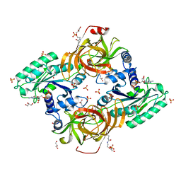
3KO0
 | | Structure of the tfp-ca2+-bound activated form of the s100a4 Metastasis factor | | 分子名称: | 10-[3-(4-METHYL-PIPERAZIN-1-YL)-PROPYL]-2-TRIFLUOROMETHYL-10H-PHENOTHIAZINE, CALCIUM ION, Protein S100-A4 | | 著者 | Malashkevich, V.N, Dulyaninova, N.G, Knight, D, Almo, S.C, Bresnick, A.R. | | 登録日 | 2009-11-12 | | 公開日 | 2010-05-26 | | 最終更新日 | 2024-02-21 | | 実験手法 | X-RAY DIFFRACTION (2.3 Å) | | 主引用文献 | Phenothiazines inhibit S100A4 function by inducing protein oligomerization.
Proc.Natl.Acad.Sci.USA, 107, 2010
|
|
4MMZ
 | | Integrin AlphaVBeta3 ectodomain bound to an antagonistic tenth domain of Fibronectin | | 分子名称: | 2-acetamido-2-deoxy-beta-D-glucopyranose, 2-acetamido-2-deoxy-beta-D-glucopyranose-(1-4)-2-acetamido-2-deoxy-beta-D-glucopyranose, Fibronectin, ... | | 著者 | van Agthoven, J, Xiong, J, Arnaout, M.A. | | 登録日 | 2013-09-09 | | 公開日 | 2014-03-26 | | 最終更新日 | 2023-09-20 | | 実験手法 | X-RAY DIFFRACTION (3.1 Å) | | 主引用文献 | Structural basis for pure antagonism of integrin alpha V beta 3 by a high-affinity form of fibronectin.
Nat.Struct.Mol.Biol., 21, 2014
|
|
5V6B
 | | Crystal structure of GIPC1 | | 分子名称: | PDZ domain-containing protein GIPC1 | | 著者 | Shang, G, Zhang, X. | | 登録日 | 2017-03-16 | | 公開日 | 2017-05-31 | | 最終更新日 | 2023-10-04 | | 実験手法 | X-RAY DIFFRACTION (1.9 Å) | | 主引用文献 | Structure analyses reveal a regulated oligomerization mechanism of the PlexinD1/GIPC/myosin VI complex.
Elife, 6, 2017
|
|
4ZNX
 | |
5X5P
 | | Human serum transferrin bound to ruthenium NTA | | 分子名称: | FE (III) ION, MALONATE ION, NITRILOTRIACETIC ACID, ... | | 著者 | Sun, H, Wang, M. | | 登録日 | 2017-02-17 | | 公開日 | 2018-02-21 | | 最終更新日 | 2023-11-22 | | 実験手法 | X-RAY DIFFRACTION (2.7 Å) | | 主引用文献 | Binding of ruthenium and osmium at non‐iron sites of transferrin accounts for their iron-independent cellular uptake.
J.Inorg.Biochem., 234, 2022
|
|
6QJA
 | | Organizational principles of the NuMA-Dynein interaction interface and implications for mitotic spindle functions | | 分子名称: | CHLORIDE ION, MAGNESIUM ION, Nuclear mitotic apparatus protein 1 | | 著者 | Renna, C, Rizzelli, F, Carminati, M, Gaddoni, C, Pirovano, L, Cecatiello, V, Pasqualato, S, Mapelli, M. | | 登録日 | 2019-01-23 | | 公開日 | 2020-02-05 | | 最終更新日 | 2024-05-15 | | 実験手法 | X-RAY DIFFRACTION (1.54 Å) | | 主引用文献 | Organizational Principles of the NuMA-Dynein Interaction Interface and Implications for Mitotic Spindle Functions.
Structure, 28, 2020
|
|
5XA5
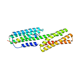 | | Crystal structure of HMP-1-HMP-2 complex | | 分子名称: | Alpha-catenin-like protein hmp-1, Beta-catenin-like protein hmp-2 | | 著者 | Shao, X, Kang, H, Weis, W.I, Hardin, J, Choi, H.J. | | 登録日 | 2017-03-11 | | 公開日 | 2017-08-30 | | 最終更新日 | 2024-03-27 | | 実験手法 | X-RAY DIFFRACTION (1.6 Å) | | 主引用文献 | Cell-cell adhesion in metazoans relies on evolutionarily conserved features of the alpha-catenin· beta-catenin-binding interface.
J.Biol.Chem., 292, 2017
|
|
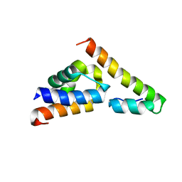
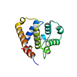
2MJF
 | | Solution structure of the complex between the yeast Rsa1 and Hit1 proteins | | 分子名称: | Protein HIT1, Ribosome assembly 1 protein | | 著者 | Quinternet, M, Roth, B, Back, R, Jacquemin, C, Manival, X. | | 登録日 | 2014-01-08 | | 公開日 | 2014-09-10 | | 最終更新日 | 2024-05-01 | | 実験手法 | SOLUTION NMR | | 主引用文献 | Protein Hit1, a novel box C/D snoRNP assembly factor, controls cellular concentration of the scaffolding protein Rsa1 by direct interaction.
Nucleic Acids Res., 42, 2014
|
|


