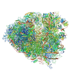
9Q7Q
 
 | | ABCE1-eRF1-RNC-AMD1C | | 分子名称: | 18S rRNA, 28S rRNA, 40S ribosomal protein S12, ... | | 著者 | Maldosevic, E, Jomaa, A. | | 登録日 | 2025-08-25 | | 公開日 | 2026-03-04 | | 最終更新日 | 2026-04-08 | | 実験手法 | ELECTRON MICROSCOPY (2.86 Å) | | 主引用文献 | Mechanism of ribosome stalling by the AMD1 C-terminal tail arrest peptide.
Sci Adv, 12, 2026
|
|
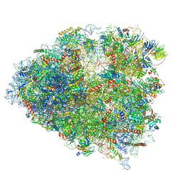
4V6X
 | | Structure of the human 80S ribosome | | 分子名称: | 18S ribosomal RNA, 28S ribosomal RNA, 40S ribosomal protein S10, ... | | 著者 | Anger, A.M, Armache, J.-P, Berninghausen, O, Habeck, M, Subklewe, M, Wilson, D.N, Beckmann, R. | | 登録日 | 2013-02-27 | | 公開日 | 2014-07-09 | | 最終更新日 | 2024-11-06 | | 実験手法 | ELECTRON MICROSCOPY (5 Å) | | 主引用文献 | Structures of the human and Drosophila 80S ribosome.
Nature, 497, 2013
|
|
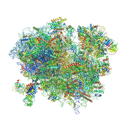
7JQC
 | | SARS-CoV-2 Nsp1, CrPV IRES and rabbit 40S ribosome complex | | 分子名称: | 40S ribosomal protein S21, 40S ribosomal protein S24, 40S ribosomal protein S26, ... | | 著者 | Yuan, S, Xiong, Y. | | 登録日 | 2020-08-10 | | 公開日 | 2020-12-02 | | 最終更新日 | 2025-05-21 | | 実験手法 | ELECTRON MICROSCOPY (3.3 Å) | | 主引用文献 | Nonstructural Protein 1 of SARS-CoV-2 Is a Potent Pathogenicity Factor Redirecting Host Protein Synthesis Machinery toward Viral RNA.
Mol.Cell, 80, 2020
|
|
8VFT
 | |
7JQB
 | | SARS-CoV-2 Nsp1 and rabbit 40S ribosome complex | | 分子名称: | 40S ribosomal protein S21, 40S ribosomal protein S24, 40S ribosomal protein S26, ... | | 著者 | Yuan, S, Xiong, Y. | | 登録日 | 2020-08-10 | | 公開日 | 2020-12-02 | | 最終更新日 | 2025-06-04 | | 実験手法 | ELECTRON MICROSCOPY (2.7 Å) | | 主引用文献 | Nonstructural Protein 1 of SARS-CoV-2 Is a Potent Pathogenicity Factor Redirecting Host Protein Synthesis Machinery toward Viral RNA.
Mol.Cell, 80, 2020
|
|
3JAH
 | | Structure of a mammalian ribosomal termination complex with ABCE1, eRF1(AAQ), and the UAG stop codon | | 分子名称: | 18S ribosomal RNA, 28S ribosomal RNA, 5.8S ribosomal RNA, ... | | 著者 | Brown, A, Shao, S, Murray, J, Hegde, R.S, Ramakrishnan, V. | | 登録日 | 2015-06-10 | | 公開日 | 2015-08-12 | | 最終更新日 | 2024-10-30 | | 実験手法 | ELECTRON MICROSCOPY (3.45 Å) | | 主引用文献 | Structural basis for stop codon recognition in eukaryotes.
Nature, 524, 2015
|
|
3JAG
 | | Structure of a mammalian ribosomal termination complex with ABCE1, eRF1(AAQ), and the UAA stop codon | | 分子名称: | 18S ribosomal RNA, 28S ribosomal RNA, 5.8S ribosomal RNA, ... | | 著者 | Brown, A, Shao, S, Murray, J, Hegde, R.S, Ramakrishnan, V. | | 登録日 | 2015-06-10 | | 公開日 | 2015-08-12 | | 最終更新日 | 2024-11-27 | | 実験手法 | ELECTRON MICROSCOPY (3.65 Å) | | 主引用文献 | Structural basis for stop codon recognition in eukaryotes.
Nature, 524, 2015
|
|
8VVR
 | | Post-decoding/Post-hydrolysis state obtained from Anisomycin-treated mammalian ribosomes | | 分子名称: | 18S rRNA, 28S rRNA, 5.8S rRNA, ... | | 著者 | Loerch, S, Petrossian, E, Smith, P.R, Campbell, Z.T. | | 登録日 | 2024-01-31 | | 公開日 | 2025-07-30 | | 実験手法 | ELECTRON MICROSCOPY (3.1 Å) | | 主引用文献 | Rapid purification reveals structural insights into transient ribosome states
To Be Published
|
|
8VVP
 | | Codon sampling state obtained from Anisomycin-treated mammalian ribosomes | | 分子名称: | 18S rRNA, 28S rRNA, 5.8S rRNA, ... | | 著者 | Loerch, S, Petrossian, E, Smith, P.R, Campbell, Z.T. | | 登録日 | 2024-01-31 | | 公開日 | 2025-07-30 | | 実験手法 | ELECTRON MICROSCOPY (2.9 Å) | | 主引用文献 | Rapid purification reveals structural insights into transient ribosome states
To Be Published
|
|
8VVQ
 | | Codon sampling state of elongation inhibitor-treated mammalian ribosomes obtained from merged datasets | | 分子名称: | 18S rRNA, 28S rRNA, 5.8S rRNA, ... | | 著者 | Loerch, S, Petrossian, E, Smith, P.R, Campbell, Z.T. | | 登録日 | 2024-01-31 | | 公開日 | 2025-07-30 | | 実験手法 | ELECTRON MICROSCOPY (2.7 Å) | | 主引用文献 | Rapid purification reveals structural insights into transient ribosome states
To Be Published
|
|
9QSA
 | |
9QOH
 | |
9QQL
 | |
8VVT
 | | Mammalian ribosomes bound to Anisomycin in the rotated conformation | | 分子名称: | 18S rRNA, 28S rRNA, 5.8S rRNA, ... | | 著者 | Loerch, S, Petrossian, E, Smith, P.R, Campbell, Z.T. | | 登録日 | 2024-01-31 | | 公開日 | 2025-07-30 | | 実験手法 | ELECTRON MICROSCOPY (2.9 Å) | | 主引用文献 | Rapid purification reveals structural insights into transient ribosome states
To Be Published
|
|
8VVU
 | | Anisomycin-bound mammalian ribosome with partially accommodated A-site tRNA | | 分子名称: | 18S rRNA, 28S rRNA, 5.8S rRNA, ... | | 著者 | Loerch, S, Petrossian, E, Smith, P.R, Campbell, Z.T. | | 登録日 | 2024-01-31 | | 公開日 | 2025-07-30 | | 実験手法 | ELECTRON MICROSCOPY (3.8 Å) | | 主引用文献 | Rapid purification reveals structural insights into transient ribosome states
To Be Published
|
|
8VVS
 | | Post-decoding post-hydrolysis state obtained from merged datasets of elongation inhibitor-treated mammalian ribosomes | | 分子名称: | 18S rRNA, 28S rRNA, 5.8S rRNA, ... | | 著者 | Loerch, S, Petrossian, E, Smith, P.R, Campbell, Z.T. | | 登録日 | 2024-01-31 | | 公開日 | 2025-07-30 | | 実験手法 | ELECTRON MICROSCOPY (3.1 Å) | | 主引用文献 | Rapid purification reveals structural insights into transient ribosome states
To Be Published
|
|
9RHU
 | | Rabbit 80S ribosome in complex with eRF1-AAQ, stalled at the Stop codon in mutated F2A sequence | | 分子名称: | 18S rRNA, 28S rRNA, 40S ribosomal protein S2, ... | | 著者 | Li, X, Zuber, P.K, Loughran, G, Bhatt, P.R, Alquraish, F, Ramakrishnan, V, Firth, A.E, Atkins, J.F. | | 登録日 | 2025-06-10 | | 公開日 | 2026-01-21 | | 最終更新日 | 2026-02-18 | | 実験手法 | ELECTRON MICROSCOPY (2.65 Å) | | 主引用文献 | Molecular architecture and diversity of StopGo/2A translational recoding.
Proc.Natl.Acad.Sci.USA, 123, 2026
|
|
9QQB
 | | Quaternary complex of a translating ribosome, NAC, NMT1, and NatA | | 分子名称: | 18S rRNA, 28S rRNA, 40S ribosomal protein S19, ... | | 著者 | Echeverria, B, Jaskolowski, M, Scaiola, A, Ban, N. | | 登録日 | 2025-03-31 | | 公開日 | 2025-07-16 | | 最終更新日 | 2025-07-30 | | 実験手法 | ELECTRON MICROSCOPY (3.43 Å) | | 主引用文献 | Mechanism of cotranslational protein N-myristoylation in human cells.
Mol.Cell, 85, 2025
|
|
7A01
 | | The Halastavi arva virus intergenic region IRES promotes translation by the simplest possible initiation mechanism | | 分子名称: | 18S RIBOSOMAL RNA, 28S RIBOSOMAL RNA, 40S RIBOSOMAL PROTEIN ES21, ... | | 著者 | Abaeva, I, Vicens, Q, Bochler, A, Soufari, H, Simonetti, A, Pestova, T.V, Hashem, Y, Hellen, C.U.T. | | 登録日 | 2020-08-05 | | 公開日 | 2020-12-30 | | 最終更新日 | 2025-12-17 | | 実験手法 | ELECTRON MICROSCOPY (3.6 Å) | | 主引用文献 | The Halastavi arva Virus Intergenic Region IRES Promotes Translation by the Simplest Possible Initiation Mechanism.
Cell Rep, 33, 2020
|
|
6P5K
 | | Structure of a mammalian 80S ribosome in complex with the Israeli Acute Paralysis Virus IRES (Class 3) | | 分子名称: | 18S rRNA, 28S rRNA, 5.8S rRNA, ... | | 著者 | Acosta-Reyes, F.J, Neupane, R, Frank, J, Fernandez, I.S. | | 登録日 | 2019-05-30 | | 公開日 | 2019-09-18 | | 最終更新日 | 2024-11-20 | | 実験手法 | ELECTRON MICROSCOPY (3.1 Å) | | 主引用文献 | The Israeli acute paralysis virus IRES captures host ribosomes by mimicking a ribosomal state with hybrid tRNAs.
Embo J., 38, 2019
|
|
9QQP
 | |
6P5N
 | | Structure of a mammalian 80S ribosome in complex with a single translocated Israeli Acute Paralysis Virus IRES and eRF1 | | 分子名称: | 18S rRNA, 28S rRNA, 5.8S rRNA, ... | | 著者 | Acosta-Reyes, F.J, Neupane, R, Frank, J, Fernandez, I.S. | | 登録日 | 2019-05-30 | | 公開日 | 2019-09-25 | | 最終更新日 | 2024-10-23 | | 実験手法 | ELECTRON MICROSCOPY (3.2 Å) | | 主引用文献 | The Israeli acute paralysis virus IRES captures host ribosomes by mimicking a ribosomal state with hybrid tRNAs.
Embo J., 38, 2019
|
|
8VVV
 | | Mammalian ribosomes bound to Anisomycin in the nonrotated conformation | | 分子名称: | 18S rRNA, 28S rRNA, 5.8S rRNA, ... | | 著者 | Loerch, S, Petrossian, E, Smith, P.R, Campbell, Z.T. | | 登録日 | 2024-01-31 | | 公開日 | 2025-07-30 | | 実験手法 | ELECTRON MICROSCOPY (3.1 Å) | | 主引用文献 | Rapid purification reveals structural insights into transient ribosome states
To Be Published
|
|
6R6G
 | | Structure of XBP1u-paused ribosome nascent chain complex with SRP. | | 分子名称: | 18S ribosomal RNA, 28S ribosomal RNA, 40S ribosomal protein S12, ... | | 著者 | Shanmuganathan, V, Cheng, J, Braunger, K, Berninghausen, O, Beatrix, B, Beckmann, R. | | 登録日 | 2019-03-27 | | 公開日 | 2019-07-10 | | 最終更新日 | 2024-11-13 | | 実験手法 | ELECTRON MICROSCOPY (3.7 Å) | | 主引用文献 | Structural and mutational analysis of the ribosome-arresting human XBP1u.
Elife, 8, 2019
|
|
9QQA
 | | Ternary complex of translating ribosome, NAC and NMT1 | | 分子名称: | 18S rRNA, 28S rRNA, 40S ribosomal protein S19, ... | | 著者 | Echeverria, B, Jaskolowski, M, Scaiola, A, Ban, N. | | 登録日 | 2025-03-31 | | 公開日 | 2025-07-16 | | 最終更新日 | 2025-07-30 | | 実験手法 | ELECTRON MICROSCOPY (2.8 Å) | | 主引用文献 | Mechanism of cotranslational protein N-myristoylation in human cells.
Mol.Cell, 85, 2025
|
|


