3LGV
 
 | |
3LGW
 | |
3LGX
 | |
3LGY
 | |
3LGZ
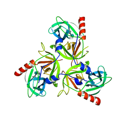
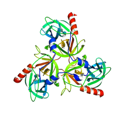
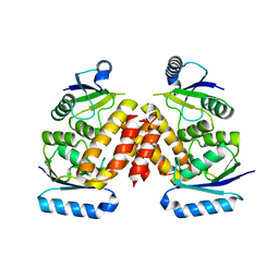
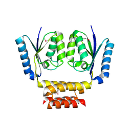
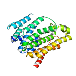 | | Crystal structure of dehydrosqualene synthase Y129A from S. aureus complexed with presqualene pyrophosphate | | 分子名称: | Dehydrosqualene synthase, MAGNESIUM ION, {(1R,2R,3R)-2-[(3E)-4,8-dimethylnona-3,7-dien-1-yl]-2-methyl-3-[(1E,5E)-2,6,10-trimethylundeca-1,5,9-trien-1-yl]cyclopropyl}methyl trihydrogen diphosphate | | 著者 | Lin, F.-Y, Liu, Y.-L, Liu, C.-I, Wang, A.H.J, Oldfield, E. | | 登録日 | 2010-01-21 | | 公開日 | 2010-12-22 | | 最終更新日 | 2023-09-06 | | 実験手法 | X-RAY DIFFRACTION (2.41 Å) | | 主引用文献 | Mechanism of action and inhibition of dehydrosqualene synthase.
Proc.Natl.Acad.Sci.USA, 107, 2010
|
|
3LH0
 | |
3LH1
 | |
3LH2
 | |
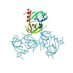
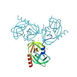
3LH3
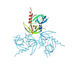
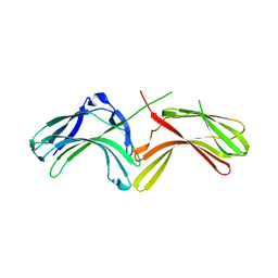
 | | DFP modified DegS delta PDZ | | 分子名称: | Protease degS | | 著者 | Sohn, J, Grant, R.A, Sauer, R.T. | | 登録日 | 2010-01-21 | | 公開日 | 2010-08-25 | | 最終更新日 | 2025-03-26 | | 実験手法 | X-RAY DIFFRACTION (2.35 Å) | | 主引用文献 | Allostery is an intrinsic property of the protease domain of DegS: implications for enzyme function and evolution.
J.Biol.Chem., 285, 2010
|
|
3LH4
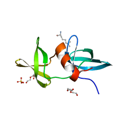
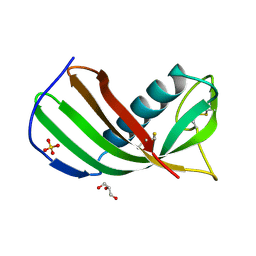 | | Crystal Structure of Sialostatin L2 | | 分子名称: | GLYCEROL, SULFATE ION, Secreted cystatin | | 著者 | Andersen, J.F, Kotsyfakis, M, Salat, J, Horka, H. | | 登録日 | 2010-01-21 | | 公開日 | 2010-09-29 | | 最終更新日 | 2024-11-27 | | 実験手法 | X-RAY DIFFRACTION (1.8 Å) | | 主引用文献 | The crystal structures of two salivary cystatins from the tick Ixodes scapularis and the effect of these inhibitors on the establishment of Borrelia burgdorferi infection in a murine model.
Mol.Microbiol., 77, 2010
|
|
3LH5
 | |
3LH8
 | |
3LH9
 | |
3LHA
 | |
3LHB
 | |
3LHC
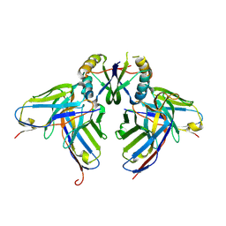
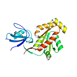
 | | Crystal structure of cyanovirin-n swapping domain b mutant | | 分子名称: | Cyanovirin-N, PHOSPHATE ION, SODIUM ION | | 著者 | Matei, E, Zheng, A, Furey, W, Rose, J, Aiken, C, Gronenborn, A.M. | | 登録日 | 2010-01-21 | | 公開日 | 2010-02-09 | | 最終更新日 | 2024-10-09 | | 実験手法 | X-RAY DIFFRACTION (1.34 Å) | | 主引用文献 | Anti-HIV activity of defective cyanovirin-N mutants is restored by dimerization.
J.Biol.Chem., 285, 2010
|
|
3LHD
 | | Crystal structure of P. abyssi tRNA m1A58 methyltransferase in complex with S-adenosyl-L-homocysteine | | 分子名称: | S-ADENOSYL-L-HOMOCYSTEINE, SAM-dependent methyltransferase, putative | | 著者 | Guelorget, A, Golinelli-Pimpaneau, B, Wouters, J, Barbey, C. | | 登録日 | 2010-01-22 | | 公開日 | 2010-05-19 | | 最終更新日 | 2024-10-16 | | 実験手法 | X-RAY DIFFRACTION (2.59 Å) | | 主引用文献 | Insights into the hyperthermostability and unusual region-specificity of archaeal Pyrococcus abyssi tRNA m1A57/58 methyltransferase.
Nucleic Acids Res., 38, 2010
|
|
3LHE
 | | The crystal structure of the C-terminal domain of a GntR family transcriptional regulator from Bacillus anthracis str. Sterne | | 分子名称: | CHLORIDE ION, GLYCEROL, GntR family Transcriptional regulator | | 著者 | Tan, K, Chhor, G, Clancy, S, Joachimiak, A, Midwest Center for Structural Genomics (MCSG) | | 登録日 | 2010-01-22 | | 公開日 | 2010-02-02 | | 最終更新日 | 2024-10-16 | | 実験手法 | X-RAY DIFFRACTION (1.62 Å) | | 主引用文献 | The crystal structure of the C-terminal domain of a GntR family transcriptional regulator from Bacillus anthracis str. Sterne
To be Published
|
|
3LHF
 | | The Crystal Structure of a Serine Recombinase from Sulfolobus solfataricus to 2.3A | | 分子名称: | Serine Recombinase | | 著者 | Stein, A.J, Osipiuk, J, Marshall, N, Bearden, J, Davidoff, J, Joachimiak, A, Midwest Center for Structural Genomics (MCSG) | | 登録日 | 2010-01-22 | | 公開日 | 2010-03-16 | | 最終更新日 | 2024-11-27 | | 実験手法 | X-RAY DIFFRACTION (2.3 Å) | | 主引用文献 | The Crystal Structure of a Serine Recombinase from Sulfolobus solfataricus to 2.3A
To be Published
|
|
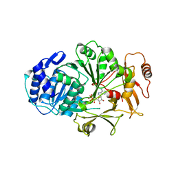
3LHG
 | | Bace1 in complex with the aminohydantoin Compound 4g | | 分子名称: | (5S)-2-amino-5-(2',5'-difluorobiphenyl-3-yl)-3-methyl-5-pyridin-4-yl-3,5-dihydro-4H-imidazol-4-one, Beta-secretase 1 | | 著者 | Olland, A.M. | | 登録日 | 2010-01-22 | | 公開日 | 2010-04-21 | | 最終更新日 | 2024-10-09 | | 実験手法 | X-RAY DIFFRACTION (2.1 Å) | | 主引用文献 | Pyridinyl aminohydantoins as small molecule BACE1 inhibitors.
Bioorg.Med.Chem.Lett., 20, 2010
|
|
3LHH
 | | The crystal structure of CBS domain protein from Shewanella oneidensis MR-1. | | 分子名称: | ADENOSINE MONOPHOSPHATE, CBS domain protein | | 著者 | Tan, K, Kagan, O, Savchenko, A, Edwards, A, Joachimiak, A, Midwest Center for Structural Genomics (MCSG) | | 登録日 | 2010-01-22 | | 公開日 | 2010-02-02 | | 最終更新日 | 2024-11-20 | | 実験手法 | X-RAY DIFFRACTION (2.1 Å) | | 主引用文献 | The crystal structure of CBS domain protein from Shewanella oneidensis MR-1.
To be Published
|
|
3LHI
 | |
3LHJ
 | | Crystal Structure of p38a Mitogen-Activated Protein Kinase in Complex with a Pyrazolopyridinone Inhibitor. | | 分子名称: | Mitogen-activated protein kinase 14, N-cyclopropyl-3-[1-(2,4-difluorophenyl)-7-methyl-6-oxo-6,7-dihydro-1H-pyrazolo[3,4-b]pyridin-5-yl]-4-methylbenzamide | | 著者 | Mohr, C, Jordan, S. | | 登録日 | 2010-01-22 | | 公開日 | 2010-04-14 | | 最終更新日 | 2024-02-21 | | 実験手法 | X-RAY DIFFRACTION (3.31 Å) | | 主引用文献 | Discovery and evaluation of 7-alkyl-1,5-bis-aryl-pyrazolopyridinones as highly potent, selective, and orally efficacious inhibitors of p38alpha mitogen-activated protein kinase.
J.Med.Chem., 53, 2010
|
|
3LHK
 | |
3LHL
 | | Crystal structure of a putative agmatinase from Clostridium difficile | | 分子名称: | (4S)-2-METHYL-2,4-PENTANEDIOL, MANGANESE (II) ION, PHOSPHATE ION, ... | | 著者 | Palani, K, Burley, S.K, Swaminathan, S, New York SGX Research Center for Structural Genomics (NYSGXRC) | | 登録日 | 2010-01-22 | | 公開日 | 2010-02-23 | | 最終更新日 | 2024-10-16 | | 実験手法 | X-RAY DIFFRACTION (2.3 Å) | | 主引用文献 | Crystal structure of a putative agmatinase from Clostridium difficile
To be Published
|
|


