6RZU
 
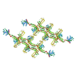
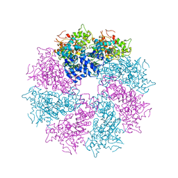
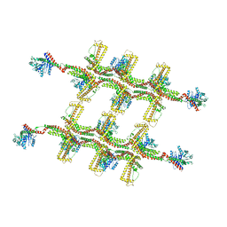 | | Structure of s-Mgm1 decorating the outer surface of tubulated lipid membranes in the GTPgammaS bound state | | 分子名称: | Putative mitochondrial dynamin protein | | 著者 | Faelber, K, Dietrich, L, Noel, J.K, Sanchez, R, Kudryashev, M, Kuelbrandt, W, Daumke, O. | | 登録日 | 2019-06-13 | | 公開日 | 2019-07-24 | | 最終更新日 | 2020-11-18 | | 実験手法 | ELECTRON MICROSCOPY (14.7 Å) | | 主引用文献 | Structure and assembly of the mitochondrial membrane remodelling GTPase Mgm1.
Nature, 571, 2019
|
|
5KPL
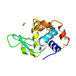
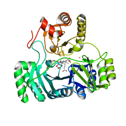
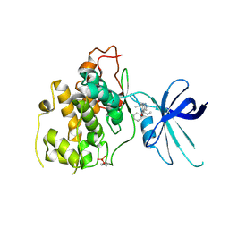 | | Glycogen Synthase Kinase 3 beta Complexed with BRD0705 | | 分子名称: | (4~{S})-4-ethyl-7,7-dimethyl-4-phenyl-2,6,8,9-tetrahydropyrazolo[3,4-b]quinolin-5-one, Glycogen synthase kinase-3 beta | | 著者 | Lakshminarasimhan, D, White, A, Nadupalli, A, Suto, R.K. | | 登録日 | 2016-07-04 | | 公開日 | 2018-03-14 | | 最終更新日 | 2018-03-21 | | 実験手法 | X-RAY DIFFRACTION (2.6 Å) | | 主引用文献 | Exploiting an Asp-Glu "switch" in glycogen synthase kinase 3 to design paralog-selective inhibitors for use in acute myeloid leukemia.
Sci Transl Med, 10, 2018
|
|
5KPK
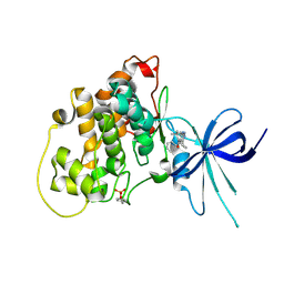 | | Glycogen Synthase Kinase 3 beta Complexed with BRD0209 | | 分子名称: | (4~{S})-3-cyclopropyl-4,7,7-trimethyl-4-phenyl-2,6,8,9-tetrahydropyrazolo[3,4-b]quinolin-5-one, Glycogen synthase kinase-3 beta | | 著者 | Lakshminarasimhan, D, White, A, Nadupalli, A, Suto, R.K. | | 登録日 | 2016-07-04 | | 公開日 | 2018-03-14 | | 最終更新日 | 2018-03-21 | | 実験手法 | X-RAY DIFFRACTION (2.4 Å) | | 主引用文献 | Exploiting an Asp-Glu "switch" in glycogen synthase kinase 3 to design paralog-selective inhibitors for use in acute myeloid leukemia.
Sci Transl Med, 10, 2018
|
|
6R52
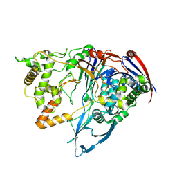
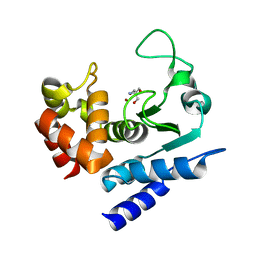 | | Crystal structure of PPEP-1(K101A) | | 分子名称: | 2-AMINO-2-HYDROXYMETHYL-PROPANE-1,3-DIOL, Pro-Pro endopeptidase, ZINC ION | | 著者 | Pichlo, C, Baumann, U. | | 登録日 | 2019-03-24 | | 公開日 | 2019-06-12 | | 最終更新日 | 2024-01-24 | | 実験手法 | X-RAY DIFFRACTION (2.022 Å) | | 主引用文献 | Molecular determinants of the mechanism and substrate specificity ofClostridium difficileproline-proline endopeptidase-1.
J.Biol.Chem., 294, 2019
|
|
6RZT
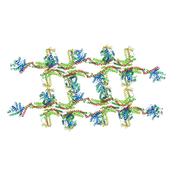
 | | Structure of s-Mgm1 decorating the outer surface of tubulated lipid membranes | | 分子名称: | Putative mitochondrial dynamin protein | | 著者 | Faelber, K, Dietrich, L, Noel, J.K, Sanchez, R, Kudryashev, M, Kuehlbrandt, W, Daumke, O. | | 登録日 | 2019-06-13 | | 公開日 | 2019-07-24 | | 最終更新日 | 2020-11-18 | | 実験手法 | ELECTRON MICROSCOPY (14.7 Å) | | 主引用文献 | Structure and assembly of the mitochondrial membrane remodelling GTPase Mgm1.
Nature, 571, 2019
|
|
6Q88
 | | RT structure of HEWL at 5 kGy | | 分子名称: | CHLORIDE ION, DI(HYDROXYETHYL)ETHER, Lysozyme C | | 著者 | de la Mora, E, Coquelle, N, Bury, C.S, Rosenthal, M, Garman, E.F, Burghammer, M, Colletier, J.P, Weik, M. | | 登録日 | 2018-12-14 | | 公開日 | 2020-01-15 | | 最終更新日 | 2024-01-24 | | 実験手法 | X-RAY DIFFRACTION (1.74007058 Å) | | 主引用文献 | Radiation damage and dose limits in serial synchrotron crystallography at cryo- and room temperatures.
Proc.Natl.Acad.Sci.USA, 117, 2020
|
|
6RZV
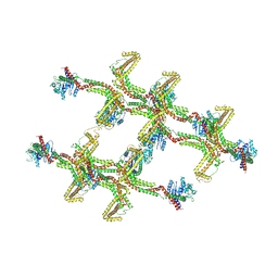
 | | Structure of s-Mgm1 decorating the inner surface of tubulated lipid membranes | | 分子名称: | Putative mitochondrial dynamin protein | | 著者 | Faelber, K, Dietrich, L, Noel, J.K, Sanchez, R, Kudryashev, M, Kuelbrandt, W, Daumke, O. | | 登録日 | 2019-06-13 | | 公開日 | 2019-07-24 | | 最終更新日 | 2020-11-18 | | 実験手法 | ELECTRON MICROSCOPY (20.6 Å) | | 主引用文献 | Structure and assembly of the mitochondrial membrane remodelling GTPase Mgm1.
Nature, 571, 2019
|
|
6RZW
 | | Structure of s-Mgm1 decorating the inner surface of tubulated lipid membranes in the GTPgammaS bound state | | 分子名称: | Putative mitochondrial dynamin protein | | 著者 | Faelber, K, Dietrich, L, Noel, J.K, Sanchez, R, Kudryashev, M, Kuelbrandt, W, Daumke, O. | | 登録日 | 2019-06-13 | | 公開日 | 2019-07-24 | | 最終更新日 | 2020-11-18 | | 実験手法 | ELECTRON MICROSCOPY (18.799999 Å) | | 主引用文献 | Structure and assembly of the mitochondrial membrane remodelling GTPase Mgm1.
Nature, 571, 2019
|
|
6HFP
 | |
6HFI
 | |
8VUV
 | |
3LU7
 | | Human serum albumin in complex with compound 2 | | 分子名称: | 4-[(1R,2R)-2-{[(5-fluoro-1H-indol-2-yl)carbonyl]amino}-2,3-dihydro-1H-inden-1-yl]butanoic acid, PHOSPHATE ION, Serum albumin | | 著者 | Buttar, D, Colclough, N, Gerhardt, S, MacFaul, P.A, Phillips, S.D, Plowright, A, Whittamore, P, Tam, K, Maskos, K, Steinbacher, S, Steuber, H. | | 登録日 | 2010-02-17 | | 公開日 | 2010-10-27 | | 最終更新日 | 2011-07-13 | | 実験手法 | X-RAY DIFFRACTION (2.8 Å) | | 主引用文献 | A combined spectroscopic and crystallographic approach to probing drug-human serum albumin interactions
Bioorg.Med.Chem., 18, 2010
|
|
1E6G
 | | A-SPECTRIN SH3 DOMAIN A11V, V23L, M25I, V53I, V58L MUTANT | | 分子名称: | SPECTRIN ALPHA CHAIN, SULFATE ION | | 著者 | Vega, M.C, Serrano, L. | | 登録日 | 2000-08-15 | | 公開日 | 2002-05-23 | | 最終更新日 | 2023-12-13 | | 実験手法 | X-RAY DIFFRACTION (2.3 Å) | | 主引用文献 | Conformational Strain in the Hydrophobic Core and its Implications for Protein Folding and Design
Nat.Struct.Biol., 9, 2002
|
|
4XGC
 | | Crystal structure of the eukaryotic origin recognition complex | | 分子名称: | CHLORIDE ION, Origin recognition complex subunit 1, Origin recognition complex subunit 2, ... | | 著者 | Bleichert, F, Botchan, M.R, Berger, J.M. | | 登録日 | 2014-12-30 | | 公開日 | 2015-04-01 | | 最終更新日 | 2019-12-04 | | 実験手法 | X-RAY DIFFRACTION (3.5 Å) | | 主引用文献 | Crystal structure of the eukaryotic origin recognition complex.
Nature, 519, 2015
|
|
3L91
 | | Structure of Pseudomonas aerugionsa PvdQ bound to octanoate | | 分子名称: | 1,2-ETHANEDIOL, Acyl-homoserine lactone acylase pvdQ subunit alpha, Acyl-homoserine lactone acylase pvdQ subunit beta, ... | | 著者 | Drake, E.J, Gulick, A.M. | | 登録日 | 2010-01-04 | | 公開日 | 2011-01-19 | | 最終更新日 | 2023-09-06 | | 実験手法 | X-RAY DIFFRACTION (1.66 Å) | | 主引用文献 | Structural characterization and high-throughput screening of inhibitors of PvdQ, an NTN hydrolase involved in pyoverdine synthesis.
Acs Chem.Biol., 6, 2011
|
|
1E7O
 | | A-SPECTRIN SH3 DOMAIN A11V, V23L, M25V, V44I, V58L MUTATIONS | | 分子名称: | GLYCEROL, SPECTRIN ALPHA CHAIN | | 著者 | Vega, M.C, Serrano, L. | | 登録日 | 2000-08-31 | | 公開日 | 2003-05-21 | | 最終更新日 | 2023-12-13 | | 実験手法 | X-RAY DIFFRACTION (3.2 Å) | | 主引用文献 | A Thermodynamic and Kinetic Analysis of the Folding Pathway of an SH3 Domain Entropically Stabilised by a Redesigned Hydrophobic Core
J.Mol.Biol., 328, 2003
|
|
3LQR
 | | Structure of CED-4:CED-3 complex | | 分子名称: | ADENOSINE-5'-TRIPHOSPHATE, Cell death protein 4, MAGNESIUM ION | | 著者 | Qi, S, Pang, Y, Shi, Y, Yan, N. | | 登録日 | 2010-02-09 | | 公開日 | 2010-04-28 | | 最終更新日 | 2023-11-01 | | 実験手法 | X-RAY DIFFRACTION (3.896 Å) | | 主引用文献 | Crystal structure of the Caenorhabditis elegans apoptosome reveals an octameric assembly of CED-4.
Cell(Cambridge,Mass.), 141, 2010
|
|
8GEP
 | | SULFITE REDUCTASE HEMOPROTEIN NITRATE COMPLEX | | 分子名称: | IRON/SULFUR CLUSTER, NITRATE ION, POTASSIUM ION, ... | | 著者 | Crane, B.R, Getzoff, E.D. | | 登録日 | 1997-07-11 | | 公開日 | 1998-01-14 | | 最終更新日 | 2023-12-27 | | 実験手法 | X-RAY DIFFRACTION (2.2 Å) | | 主引用文献 | Probing the catalytic mechanism of sulfite reductase by X-ray crystallography: structures of the Escherichia coli hemoprotein in complex with substrates, inhibitors, intermediates, and products.
Biochemistry, 36, 1997
|
|
3ME4
 | | Crystal structure of mouse RANK | | 分子名称: | ACETATE ION, CHLORIDE ION, GLYCEROL, ... | | 著者 | Walter, S.W, Liu, C, Zhu, X, Wu, Y, Owens, R.J, Stuart, D.I, Gao, B, Ren, J. | | 登録日 | 2010-03-31 | | 公開日 | 2010-06-02 | | 最終更新日 | 2023-11-01 | | 実験手法 | X-RAY DIFFRACTION (2.01 Å) | | 主引用文献 | Structural and Functional Insights of RANKL-RANK Interaction and Signaling.
J.Immunol., 2010
|
|
4XSJ
 | | Crystal structure of the N-terminal domain of the human mitochondrial calcium uniporter fused with T4 lysozyme | | 分子名称: | Lysozyme,Calcium uniporter protein, mitochondrial, SULFATE ION | | 著者 | Lee, Y, Min, C.K, Kim, T.G, Song, H.K, Lim, Y, Kim, D, Shin, K, Kang, M, Kang, J.Y, Youn, H.-S, Lee, J.-G, An, J.Y, Park, K.R, Lim, J.J, Kim, J.H, Kim, J.H, Park, Z.Y, Kim, Y.-S, Wang, J, Kim, D.H, Eom, S.H. | | 登録日 | 2015-01-22 | | 公開日 | 2015-09-16 | | 最終更新日 | 2023-11-08 | | 実験手法 | X-RAY DIFFRACTION (1.8 Å) | | 主引用文献 | Structure and function of the N-terminal domain of the human mitochondrial calcium uniporter.
Embo Rep., 16, 2015
|
|
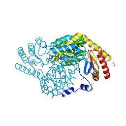
3ML0
 | | Thermostable Penicillin G acylase from Alcaligenes faecalis in tetragonal form | | 分子名称: | CALCIUM ION, Penicillin G acylase, alpha subunit, ... | | 著者 | Varshney, N.K, Kumar, R.S, Ignatova, Z, Dodson, E, Suresh, C.G. | | 登録日 | 2010-04-16 | | 公開日 | 2010-05-05 | | 最終更新日 | 2023-11-01 | | 実験手法 | X-RAY DIFFRACTION (3.5 Å) | | 主引用文献 | Crystallization and X-ray structure analysis of a thermostable penicillin G acylase from Alcaligenes faecalis.
Acta Crystallogr.,Sect.F, 68, 2012
|
|
8GUH
 | | Serine Palmitoyltransferase from Sphingobacterium multivorum complexed with Tris | | 分子名称: | 1,2-ETHANEDIOL, Serine palmitoyltransferase, [4-[[[2-(hydroxymethyl)-1,3-bis(oxidanyl)propan-2-yl]amino]methyl]-6-methyl-5-oxidanyl-pyridin-3-yl]methyl dihydrogen phosphate | | 著者 | Murakami, T, Takahashi, A, Katayama, A, Miyahara, I, Kamiya, N, Ikushiro, H, Yano, T. | | 登録日 | 2022-09-12 | | 公開日 | 2023-07-19 | | 最終更新日 | 2023-11-29 | | 実験手法 | X-RAY DIFFRACTION (1.65 Å) | | 主引用文献 | Crystal structure of Sphingobacterium multivorum serine palmitoyltransferase complexed with tris(hydroxymethyl)aminomethane.
Acta Crystallogr.,Sect.F, 78, 2022
|
|
8JRU
 | | Cryo-EM structure of the glucagon receptor bound to beta-arrestin 1 in ligand-free state | | 分子名称: | Beta-arrestin 1 and single-chain fragment variable 30 (scFv30), HA signal peptide,HPC4 purification tag,Glucagon receptor,C-terminal tail of Vasopressin V2 receptor, Nanobody 32, ... | | 著者 | Chen, K, Zhang, C, Lin, S, Zhao, Q, Wu, B. | | 登録日 | 2023-06-17 | | 公開日 | 2023-08-16 | | 最終更新日 | 2023-09-13 | | 実験手法 | ELECTRON MICROSCOPY (3.5 Å) | | 主引用文献 | Tail engagement of arrestin at the glucagon receptor.
Nature, 620, 2023
|
|
8JRV
 | | Cryo-EM structure of the glucagon receptor bound to glucagon and beta-arrestin 1 | | 分子名称: | Beta-arrestin 1 and single-chain fragment variable 30 (scFv30), Glucagon, HA signal peptide,HPC4 purification tag,Glucagon receptor,C-terminal tail of Vasopressin V2 receptor, ... | | 著者 | Chen, K, Zhang, C, Lin, S, Zhao, Q, Wu, B. | | 登録日 | 2023-06-17 | | 公開日 | 2023-08-16 | | 最終更新日 | 2023-09-13 | | 実験手法 | ELECTRON MICROSCOPY (3.3 Å) | | 主引用文献 | Tail engagement of arrestin at the glucagon receptor.
Nature, 620, 2023
|
|
4YLK
 | | Crystal structure of DYRK1A in complex with 10-Chloro-substituted 11H-indolo[3,2-c]quinolone-6-carboxylic acid inhibitor 5s | | 分子名称: | 1,2-ETHANEDIOL, 10-chloro-2-iodo-11H-indolo[3,2-c]quinoline-6-carboxylic acid, Dual specificity tyrosine-phosphorylation-regulated kinase 1A, ... | | 著者 | Chaikuad, A, Falke, H, Krojer, T, von Delft, F, Arrowsmith, C.H, Edwards, A.M, Bountra, C, Kunick, C, Knapp, S, Structural Genomics Consortium (SGC) | | 登録日 | 2015-03-05 | | 公開日 | 2015-03-25 | | 最終更新日 | 2023-11-15 | | 実験手法 | X-RAY DIFFRACTION (1.4 Å) | | 主引用文献 | 10-Iodo-11H-indolo[3,2-c]quinoline-6-carboxylic Acids Are Selective Inhibitors of DYRK1A.
J.Med.Chem., 58, 2015
|
|


