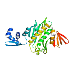
4WVS
 
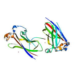
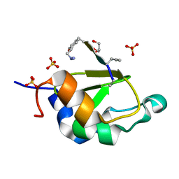 | | Crystal structure of XIAP-BIR2 domain complexed with (S)-3-(4-methoxyphenyl)-2-((S)-2-((S)-1-((S)-2-((S)-2-(methylamino)propanamido)pent-4-ynoyl)pyrrolidine-2-carboxamido)-3-phenylpropanamido)propanoic acid | | 分子名称: | 3,11-DIFLUORO-6,8,13-TRIMETHYL-8H-QUINO[4,3,2-KL]ACRIDIN-13-IUM, E3 ubiquitin-protein ligase XIAP, GLYCEROL, ... | | 著者 | Pokross, M.E. | | 登録日 | 2014-11-07 | | 公開日 | 2015-05-06 | | 最終更新日 | 2023-11-15 | | 実験手法 | X-RAY DIFFRACTION (2.09 Å) | | 主引用文献 | The Discovery of Macrocyclic XIAP Antagonists from a DNA-Programmed Chemistry Library, and Their Optimization To Give Lead Compounds with in Vivo Antitumor Activity.
J.Med.Chem., 58, 2015
|
|
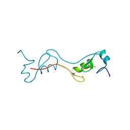
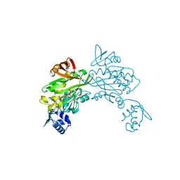
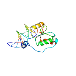
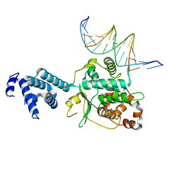
6CG0
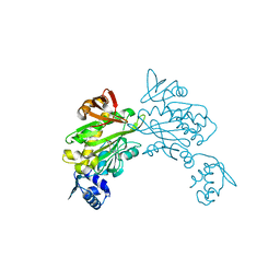
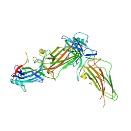
 | | Cryo-EM structure of mouse RAG1/2 HFC complex (3.17 A) | | 分子名称: | CALCIUM ION, DNA (30-MER), DNA (41-MER), ... | | 著者 | Chen, X, Kim, M, Chuenchor, W, Cui, Y, Zhang, X, Zhou, Z.H, Gellert, M, Yang, W. | | 登録日 | 2018-02-19 | | 公開日 | 2018-04-25 | | 最終更新日 | 2024-03-13 | | 実験手法 | ELECTRON MICROSCOPY (3.17 Å) | | 主引用文献 | Cracking the DNA Code for V(D)J Recombination.
Mol. Cell, 70, 2018
|
|
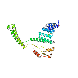
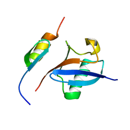
4PPE
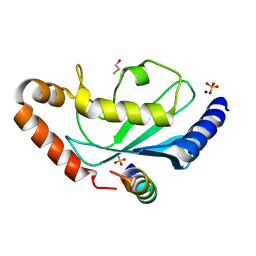
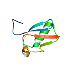
 | | human RNF4 RING domain | | 分子名称: | E3 ubiquitin-protein ligase RNF4, ZINC ION | | 著者 | Perry, J.J, Arvai, A.S, Hitomi, C, Tainer, J.A. | | 登録日 | 2014-02-26 | | 公開日 | 2014-03-26 | | 最終更新日 | 2024-02-28 | | 実験手法 | X-RAY DIFFRACTION (2 Å) | | 主引用文献 | RNF4 interacts with both SUMO and nucleosomes to promote the DNA damage response.
Embo Rep., 15, 2014
|
|
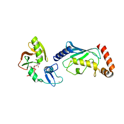
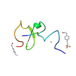
2C2L
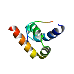
 | | Crystal structure of the CHIP U-box E3 ubiquitin ligase | | 分子名称: | CARBOXY TERMINUS OF HSP70-INTERACTING PROTEIN, HSP90, NICKEL (II) ION, ... | | 著者 | Zhang, M, Roe, S.M, Pearl, L.H. | | 登録日 | 2005-09-29 | | 公開日 | 2005-11-23 | | 最終更新日 | 2024-05-01 | | 実験手法 | X-RAY DIFFRACTION (3.3 Å) | | 主引用文献 | Chaperoned Ubiquitylation-Crystal Structures of the Chip U Box E3 Ubiquitin Ligase and a Chip-Ubc13-Uev1A Complex
Mol.Cell, 20, 2005
|
|
4QPL
 | | Crystal structure of RNF146(RING-WWE)/UbcH5a/iso-ADPr complex | | 分子名称: | 2'-O-(5-O-phosphono-alpha-D-ribofuranosyl)adenosine 5'-(dihydrogen phosphate), E3 ubiquitin-protein ligase RNF146, Ubiquitin-conjugating enzyme E2 D1, ... | | 著者 | Wang, Z, DaRosa, P.A, Klevit, R.E, Xu, W. | | 登録日 | 2014-06-23 | | 公開日 | 2014-10-15 | | 最終更新日 | 2024-02-28 | | 実験手法 | X-RAY DIFFRACTION (1.9 Å) | | 主引用文献 | Allosteric activation of the RNF146 ubiquitin ligase by a poly(ADP-ribosyl)ation signal.
Nature, 517, 2015
|
|
3RKY
 | |
2YBF
 | | Complex of Rad18 (Rad6 binding domain) with Rad6b | | 分子名称: | BETA-MERCAPTOETHANOL, E3 UBIQUITIN-PROTEIN LIGASE RAD18, SODIUM ION, ... | | 著者 | Hibbert, R.G, Sixma, T.K. | | 登録日 | 2011-03-08 | | 公開日 | 2011-04-20 | | 最終更新日 | 2023-12-20 | | 実験手法 | X-RAY DIFFRACTION (2 Å) | | 主引用文献 | E3 Ligase Rad18 Promotes Monoubiquitination Rather Than Ubiquitin Chain Formation by E2 Enzyme Rad6.
Proc.Natl.Acad.Sci.USA, 108, 2011
|
|
5CKI
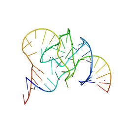 | | Crystal structure of 9DB1* deoxyribozyme (Cobalt hexammine soaked crystals) | | 分子名称: | COBALT (II) ION, DNA (44-MER), MAGNESIUM ION, ... | | 著者 | Ponce-Salvatierra, A, Hoebartner, C, Pena, V. | | 登録日 | 2015-07-15 | | 公開日 | 2016-01-13 | | 最終更新日 | 2024-05-08 | | 実験手法 | X-RAY DIFFRACTION (2.985 Å) | | 主引用文献 | Crystal structure of a DNA catalyst.
Nature, 529, 2016
|
|
3HQM
 | |
3HSV
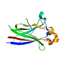
 | | Structures of SPOP-Substrate Complexes: Insights into Molecular Architectures of BTB-Cul3 Ubiquitin Ligases: SPOPMATHx-MacroH2ASBCpep2 | | 分子名称: | Core histone macro-H2A.1, SULFATE ION, Speckle-type POZ protein, ... | | 著者 | Zhuang, M, Schulman, B.A, Miller, D. | | 登録日 | 2009-06-10 | | 公開日 | 2009-10-20 | | 最終更新日 | 2023-09-06 | | 実験手法 | X-RAY DIFFRACTION (1.43 Å) | | 主引用文献 | Structures of SPOP-substrate complexes: insights into molecular architectures of BTB-Cul3 ubiquitin ligases.
Mol.Cell, 36, 2009
|
|
3RD2
 | |
8HSV
 | | The structure of rat beta-arrestin1 in complex with a rat Mdm2 peptide | | 分子名称: | Beta-arrestin-1, SULFATE ION, peptide from E3 ubiquitin-protein ligase Mdm2 | | 著者 | Yun, Y, Yoon, H.J, Choi, Y, Lee, H.H. | | 登録日 | 2022-12-20 | | 公開日 | 2023-07-19 | | 最終更新日 | 2024-05-29 | | 実験手法 | X-RAY DIFFRACTION (3 Å) | | 主引用文献 | GPCR targeting of E3 ubiquitin ligase MDM2 by inactive beta-arrestin.
Proc.Natl.Acad.Sci.USA, 120, 2023
|
|
3NTW
 | |
3RKX
 | |
3RKW
 | |
3GNB
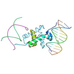 | | Crystal structure of the RAG1 nonamer-binding domain with DNA | | 分子名称: | 5'-D(*AP*AP*TP*TP*TP*TP*CP*AP*GP*AP*AP*AP*CP*C)-3', 5'-D(*AP*GP*GP*TP*TP*TP*CP*TP*GP*AP*AP*AP*AP*C)-3', V(D)J recombination-activating protein 1 | | 著者 | Yin, F.F, Bailey, S, Innis, C.A, Steitz, T.A, Schatz, D.G. | | 登録日 | 2009-03-16 | | 公開日 | 2009-04-28 | | 最終更新日 | 2023-09-06 | | 実験手法 | X-RAY DIFFRACTION (3 Å) | | 主引用文献 | Structure of the RAG1 nonamer binding domain with DNA reveals a dimer that mediates DNA synapsis.
Nat.Struct.Mol.Biol., 16, 2009
|
|
3GNA
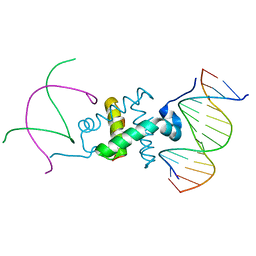 | | Crystal structure of the RAG1 nonamer-binding domain with DNA | | 分子名称: | 5'-D(*AP*CP*TP*TP*AP*AP*CP*AP*AP*AP*AP*AP*CP*C)-3', 5'-D(*TP*GP*GP*TP*TP*TP*TP*TP*GP*TP*TP*AP*AP*G)-3', V(D)J recombination-activating protein 1 | | 著者 | Yin, F.F, Bailey, S, Innis, C.A, Steitz, T.A, Schatz, D.G. | | 登録日 | 2009-03-16 | | 公開日 | 2009-04-28 | | 最終更新日 | 2024-02-21 | | 実験手法 | X-RAY DIFFRACTION (2.4 Å) | | 主引用文献 | Structure of the RAG1 nonamer binding domain with DNA reveals a dimer that mediates DNA synapsis.
Nat.Struct.Mol.Biol., 16, 2009
|
|
2K7F
 | | HADDOCK calculated model of the complex between the BRCT region of RFC p140 and dsDNA | | 分子名称: | 5'-D(P*DCP*DGP*DAP*DCP*DCP*DTP*DCP*DGP*DAP*DGP*DAP*DTP*DCP*DA)-3', 5'-D(P*DCP*DTP*DCP*DGP*DAP*DGP*DGP*DTP*DCP*DG)-3', Replication factor C subunit 1 | | 著者 | Kobayashi, M, Ab, E, Bonvin, A, Siegal, G. | | 登録日 | 2008-08-10 | | 公開日 | 2009-09-08 | | 最終更新日 | 2024-05-01 | | 実験手法 | SOLUTION NMR | | 主引用文献 | Structure of the DNA-bound BRCA1 C-terminal region from human replication factor C p140 and model of the protein-DNA complex.
J.Biol.Chem., 285, 2010
|
|
3FL2
 | | Crystal structure of the ring domain of the E3 ubiquitin-protein ligase UHRF1 | | 分子名称: | E3 ubiquitin-protein ligase UHRF1, ZINC ION | | 著者 | Walker, J.R, Avvakumov, G.V, Xue, S, Li, Y, Bountra, C, Weigelt, J, Arrowsmith, C.H, Edwards, A.M, Bochkarev, A, Dhe-Paganon, S, Structural Genomics Consortium (SGC) | | 登録日 | 2008-12-18 | | 公開日 | 2009-01-20 | | 最終更新日 | 2023-09-06 | | 実験手法 | X-RAY DIFFRACTION (1.75 Å) | | 主引用文献 | Structure of the Ring Domain of the E3 Ubiquitin-Protein Ligase Uhrf1
To be Published
|
|
3SQI
 | | DNA binding domain of Ndc10 | | 分子名称: | DNA (5'-D(P*AP*AP*AP*TP*TP*TP*TP*AP*TP*AP*AP*AP*TP*TP*A)-3'), DNA (5'-D(P*TP*TP*AP*AP*TP*TP*TP*AP*TP*AP*AP*AP*AP*TP*T)-3'), KLLA0E03807p | | 著者 | Cho, U.S, Harrison, S.C. | | 登録日 | 2011-07-05 | | 公開日 | 2011-12-07 | | 最終更新日 | 2024-02-28 | | 実験手法 | X-RAY DIFFRACTION (2.8245 Å) | | 主引用文献 | Ndc10 is a platform for inner kinetochore assembly in budding yeast.
Nat.Struct.Mol.Biol., 19, 2011
|
|
2MRE
 | | NMR structure of the Rad18-UBZ/ubiquitin complex | | 分子名称: | E3 ubiquitin-protein ligase RAD18, Polyubiquitin-C, ZINC ION | | 著者 | Rizzo, A.A, Salerno, P.E, Bezsonova, I, Korzhnev, D.M. | | 登録日 | 2014-07-03 | | 公開日 | 2014-10-22 | | 最終更新日 | 2024-05-01 | | 実験手法 | SOLUTION NMR | | 主引用文献 | NMR Structure of the Human Rad18 Zinc Finger in Complex with Ubiquitin Defines a Class of UBZ Domains in Proteins Linked to the DNA Damage Response.
Biochemistry, 53, 2014
|
|
6IIW
 | |
6BRQ
 | | Crystal structure of rice ASK1-D3 ubiquitin ligase complex crystal form 3 | | 分子名称: | F-box/LRR-repeat MAX2 homolog, SKP1-like protein 1A | | 著者 | Shabek, N, Zheng, N, Mao, H, Hinds, T.R, Ticchiarelli, F, Leyser, O. | | 登録日 | 2017-11-30 | | 公開日 | 2018-11-21 | | 最終更新日 | 2024-03-13 | | 実験手法 | X-RAY DIFFRACTION (2.99 Å) | | 主引用文献 | Structural plasticity of D3-D14 ubiquitin ligase in strigolactone signalling.
Nature, 563, 2018
|
|
6BRO
 | |
4K95
 | | Crystal Structure of Parkin | | 分子名称: | E3 ubiquitin-protein ligase parkin, ZINC ION | | 著者 | Seirafi, M, Menade, M, Sauve, V, Kozlov, G, Trempe, J.-F, Nagar, B, Gehring, K. | | 登録日 | 2013-04-19 | | 公開日 | 2013-05-15 | | 最終更新日 | 2023-09-20 | | 実験手法 | X-RAY DIFFRACTION (6.499 Å) | | 主引用文献 | Structure of parkin reveals mechanisms for ubiquitin ligase activation.
Science, 340, 2013
|
|


