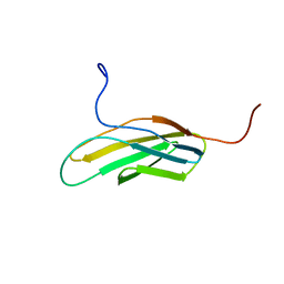
173D
 
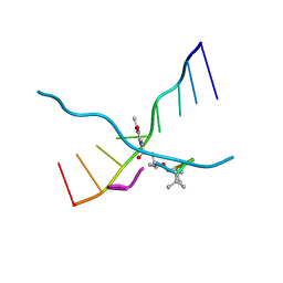 | | MULTIPLE BINDING MODES OF ANTICANCER DRUG ACTINOMYCIN D: X-RAY, MOLECULAR MODELING, AND SPECTROSCOPIC STUDIES OF D(GAAGCTTC)2-ACTINOMYCIN D COMPLEXES AND ITS HOST DNA | | 分子名称: | ACTINOMYCIN D, DNA (5'-D(*GP*AP*AP*GP*CP*TP*TP*C)-3') | | 著者 | Kamitori, S, Takusagawa, F. | | 登録日 | 1994-04-18 | | 公開日 | 1994-10-15 | | 最終更新日 | 2024-07-10 | | 実験手法 | X-RAY DIFFRACTION (3 Å) | | 主引用文献 | Multiple Binding Modes of Anticancer Drug Actinomycin D: X-Ray, Molecular Modeling, and Spectroscopic Studies of D(Gaagcttc)2-Actinomycin D Complexes and its Host DNA
J.Am.Chem.Soc., 116, 1994
|
|
4RKG
 | |
1BID
 | |
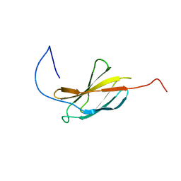
1X59
 | | Solution structures of the WHEP-TRS domain of human histidyl-tRNA synthetase | | 分子名称: | Histidyl-tRNA synthetase | | 著者 | Nameki, N, Sasagawa, A, Tomizawa, T, Koshiba, S, Inoue, M, Kigawa, T, Yokoyama, S, RIKEN Structural Genomics/Proteomics Initiative (RSGI) | | 登録日 | 2005-05-15 | | 公開日 | 2005-11-15 | | 最終更新日 | 2024-05-29 | | 実験手法 | SOLUTION NMR | | 主引用文献 | Solution structures of the WHEP-TRS domain of human histidyl-tRNA synthetase
To be Published
|
|
1AN5
 | | E. COLI THYMIDYLATE SYNTHASE IN COMPLEX WITH CB3717 | | 分子名称: | 10-PROPARGYL-5,8-DIDEAZAFOLIC ACID, PHOSPHATE ION, THYMIDYLATE SYNTHASE | | 著者 | Stout, T.J, Sage, C.R, Stroud, R.M. | | 登録日 | 1997-06-26 | | 公開日 | 1998-07-01 | | 最終更新日 | 2023-08-02 | | 実験手法 | X-RAY DIFFRACTION (2.6 Å) | | 主引用文献 | The additivity of substrate fragments in enzyme-ligand binding.
Structure, 6, 1998
|
|
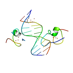
4RKH
 | | Structure of the MSL2 CXC domain bound with a specific MRE sequence | | 分子名称: | DNA (5'-D(*AP*TP*CP*CP*AP*TP*CP*TP*CP*GP*CP*TP*CP*AP*T)-3'), DNA (5'-D(*AP*TP*GP*AP*GP*CP*GP*AP*GP*AP*TP*GP*GP*AP*T)-3'), E3 ubiquitin-protein ligase msl-2, ... | | 著者 | Zheng, S, Ye, K. | | 登録日 | 2014-10-13 | | 公開日 | 2015-01-21 | | 最終更新日 | 2024-03-20 | | 実験手法 | X-RAY DIFFRACTION (2 Å) | | 主引用文献 | Structural basis of X chromosome DNA recognition by the MSL2 CXC domain during Drosophila dosage compensation.
Genes Dev., 28, 2014
|
|
1AOB
 | | E. COLI THYMIDYLATE SYNTHASE COMPLEXED WITH DDURD | | 分子名称: | 2'-5'DIDEOXYURIDINE, FORMIC ACID, PHOSPHATE ION, ... | | 著者 | Stout, T.J, Sage, C.R, Stroud, R.M. | | 登録日 | 1997-06-30 | | 公開日 | 1998-07-01 | | 最終更新日 | 2023-08-02 | | 実験手法 | X-RAY DIFFRACTION (2.1 Å) | | 主引用文献 | The additivity of substrate fragments in enzyme-ligand binding.
Structure, 6, 1998
|
|
1BDU
 | |
1V9Y
 | | Crystal Structure of the heme PAS sensor domain of Ec DOS (ferric form) | | 分子名称: | Heme pas sensor protein, PROTOPORPHYRIN IX CONTAINING FE | | 著者 | Kurokawa, H, Lee, D.S, Watanabe, M, Sagami, I, Mikami, B, Raman, C.S, Shimizu, T. | | 登録日 | 2004-02-04 | | 公開日 | 2004-05-25 | | 最終更新日 | 2023-12-27 | | 実験手法 | X-RAY DIFFRACTION (1.32 Å) | | 主引用文献 | A redox-controlled molecular switch revealed by the crystal structure of a bacterial heme PAS sensor.
J.Biol.Chem., 279, 2004
|
|
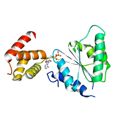
1B6C
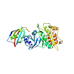 | | CRYSTAL STRUCTURE OF THE CYTOPLASMIC DOMAIN OF THE TYPE I TGF-BETA RECEPTOR IN COMPLEX WITH FKBP12 | | 分子名称: | FK506-BINDING PROTEIN, SULFATE ION, TGF-B SUPERFAMILY RECEPTOR TYPE I | | 著者 | Huse, M, Chen, Y.-G, Massague, J, Kuriyan, J. | | 登録日 | 1999-01-13 | | 公開日 | 1999-06-15 | | 最終更新日 | 2024-02-07 | | 実験手法 | X-RAY DIFFRACTION (2.6 Å) | | 主引用文献 | Crystal structure of the cytoplasmic domain of the type I TGF beta receptor in complex with FKBP12.
Cell(Cambridge,Mass.), 96, 1999
|
|
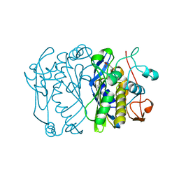
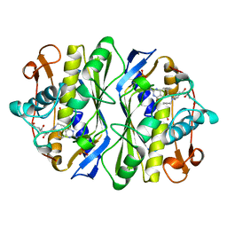
1RG9
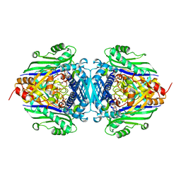 | | S-Adenosylmethionine synthetase complexed with SAM and PPNP | | 分子名称: | (DIPHOSPHONO)AMINOPHOSPHONIC ACID, MAGNESIUM ION, POTASSIUM ION, ... | | 著者 | Komoto, J, Yamada, T, Takata, Y, Markham, G.D, Takusagawa, F. | | 登録日 | 2003-11-11 | | 公開日 | 2004-03-02 | | 最終更新日 | 2024-02-14 | | 実験手法 | X-RAY DIFFRACTION (2.5 Å) | | 主引用文献 | Crystal structure of the S-adenosylmethionine synthetase ternary complex: a novel catalytic mechanism of s-adenosylmethionine synthesis from ATP and MET.
Biochemistry, 43, 2004
|
|
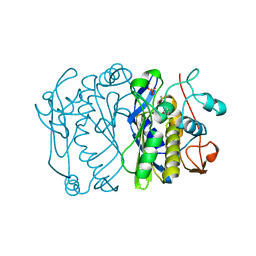
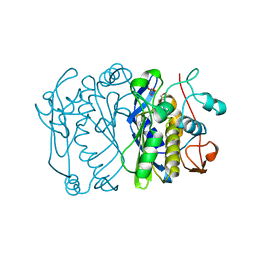
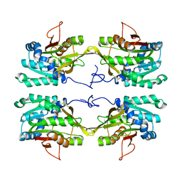
1B3R
 | | RAT LIVER S-ADENOSYLHOMOCYSTEIN HYDROLASE | | 分子名称: | NICOTINAMIDE-ADENINE-DINUCLEOTIDE, PROTEIN (S-ADENOSYLHOMOCYSTEINE HYDROLASE) | | 著者 | Hu, Y, Komoto, J, Huang, Y, Takusagawa, F, Gomi, T, Ogawa, H, Takata, Y, Fujioka, M. | | 登録日 | 1998-12-14 | | 公開日 | 1998-12-23 | | 最終更新日 | 2024-04-03 | | 実験手法 | X-RAY DIFFRACTION (2.8 Å) | | 主引用文献 | Crystal structure of S-adenosylhomocysteine hydrolase from rat liver.
Biochemistry, 38, 1999
|
|
1NBH
 | |
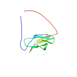
1X5F
 | | The solution structure of the first fibronectin type III domain of human Neogenin | | 分子名称: | Neogenin | | 著者 | Tochio, N, Sasagawa, A, Koshiba, S, Inoue, M, Kigawa, T, Yokoyama, S, RIKEN Structural Genomics/Proteomics Initiative (RSGI) | | 登録日 | 2005-05-15 | | 公開日 | 2005-11-15 | | 最終更新日 | 2024-05-29 | | 実験手法 | SOLUTION NMR | | 主引用文献 | The solution structure of the first fibronectin type III domain of human Neogenin
To be Published
|
|
1UXC
 | | FRUCTOSE REPRESSOR DNA-BINDING DOMAIN, NMR, MINIMIZED STRUCTURE | | 分子名称: | FRUCTOSE REPRESSOR | | 著者 | Penin, F, Geourjon, C, Montserret, R, Bockmann, A, Lesage, A, Yang, Y, Bonod-Bidaud, C, Cortay, J.C, Negre, D, Cozzone, A.J, Deleage, G. | | 登録日 | 1996-12-26 | | 公開日 | 1997-04-21 | | 最終更新日 | 2024-05-01 | | 実験手法 | SOLUTION NMR | | 主引用文献 | Three-dimensional structure of the DNA-binding domain of the fructose repressor from Escherichia coli by 1H and 15N NMR.
J.Mol.Biol., 270, 1997
|
|
1V9Z
 | | Crystal Structure of the heme PAS sensor domain of Ec DOS (Ferrous Form) | | 分子名称: | Heme pas sensor protein, PROTOPORPHYRIN IX CONTAINING FE | | 著者 | Kurokawa, H, Lee, D.S, Watanabe, M, Sagami, I, Mikami, B, Raman, C.S, Shimizu, T. | | 登録日 | 2004-02-04 | | 公開日 | 2004-05-25 | | 最終更新日 | 2023-12-27 | | 実験手法 | X-RAY DIFFRACTION (1.9 Å) | | 主引用文献 | A redox-controlled molecular switch revealed by the crystal structure of a bacterial heme PAS sensor.
J.Biol.Chem., 279, 2004
|
|
172D
 | |
1X5H
 | | The solution structure of the third fibronectin type III domain of human Neogenin | | 分子名称: | Neogenin | | 著者 | Tochio, N, Sasagawa, A, Koshiba, S, Inoue, M, Kigawa, T, Yokoyama, S, RIKEN Structural Genomics/Proteomics Initiative (RSGI) | | 登録日 | 2005-05-15 | | 公開日 | 2005-11-15 | | 最終更新日 | 2024-05-29 | | 実験手法 | SOLUTION NMR | | 主引用文献 | The solution structure of the third fibronectin type III domain of human Neogenin
To be Published
|
|
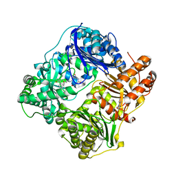
2QZ4
 | | Human paraplegin, AAA domain in complex with ADP | | 分子名称: | ADENOSINE-5'-DIPHOSPHATE, Paraplegin | | 著者 | Karlberg, T, Lehtio, L, Arrowsmith, C.H, Berglund, H, Busam, R.D, Collins, R, Dahlgren, L.G, Edwards, A, Flodin, S, Flores, A, Graslund, S, Hammarstrom, M, Herman, M.D, Johansson, I, Kallas, A, Kotenyova, T, Moche, M, Nilsson, M.E, Nordlund, P, Nyman, T, Persson, J, Sagemark, C, Sundstrom, M, Thorsell, A.G, Tresauges, L, Van Den Berg, S, Weigelt, J, Welin, M, Holmberg-Schiavone, L, Structural Genomics Consortium (SGC) | | 登録日 | 2007-08-16 | | 公開日 | 2007-09-11 | | 最終更新日 | 2023-08-30 | | 実験手法 | X-RAY DIFFRACTION (2.22 Å) | | 主引用文献 | Crystal Structure of the ATPase Domain of the Human AAA+ Protein Paraplegin/SPG7.
Plos One, 4, 2009
|
|
1X5A
 | | The solution structure of the second fibronectin type III domain of mouse Ephrin type-A receptor 1 | | 分子名称: | Ephrin type-A receptor 1 | | 著者 | Tochio, N, Sasagawa, A, Koshiba, S, Inoue, M, Kigawa, T, Yokoyama, S, RIKEN Structural Genomics/Proteomics Initiative (RSGI) | | 登録日 | 2005-05-15 | | 公開日 | 2005-11-15 | | 最終更新日 | 2024-05-29 | | 実験手法 | SOLUTION NMR | | 主引用文献 | The solution structure of the second fibronectin type III domain of mouse Ephrin type-A receptor 1
To be Published
|
|
1X5J
 | | The solution structure of the fifth fibronectin type III domain of human Neogenin | | 分子名称: | Neogenin | | 著者 | Tochio, N, Sasagawa, A, Koshiba, S, Inoue, M, Kigawa, T, Yokoyama, S, RIKEN Structural Genomics/Proteomics Initiative (RSGI) | | 登録日 | 2005-05-15 | | 公開日 | 2005-11-15 | | 最終更新日 | 2024-05-29 | | 実験手法 | SOLUTION NMR | | 主引用文献 | The solution structure of the fifth fibronectin type III domain of human Neogenin
To be Published
|
|
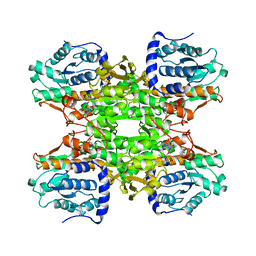
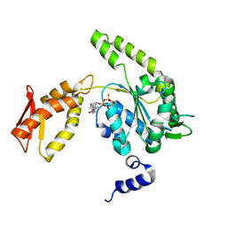
1RY8
 | | Prostaglandin F synthase complexed with NADPH and rutin | | 分子名称: | Aldo-keto reductase family 1 member C3, NADPH DIHYDRO-NICOTINAMIDE-ADENINE-DINUCLEOTIDE PHOSPHATE, RUTIN | | 著者 | Komoto, J, Yamada, T, Watanabe, K, Takusagawa, F. | | 登録日 | 2003-12-19 | | 公開日 | 2004-11-02 | | 最終更新日 | 2023-08-23 | | 実験手法 | X-RAY DIFFRACTION (1.69 Å) | | 主引用文献 | Crystal structure of human prostaglandin F synthase (AKR1C3).
Biochemistry, 43, 2004
|
|
2JCN
 | | The crystal structure of BAK1 - a mitochondrial apoptosis regulator | | 分子名称: | BCL-2 HOMOLOGOUS ANTAGONIST/KILLER, SULFATE ION | | 著者 | Moche, M, Stenmark, P, Arrowsmith, C, Berglund, H, Busam, R, Collins, R, Edwards, A, Ericsson, U.B, Flodin, S, Flores, A, Graslund, S, Hammarstrom, M, Hallberg, B.M, Holmberg Schiavone, L, Johansson, I, Karlberg, T, Kosinska, U, Kotenyova, T, Lundgren, S, Nilsson, M.E, Nyman, T, Ogg, D, Persson, C, Sagemark, J, Sundstrom, M, Uppenberg, J, Upsten, M, Thorsell, A.G, van den Berg, S, Weigelt, J, Nordlund, P, Structural Genomics Consortium (SGC) | | 登録日 | 2006-12-27 | | 公開日 | 2007-01-04 | | 最終更新日 | 2018-06-13 | | 実験手法 | X-RAY DIFFRACTION (1.8 Å) | | 主引用文献 | The Crystal Structure of Bak1 - an Apoptosis Trigger in the Mitochondrial Outer Membrane
To be Published
|
|
4LTE
 | | Structure of Cysteine-free Human Insulin Degrading Enzyme in Complex with Macrocyclic Inhibitor | | 分子名称: | 2,6-DIAMINO-HEXANOIC ACID AMIDE, 4-(2-HYDROXYETHYL)-1-PIPERAZINE ETHANESULFONIC ACID, FUMARIC ACID, ... | | 著者 | Foda, Z.H, Seeliger, M.A, Saghatelian, A, Liu, D.R. | | 登録日 | 2013-07-23 | | 公開日 | 2014-05-21 | | 最終更新日 | 2024-04-24 | | 実験手法 | X-RAY DIFFRACTION (2.705 Å) | | 主引用文献 | Anti-diabetic activity of insulin-degrading enzyme inhibitors mediated by multiple hormones.
Nature, 511, 2014
|
|
4LJ5
 | | ClpB NBD2 from T. thermophilus in complex with ADP | | 分子名称: | ADENOSINE-5'-DIPHOSPHATE, Chaperone protein ClpB | | 著者 | Zeymer, C, Barends, T.R.M, Werbeck, N.D, Schlichting, I, Reinstein, J. | | 登録日 | 2013-07-04 | | 公開日 | 2014-02-12 | | 最終更新日 | 2023-11-08 | | 実験手法 | X-RAY DIFFRACTION (2.4 Å) | | 主引用文献 | Elements in nucleotide sensing and hydrolysis of the AAA+ disaggregation machine ClpB: a structure-based mechanistic dissection of a molecular motor
Acta Crystallogr.,Sect.D, 70, 2014
|
|


