1EIE
 
 | |
1EID
 | |
1EOW
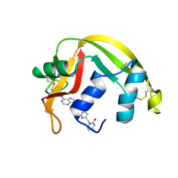
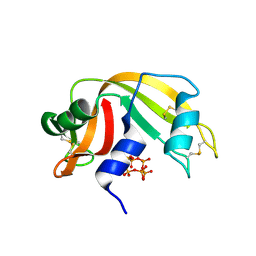
 | | CRYSTAL STRUCTURE OF RIBONUCLEASE A COMPLEXED WITH URIDYLYL(2',5')GUANOSINE (NON-PRODUCTIVE BINDING) | | 分子名称: | RIBONUCLEASE PANCREATIC, SULFATE ION, URIDYLYL-2'-5'-PHOSPHO-GUANOSINE | | 著者 | Vitagliano, L, Merlino, A, Zagari, A, Mazzarella, L. | | 登録日 | 2000-03-24 | | 公開日 | 2000-11-17 | | 最終更新日 | 2011-07-13 | | 実験手法 | X-RAY DIFFRACTION (2 Å) | | 主引用文献 | Productive and nonproductive binding to ribonuclease A: X-ray structure of two complexes with uridylyl(2',5')guanosine.
Protein Sci., 9, 2000
|
|
7P4R
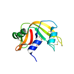
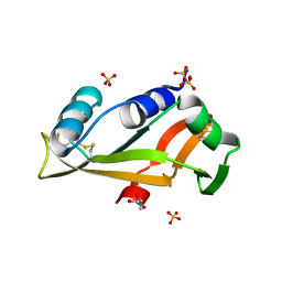
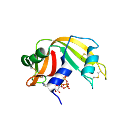
 | | Ultra High Resolution X-ray Structure of Orthorhombic Bovine Pancreatic Ribonuclease at 100K | | 分子名称: | ETHANOL, Ribonuclease pancreatic, SULFATE ION | | 著者 | Lisgarten, D.R, Palmer, R.A, Cooper, J.B, Naylor, C.E, Howlin, B.J, Lisgarten, J.N, Najmudin, S, Lobley, C.M.C. | | 登録日 | 2021-07-12 | | 公開日 | 2022-07-27 | | 最終更新日 | 2024-01-31 | | 実験手法 | X-RAY DIFFRACTION (0.85 Å) | | 主引用文献 | Ultra-high resolution X-ray structure of orthorhombic bovine pancreatic Ribonuclease A at 100K.
BMC Chem, 17, 2023
|
|
1EIC
 | |
7PNJ
 | |
7PNR
 | |
7PNI
 | |
8F5X
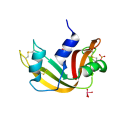
 | | Crystal structure of human eosinophil-derived neurotoxin (EDN, ribonuclease 2) in complex with 5'-adenosine monophosphate (AMP) | | 分子名称: | 1,2-ETHANEDIOL, ADENOSINE MONOPHOSPHATE, Non-secretory ribonuclease, ... | | 著者 | Tran, T.T.Q, Pham, N.T.H, Calmettes, C, Doucet, N. | | 登録日 | 2022-11-15 | | 公開日 | 2023-11-29 | | 最終更新日 | 2024-05-29 | | 実験手法 | X-RAY DIFFRACTION (1.7 Å) | | 主引用文献 | Ancestral sequence reconstruction dissects structural and functional differences among eosinophil ribonucleases.
J.Biol.Chem., 300, 2024
|
|
7NPM
 | |
8FHM
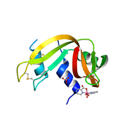
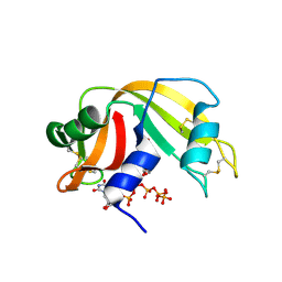
 | | RNase A-Uridine 5'-Hexaphosphate (RNaseA.p6U) | | 分子名称: | 5'-O-[(S)-hydroxy{[(S)-hydroxy{[(R)-hydroxy{[(S)-hydroxy{[(R)-hydroxy(phosphonooxy)phosphoryl]oxy}phosphoryl]oxy}phosphoryl]oxy}phosphoryl]oxy}phosphoryl]uridine, Ribonuclease pancreatic | | 著者 | Park, G, Cummins, C. | | 登録日 | 2022-12-14 | | 公開日 | 2023-12-20 | | 最終更新日 | 2024-08-07 | | 実験手法 | SOLUTION SCATTERING (1.79 Å), X-RAY DIFFRACTION | | 主引用文献 | Pentaphosphorylation via the Anhydride of Dihydrogen Pentametaphosphate: Access to Nucleoside Hexa- and Heptaphosphates and Study of Their Interaction with Ribonuclease A.
Acs Cent.Sci., 10, 2024
|
|
7OR6
 | |
7ORD
 | |
7P8R
 | |
8GGG
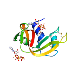 | | RNase A-Adenosine 5'-Hexaphosphate (RNaseA.p6A) | | 分子名称: | GLYCEROL, Ribonuclease pancreatic, adenosine 5'-hexaphosphate | | 著者 | Park, G, Cummins, C. | | 登録日 | 2023-03-08 | | 公開日 | 2024-03-13 | | 最終更新日 | 2024-08-07 | | 実験手法 | X-RAY DIFFRACTION (1.86 Å) | | 主引用文献 | Pentaphosphorylation via the Anhydride of Dihydrogen Pentametaphosphate: Access to Nucleoside Hexa- and Heptaphosphates and Study of Their Interaction with Ribonuclease A.
Acs Cent.Sci., 10, 2024
|
|
8GC9
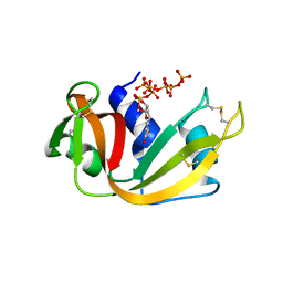 | | RNase A-Uridine 5'-Heptaphosphate (RNase A.p7U) | | 分子名称: | Ribonuclease pancreatic, uridine 5'-heptaphosphate | | 著者 | Park, G, Cummins, C. | | 登録日 | 2023-03-01 | | 公開日 | 2024-03-06 | | 最終更新日 | 2024-08-07 | | 実験手法 | X-RAY DIFFRACTION (1.85 Å) | | 主引用文献 | Pentaphosphorylation via the Anhydride of Dihydrogen Pentametaphosphate: Access to Nucleoside Hexa- and Heptaphosphates and Study of Their Interaction with Ribonuclease A.
Acs Cent.Sci., 10, 2024
|
|
5E13
 | | Crystal structure of Eosinophil-derived neurotoxin in complex with the triazole double-headed ribonucleoside 11c | | 分子名称: | 3'-{4-[(4-amino-2-oxopyrimidin-1(2H)-yl)methyl]-1H-1,2,3-triazol-1-yl}-3'-deoxyadenosine, Non-secretory ribonuclease | | 著者 | Chatzileontiadou, D.S.M, Stravodimos, G.A, Kantsadi, A.L, Leonidas, D.D. | | 登録日 | 2015-09-29 | | 公開日 | 2015-11-18 | | 最終更新日 | 2024-01-10 | | 実験手法 | X-RAY DIFFRACTION (1.34 Å) | | 主引用文献 | Triazole double-headed ribonucleosides as inhibitors of eosinophil derived neurotoxin.
Bioorg.Chem., 63, 2015
|
|
5EPZ
 | |
5EOP
 | |
5OAB
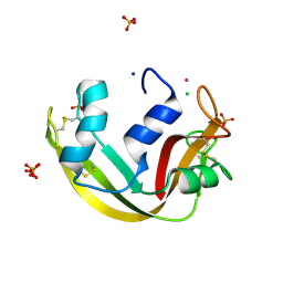 | | A novel crystal form of human RNase6 at atomic resolution | | 分子名称: | CHLORIDE ION, PHOSPHATE ION, POTASSIUM ION, ... | | 著者 | Prats-Ejarque, G, Moussaoui, M, Boix, E. | | 登録日 | 2017-06-21 | | 公開日 | 2018-08-01 | | 最終更新日 | 2024-01-17 | | 実験手法 | X-RAY DIFFRACTION (1.111 Å) | | 主引用文献 | Characterization of an RNase with two catalytic centers. Human RNase6 catalytic and phosphate-binding site arrangement favors the endonuclease cleavage of polymeric substrates.
Biochim Biophys Acta Gen Subj, 1863, 2019
|
|
5ET4
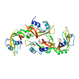 | | Structure of RNase A-K7H/R10H in complex with 3'-CMP | | 分子名称: | (4S)-2-METHYL-2,4-PENTANEDIOL, CYTIDINE-3'-MONOPHOSPHATE, Ribonuclease pancreatic | | 著者 | Blanco, J.A, Salazar, V.A, Moussaoui, M, Boix, E. | | 登録日 | 2015-11-17 | | 公開日 | 2016-11-30 | | 最終更新日 | 2024-01-10 | | 実験手法 | X-RAY DIFFRACTION (2.1 Å) | | 主引用文献 | Characterization of an RNase with two catalytic centers. Human RNase6 catalytic and phosphate-binding site arrangement favors the endonuclease cleavage of polymeric substrates.
Biochim Biophys Acta Gen Subj, 1863, 2019
|
|
6PVU
 | | RNase A in complex with hexametaphosphate | | 分子名称: | 2,4,6,8,10,12-hexahydroxy-2lambda~5~,4lambda~5~,6lambda~5~,8lambda~5~,10lambda~5~,12lambda~5~-cyclohexaphosphoxane-2,4,6,8,10,12-hexone, Ribonuclease pancreatic | | 著者 | Windsor, I.W, Sheppard, S.M, Cummins, C.C, Raines, R.T. | | 登録日 | 2019-07-21 | | 公開日 | 2019-11-06 | | 最終更新日 | 2023-10-11 | | 実験手法 | X-RAY DIFFRACTION (1.49 Å) | | 主引用文献 | Nucleoside Tetra- and Pentaphosphates Prepared Using a Tetraphosphorylation Reagent Are Potent Inhibitors of Ribonuclease A.
J.Am.Chem.Soc., 141, 2019
|
|
6PVW
 | | RNase A in complex with cp4pA | | 分子名称: | 2,4,6,8-tetrahydroxy-1,3,5,7,2lambda~5~,4lambda~5~,6lambda~5~,8lambda~5~-tetroxatetraphosphocane-2,4,6,8-tetrone, 5'-O-[(R)-hydroxy{[(4R,8S)-4,6,8-trihydroxy-2,4,6,8-tetraoxo-1,3,5,7,2lambda~5~,4lambda~5~,6lambda~5~,8lambda~5~-tetroxatetraphosphocan-2-yl]oxy}phosphoryl]adenosine, Ribonuclease pancreatic | | 著者 | Windsor, I.W, Sheppard, S.M, Cummins, C.C, Raines, R.T. | | 登録日 | 2019-07-21 | | 公開日 | 2019-11-06 | | 最終更新日 | 2023-10-11 | | 実験手法 | X-RAY DIFFRACTION (1.6 Å) | | 主引用文献 | Nucleoside Tetra- and Pentaphosphates Prepared Using a Tetraphosphorylation Reagent Are Potent Inhibitors of Ribonuclease A.
J.Am.Chem.Soc., 141, 2019
|
|
5OBC
 | | X-ray structure of the adduct formed upon reaction of ribonuclease A with the compound fac-[RuII(CO)3Cl2(N3-IM), IM=imidazole | | 分子名称: | PHOSPHATE ION, Ribonuclease pancreatic, pentakis(oxidaniumyl)-(oxidaniumylidynemethyl)ruthenium, ... | | 著者 | Pontillo, N, Ferraro, G, Merlino, A. | | 登録日 | 2017-06-26 | | 公開日 | 2017-07-26 | | 最終更新日 | 2024-01-17 | | 実験手法 | X-RAY DIFFRACTION (2.07 Å) | | 主引用文献 | Ru-Based CO releasing molecules with azole ligands: interaction with proteins and the CO release mechanism disclosed by X-ray crystallography.
Dalton Trans, 46, 2017
|
|
6PVX
 | | RNase A in complex with p5U | | 分子名称: | 5'-O-[(R)-hydroxy{[(S)-hydroxy{[(S)-hydroxy{[(S)-hydroxy(phosphonooxy)phosphoryl]oxy}phosphoryl]oxy}phosphoryl]oxy}phosphoryl]uridine, Ribonuclease pancreatic | | 著者 | Windsor, I.W, Sheppard, S.M, Cummins, C.C, Raines, R.T. | | 登録日 | 2019-07-21 | | 公開日 | 2019-11-06 | | 最終更新日 | 2023-10-11 | | 実験手法 | X-RAY DIFFRACTION (1.55 Å) | | 主引用文献 | Nucleoside Tetra- and Pentaphosphates Prepared Using a Tetraphosphorylation Reagent Are Potent Inhibitors of Ribonuclease A.
J.Am.Chem.Soc., 141, 2019
|
|


