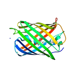
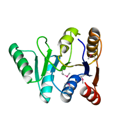
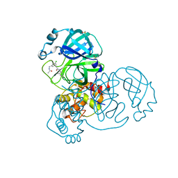
2GX2
 
 | | Crystal structural and functional analysis of GFP-like fluorescent protein Dronpa | | 分子名称: | MAGNESIUM ION, fluorescent protein Dronpa | | 著者 | Hwang, K.Y, Nam, K.-H, Park, S.-Y, Sugiyama, K. | | 登録日 | 2006-05-08 | | 公開日 | 2007-05-08 | | 最終更新日 | 2023-11-15 | | 実験手法 | X-RAY DIFFRACTION (1.8 Å) | | 主引用文献 | Structural characterization of the photoswitchable fluorescent protein Dronpa-C62S
Biochem.Biophys.Res.Commun., 354, 2007
|
|
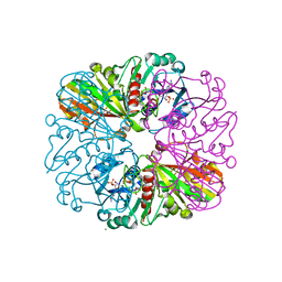
2GX0
 | |
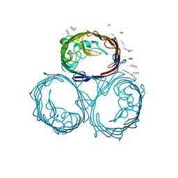
3VHT
 | | Crystal structure of GFP-Wrnip1 UBZ domain fusion protein in complex with ubiquitin | | 分子名称: | Green fluorescent protein, Green fluorescent protein,ATPase WRNIP1, Ubiquitin, ... | | 著者 | Suzuki, N, Wakatsuki, S, Kawasaki, M. | | 登録日 | 2011-09-06 | | 公開日 | 2012-10-10 | | 最終更新日 | 2023-12-06 | | 実験手法 | X-RAY DIFFRACTION (2.4 Å) | | 主引用文献 | A novel mode of ubiquitin recognition by the ubiquitin-binding zinc finger domain of WRNIP1.
Febs J., 2016
|
|
3VIC
 | | Green-fluorescent variant of the non-fluorescent chromoprotein Rtms5 | | 分子名称: | CHLORIDE ION, GFP-like non-fluorescent chromoprotein, IODIDE ION | | 著者 | Battad, J.M, Traore, D.A.K, Byres, E, Wilce, M, Devenish, R.J, Rossjohn, J, Prescott, M. | | 登録日 | 2011-09-28 | | 公開日 | 2012-06-06 | | 最終更新日 | 2023-11-15 | | 実験手法 | X-RAY DIFFRACTION (2.2 Å) | | 主引用文献 | A Green Fluorescent Protein Containing a QFG Tri-Peptide Chromophore: Optical Properties and X-Ray Crystal Structure.
Plos One, 7, 2012
|
|
3VK1
 | | Green-fluorescent variant of the non-fluorescent chromoprotein Rtms5 | | 分子名称: | CHLORIDE ION, GFP-like non-fluorescent chromoprotein, IODIDE ION | | 著者 | Battad, J.M, Traore, D.A.K, Wilce, M, Byres, M, Rossjohn, J, Devenish, R.J, Prescott, M. | | 登録日 | 2011-11-07 | | 公開日 | 2012-06-06 | | 最終更新日 | 2023-11-15 | | 実験手法 | X-RAY DIFFRACTION (2.2 Å) | | 主引用文献 | A Green Fluorescent Protein Containing a QFG Tri-Peptide Chromophore: Optical Properties and X-Ray Crystal Structure.
Plos One, 7, 2012
|
|
7Z01
 | | Z-SBTubA4 photoswitch bound to tubulin-DARPin D1 complex | | 分子名称: | 2-methoxy-5-[2-(5,6,7-trimethoxy-1,3-benzothiazol-2-yl)ethyl]phenol, CALCIUM ION, Designed Ankyrin Repeat Protein (DARPIN) D1, ... | | 著者 | Wranik, M, Weinert, T, Standfuss, J, Steinmetz, M. | | 登録日 | 2022-02-21 | | 公開日 | 2022-03-30 | | 最終更新日 | 2024-01-31 | | 実験手法 | X-RAY DIFFRACTION (1.82 Å) | | 主引用文献 | In Vivo Photocontrol of Microtubule Dynamics and Integrity, Migration and Mitosis, by the Potent GFP-Imaging-Compatible Photoswitchable Reagents SBTubA4P and SBTub2M.
J.Am.Chem.Soc., 144, 2022
|
|
7Z02
 | | Z-SBTub2M photoswitch bound to tubulin-DARPin D1 complex | | 分子名称: | 6-methyl-2-[2-(3,4,5-trimethoxyphenyl)ethyl]-1,3-benzothiazole, Designed Ankyrin Repeat Protein (DARPIN) D1, GUANOSINE-5'-DIPHOSPHATE, ... | | 著者 | Wranik, M, Weinert, T, Standfuss, J, Steinmetz, M. | | 登録日 | 2022-02-21 | | 公開日 | 2022-03-30 | | 最終更新日 | 2024-01-31 | | 実験手法 | X-RAY DIFFRACTION (2.36 Å) | | 主引用文献 | In Vivo Photocontrol of Microtubule Dynamics and Integrity, Migration and Mitosis, by the Potent GFP-Imaging-Compatible Photoswitchable Reagents SBTubA4P and SBTub2M.
J.Am.Chem.Soc., 144, 2022
|
|
8DJ8
 | |
6FSQ
 | |
6Y1G
 | | Photoconverted HcRed in its optoacoustic state | | 分子名称: | 1,2-ETHANEDIOL, GFP-like non-fluorescent chromoprotein | | 著者 | Janowski, R, Fuenzalida-Werner, J.P, Mishra, K, Stiel, A.C, Niessing, D. | | 登録日 | 2020-02-12 | | 公開日 | 2020-07-29 | | 最終更新日 | 2024-01-24 | | 実験手法 | X-RAY DIFFRACTION (2.3 Å) | | 主引用文献 | Challenging a Preconception: Optoacoustic Spectrum Differs from the Optical Absorption Spectrum of Proteins and Dyes for Molecular Imaging.
Anal.Chem., 92, 2020
|
|
6YLS
 | | mEos4b - Directionality of Optical Properties of Fluorescent Proteins | | 分子名称: | CHLORIDE ION, Green to red photoconvertible GFP-like protein EosFP, SODIUM ION | | 著者 | Myskova, J, Rybakova, O, Brynda, J, Lazar, J. | | 登録日 | 2020-04-07 | | 公開日 | 2020-12-16 | | 最終更新日 | 2024-01-24 | | 実験手法 | X-RAY DIFFRACTION (1.55 Å) | | 主引用文献 | Directionality of light absorption and emission in representative fluorescent proteins.
Proc.Natl.Acad.Sci.USA, 117, 2020
|
|
8AW4
 | |
6XZF
 | |
4DKM
 | | Crystal Structure of Amphioxus GFPc1a | | 分子名称: | Amphioxus Green Fluorescent Protein, GFPc1a | | 著者 | Deheyn, D.D, Bomati, E.K. | | 登録日 | 2012-02-03 | | 公開日 | 2013-05-15 | | 最終更新日 | 2023-12-06 | | 実験手法 | X-RAY DIFFRACTION (1.95 Å) | | 主引用文献 | Fluorescent proteins in Amphioxus have strickingly different brightness, yet
only few (but key) molecular differences
To be Published
|
|
4DKN
 | |
1GFP
 | | OMPF PORIN (MUTANT R42C) | | 分子名称: | (HYDROXYETHYLOXY)TRI(ETHYLOXY)OCTANE, MATRIX PORIN OUTER MEMBRANE PROTEIN F | | 著者 | Lou, K.-L, Schirmer, T. | | 登録日 | 1996-05-08 | | 公開日 | 1996-12-07 | | 最終更新日 | 2024-02-07 | | 実験手法 | X-RAY DIFFRACTION (2.7 Å) | | 主引用文献 | Structural and functional characterization of OmpF porin mutants selected for larger pore size. I. Crystallographic analysis.
J.Biol.Chem., 271, 1996
|
|
3GFP
 | | Structure of the C-terminal domain of the DEAD-box protein Dbp5 | | 分子名称: | DEAD box protein 5 | | 著者 | Erzberger, J.P, Dossani, Z.Y, Weirich, C.S, Weis, K, Berger, J.M. | | 登録日 | 2009-02-27 | | 公開日 | 2009-09-01 | | 最終更新日 | 2011-07-13 | | 実験手法 | X-RAY DIFFRACTION (1.8 Å) | | 主引用文献 | Structure of the C-terminus of the mRNA export factor Dbp5 reveals the interaction surface for the ATPase activator Gle1
Proc.Natl.Acad.Sci.USA, 106, 2009
|
|
4GFP
 | | 2.7 Angstrom resolution structure of 3-phosphoshikimate 1-carboxyvinyltransferase (AroA) from Coxiella burnetii in a second conformational state | | 分子名称: | 3-phosphoshikimate 1-carboxyvinyltransferase, BETA-MERCAPTOETHANOL | | 著者 | Light, S.H, Minasov, G, Krishna, S.N, Shuvalova, L, Papazisi, L, Anderson, W.F, Center for Structural Genomics of Infectious Diseases (CSGID) | | 登録日 | 2012-08-03 | | 公開日 | 2012-08-15 | | 最終更新日 | 2023-09-13 | | 実験手法 | X-RAY DIFFRACTION (2.7 Å) | | 主引用文献 | 2.7 Angstrom resolution structure of 3-phosphoshikimate 1-carboxyvinyltransferase (AroA) from Coxiella burnetii in second conformational state
TO BE PUBLISHED
|
|
8GFP
 | | Crystal structure of soluble lytic transglycosylase Cj0843 of Campylobacter jejuni in complex with N-acetyl-2,3-dehydro-2-deoxyneuraminic acid inhibitor | | 分子名称: | 2-DEOXY-2,3-DEHYDRO-N-ACETYL-NEURAMINIC ACID, CITRIC ACID, Lytic transglycosylase domain-containing protein, ... | | 著者 | van den Akker, F, Kumar, V. | | 登録日 | 2023-03-08 | | 公開日 | 2023-05-24 | | 最終更新日 | 2023-08-16 | | 実験手法 | X-RAY DIFFRACTION (2.07 Å) | | 主引用文献 | Exploring the inhibition of the soluble lytic transglycosylase Cj0843c of Campylobacter jejuni via targeting different sites with different scaffolds.
Protein Sci., 32, 2023
|
|
7GFP
 | | Group deposition SARS-CoV-2 main protease in complex with inhibitors from the COVID Moonshot -- Crystal Structure of SARS-CoV-2 main protease in complex with RAL-THA-6b94ceba-5 (Mpro-x11801) | | 分子名称: | 2-[3-(acetamidomethyl)-5-chlorophenyl]-N-(4-methylpyridin-3-yl)acetamide, 3C-like proteinase, DIMETHYL SULFOXIDE | | 著者 | Fearon, D, Aimon, A, Aschenbrenner, J.C, Balcomb, B.H, Bertram, F.K.R, Brandao-Neto, J, Dias, A, Douangamath, A, Dunnett, L, Godoy, A.S, Gorrie-Stone, T.J, Koekemoer, L, Krojer, T, Lithgo, R.M, Lukacik, P, Marples, P.G, Mikolajek, H, Nelson, E, Owen, C.D, Powell, A.J, Rangel, V.L, Skyner, R, Strain-Damerell, C.M, Thompson, W, Tomlinson, C.W.E, Wild, C, Walsh, M.A, von Delft, F. | | 登録日 | 2023-08-11 | | 公開日 | 2023-11-08 | | 最終更新日 | 2023-12-06 | | 実験手法 | X-RAY DIFFRACTION (1.769 Å) | | 主引用文献 | Open science discovery of potent noncovalent SARS-CoV-2 main protease inhibitors.
Science, 382, 2023
|
|
6GFP
 | | cyanobacterial GAPDH with NADP bound | | 分子名称: | FORMIC ACID, Glyceraldehyde-3-phosphate dehydrogenase, MAGNESIUM ION, ... | | 著者 | McFarlane, C.R, Briggs, L, Murray, J.W. | | 登録日 | 2018-05-01 | | 公開日 | 2019-05-08 | | 最終更新日 | 2024-01-17 | | 実験手法 | X-RAY DIFFRACTION (1.54 Å) | | 主引用文献 | Structural basis of light-induced redox regulation in the Calvin-Benson cycle in cyanobacteria.
Proc.Natl.Acad.Sci.USA, 116, 2019
|
|
2GFP
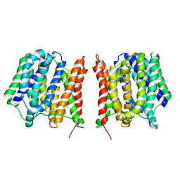 | | Structure of the Multidrug Transporter EmrD from Escherichia coli | | 分子名称: | Multidrug resistance protein D | | 著者 | Yin, Y, He, X, Szewczyk, P, Nguyen, T, Chang, G. | | 登録日 | 2006-03-22 | | 公開日 | 2006-05-16 | | 最終更新日 | 2024-02-14 | | 実験手法 | X-RAY DIFFRACTION (3.5 Å) | | 主引用文献 | Structure of the multidrug transporter EmrD from Escherichia coli
Science, 312, 2006
|
|
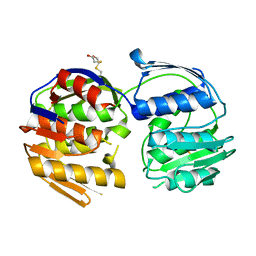
3ZTF
 | | X-ray Structure of the Cyan Fluorescent Protein mTurquoise2 (K206A mutant) | | 分子名称: | GREEN FLUORESCENT PROTEIN | | 著者 | von Stetten, D, Goedhart, J, Noirclerc-Savoye, M, Lelimousin, M, Joosen, L, Hink, M.A, van Weeren, L, Gadella, T.W.J, Royant, A. | | 登録日 | 2011-07-07 | | 公開日 | 2012-03-21 | | 最終更新日 | 2023-12-20 | | 実験手法 | X-RAY DIFFRACTION (1.31 Å) | | 主引用文献 | Structure-Guided Evolution of Cyan Fluorescent Proteins Towards a Quantum Yield of 93%
Nat.Commun, 3, 2012
|
|
4B5Y
 | | X-ray structure of the cyan fluorescent protein mTurquoise-GL (K206A mutant) in space group C222(1) | | 分子名称: | GREEN FLUORESCENT PROTEIN | | 著者 | von Stetten, D, Lelimousin, M, Oost, K, Noirclerc-Savoye, M, Gadella, T.W.J, Goedhart, J, Royant, A. | | 登録日 | 2012-08-08 | | 公開日 | 2013-08-28 | | 最終更新日 | 2023-12-20 | | 実験手法 | X-RAY DIFFRACTION (1.45 Å) | | 主引用文献 | Influence of the H148G Mutation on Fluorescence Properties of Cyan Fluorescent Proteins
To be Published
|
|
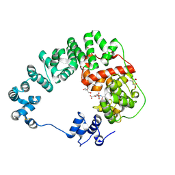
4AS8
 | | X-ray structure of the cyan fluorescent protein Cerulean cryoprotected with ethylene glycol | | 分子名称: | 1,2-ETHANEDIOL, GREEN FLUORESCENT PROTEIN, MAGNESIUM ION | | 著者 | von Stetten, D, Batot, G, Noirclerc-Savoye, M, Royant, A. | | 登録日 | 2012-04-29 | | 公開日 | 2012-10-31 | | 最終更新日 | 2023-12-20 | | 実験手法 | X-RAY DIFFRACTION (1.02 Å) | | 主引用文献 | Alteration of Fluorescent Protein Spectroscopic Properties Upon Cryoprotection
Acta Crystallogr.,Sect.D, 68, 2012
|
|


