2NLE
 
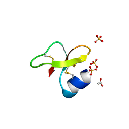 | |
2NLF
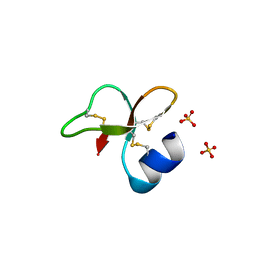 | |
2NLG
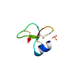 | |
2NLH
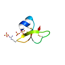 | |
2NLI
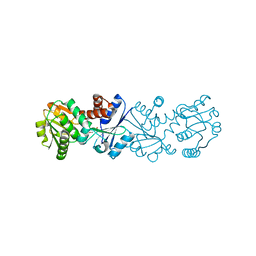
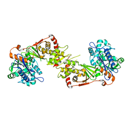
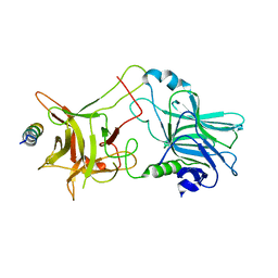
 | | Crystal Structure of the complex between L-lactate oxidase and a substrate analogue at 1.59 angstrom resolution | | 分子名称: | FLAVIN MONONUCLEOTIDE, HYDROGEN PEROXIDE, LACTIC ACID, ... | | 著者 | Furuichi, M, Suzuki, N, Balasundaresan, D, Yoshida, Y, Minagawa, H, Watanabe, Y, Kaneko, H, Waga, I, Kumar, P.K.R, Mizuno, H. | | 登録日 | 2006-10-20 | | 公開日 | 2007-10-23 | | 最終更新日 | 2023-11-15 | | 実験手法 | X-RAY DIFFRACTION (1.59 Å) | | 主引用文献 | X-ray structures of Aerococcus viridans lactate oxidase and its complex with D-lactate at pH 4.5 show an alpha-hydroxyacid oxidation mechanism
J.Mol.Biol., 378, 2008
|
|
2NLJ
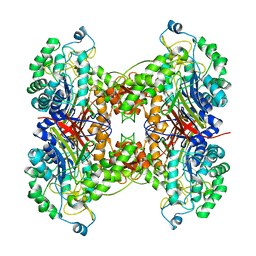
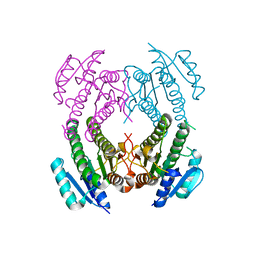
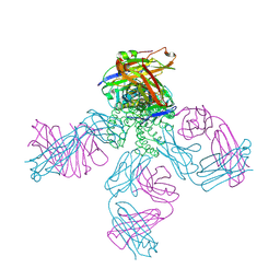 | | Potassium Channel KcsA(M96V)-Fab complex in KCl | | 分子名称: | DIACYL GLYCEROL, POTASSIUM ION, Voltage-gated potassium channel, ... | | 著者 | Lockless, S.W, Zhou, M, MacKinnon, R. | | 登録日 | 2006-10-20 | | 公開日 | 2007-05-15 | | 最終更新日 | 2023-12-27 | | 実験手法 | X-RAY DIFFRACTION (2.52 Å) | | 主引用文献 | Structural and Thermodynamic Properties of Selective Ion Binding in a K(+) Channel.
Plos Biol., 5, 2007
|
|
2NLK
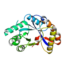
 | | Crystal structure of D1 and D2 catalytic domains of human Protein Tyrosine Phosphatase Gamma (D1+D2 PTPRG) | | 分子名称: | Protein tyrosine phosphatase, receptor type, G variant (Fragment) | | 著者 | Filippakopoulos, P, Gileadi, O, Johansson, C, Ugochukwu, E, Edwards, A, Arrowsmith, C, Sundstrom, M, von Delft, F, Knapp, S, Structural Genomics Consortium (SGC) | | 登録日 | 2006-10-20 | | 公開日 | 2006-11-21 | | 最終更新日 | 2023-08-30 | | 実験手法 | X-RAY DIFFRACTION (2.4 Å) | | 主引用文献 | Large-scale structural analysis of the classical human protein tyrosine phosphatome.
Cell(Cambridge,Mass.), 136, 2009
|
|
2NLL
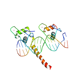 | | RETINOID X RECEPTOR-THYROID HORMONE RECEPTOR DNA-BINDING DOMAIN HETERODIMER BOUND TO THYROID RESPONSE ELEMENT DNA | | 分子名称: | DNA (5'-D(*CP*AP*GP*GP*TP*CP*AP*TP*TP*(5IU)P*CP*AP*GP*GP*TP*CP*AP*G)-3'), DNA (5'-D(*CP*TP*GP*AP*CP*CP*TP*GP*AP*AP*AP*TP*GP*AP*CP*CP*T P*G)-3'), PROTEIN (RETINOIC ACID RECEPTOR), ... | | 著者 | Rastinejad, F, Perlmann, T, Evans, R.M, Sigler, P.B. | | 登録日 | 1996-11-20 | | 公開日 | 1997-03-12 | | 最終更新日 | 2024-02-21 | | 実験手法 | X-RAY DIFFRACTION (1.9 Å) | | 主引用文献 | Structural determinants of nuclear receptor assembly on DNA direct repeats.
Nature, 375, 1995
|
|
2NLM
 | |
2NLN
 | |
2NLO
 | |
2NLP
 | |
2NLQ
 | |
2NLR
 | |
2NLS
 | |
2NLU
 | |
2NLV
 | |
2NLW
 | |
2NLX
 | |
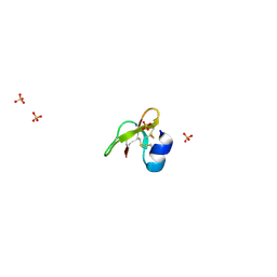
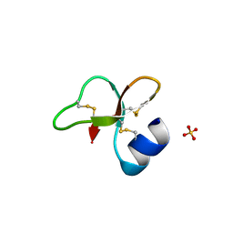
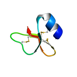
2NLY
 | | Crystal structure of protein BH1492 from Bacillus halodurans, Pfam DUF610 | | 分子名称: | Divergent polysaccharide deacetylase hypothetical protein, ZINC ION | | 著者 | Jin, X, Sauder, J.M, Wasserman, S, Smith, D, Burley, S.K, Shapiro, L, New York SGX Research Center for Structural Genomics (NYSGXRC) | | 登録日 | 2006-10-20 | | 公開日 | 2006-11-07 | | 最終更新日 | 2023-12-27 | | 実験手法 | X-RAY DIFFRACTION (2.5 Å) | | 主引用文献 | Crystal structure of hypothetical protein BH1492 from Bacillus halodurans C-125
To be Published
|
|
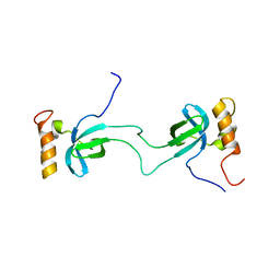
2NLZ
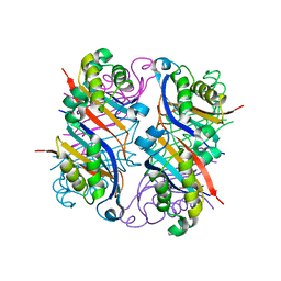
 | | Crystal structure of cephalosporin acylase from Bacillus halodurans | | 分子名称: | Cephalosporin acylase | | 著者 | Patskovsky, Y, Ramagopal, U, Sauder, J.M, Dickey, M, Adams, J.M, Ozyurt, S, Wasserman, S.R, Burley, S.K, Almo, S.C, New York SGX Research Center for Structural Genomics (NYSGXRC) | | 登録日 | 2006-10-20 | | 公開日 | 2006-11-07 | | 最終更新日 | 2023-12-27 | | 実験手法 | X-RAY DIFFRACTION (2.7 Å) | | 主引用文献 | Crystal structure of cephalosporin acylase from Bacillus halodurans
To be Published
|
|
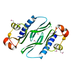
2NM0
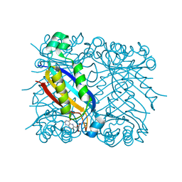
 | | Crystal Structure of SCO1815: a Beta-Ketoacyl-Acyl Carrier Protein Reductase from Streptomyces coelicolor A3(2) | | 分子名称: | Probable 3-oxacyl-(Acyl-carrier-protein) reductase | | 著者 | Khosla, C, Tang, Y, Lee, H.Y, Tang, Y, Kim, C.Y, Mathews, I.I. | | 登録日 | 2006-10-20 | | 公開日 | 2007-01-02 | | 最終更新日 | 2023-08-30 | | 実験手法 | X-RAY DIFFRACTION (1.99 Å) | | 主引用文献 | Structural and functional studies on SCO1815: a beta-ketoacyl-acyl carrier protein reductase from Streptomyces coelicolor A3(2).
Biochemistry, 45, 2006
|
|
2NM1
 | | Structure of BoNT/B in complex with its protein receptor | | 分子名称: | Botulinum neurotoxin type B, Synaptotagmin-2 | | 著者 | Jin, R, Rummel, A, Binz, T, Brunger, A.T. | | 登録日 | 2006-10-20 | | 公開日 | 2006-12-19 | | 最終更新日 | 2023-08-30 | | 実験手法 | X-RAY DIFFRACTION (2.15 Å) | | 主引用文献 | Botulinum neurotoxin B recognizes its protein receptor with high affinity and specificity.
Nature, 444, 2006
|
|
2NM2
 | |
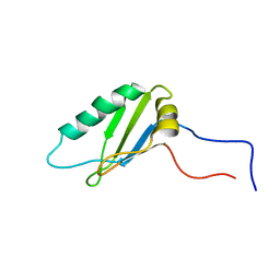
2NM3
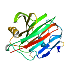
 | | Crystal structure of dihydroneopterin aldolase from S. aureus in complex with (1S,2S)-monapterin at 1.68 angstrom resolution | | 分子名称: | ACETATE ION, D-MONAPTERIN, Dihydroneopterin aldolase | | 著者 | Blaszczyk, J, Ji, X, Yan, H. | | 登録日 | 2006-10-20 | | 公開日 | 2007-09-04 | | 最終更新日 | 2023-08-30 | | 実験手法 | X-RAY DIFFRACTION (1.68 Å) | | 主引用文献 | Structural basis for the aldolase and epimerase activities of Staphylococcus aureus dihydroneopterin aldolase.
J.Mol.Biol., 368, 2007
|
|


