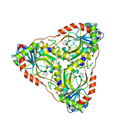
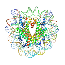
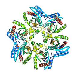
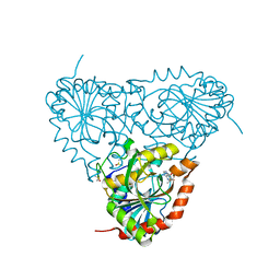
1S57
 
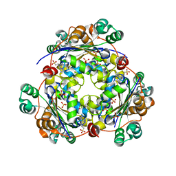 | | crystal structure of nucleoside diphosphate kinase 2 from Arabidopsis | | 分子名称: | 4-(2-HYDROXYETHYL)-1-PIPERAZINE ETHANESULFONIC ACID, Nucleoside diphosphate kinase II, SULFATE ION | | 著者 | Im, Y.J, Kim, J.-I, Shen, Y, Na, Y, Han, Y.-J, Kim, S.-H, Song, P.-S, Eom, S.H. | | 登録日 | 2004-01-20 | | 公開日 | 2004-11-30 | | 最終更新日 | 2023-10-25 | | 実験手法 | X-RAY DIFFRACTION (1.8 Å) | | 主引用文献 | Structural analysis of Arabidopsis thaliana nucleoside diphosphate kinase-2 for phytochrome-mediated light signaling
J.Mol.Biol., 343, 2004
|
|
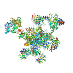
6FF4
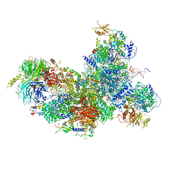 | | human Bact spliceosome core structure | | 分子名称: | 116 kDa U5 small nuclear ribonucleoprotein component, BUD13 homolog, Cell division cycle 5-like protein, ... | | 著者 | Haselbach, D, Komarov, I, Agafonov, D, Hartmuth, K, Graf, B, Kastner, B, Luehrmann, R, Stark, H. | | 登録日 | 2018-01-03 | | 公開日 | 2018-08-29 | | 最終更新日 | 2025-07-02 | | 実験手法 | ELECTRON MICROSCOPY (3.4 Å) | | 主引用文献 | Structure and Conformational Dynamics of the Human Spliceosomal BactComplex.
Cell, 172, 2018
|
|
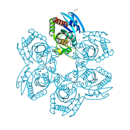
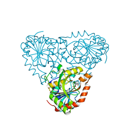
3KHS
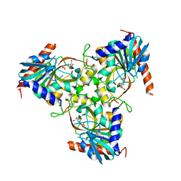 | | Crystal structure of grouper iridovirus purine nucleoside phosphorylase | | 分子名称: | 2-AMINO-2-HYDROXYMETHYL-PROPANE-1,3-DIOL, PHOSPHATE ION, Purine nucleoside phosphorylase | | 著者 | Kang, Y.N, Zhang, Y, Allan, P.W, Parker, W.B, Ting, J.W, Chang, C.Y, Ealick, S.E. | | 登録日 | 2009-10-30 | | 公開日 | 2010-02-16 | | 最終更新日 | 2024-11-06 | | 実験手法 | X-RAY DIFFRACTION (2.38 Å) | | 主引用文献 | Structure of grouper iridovirus purine nucleoside phosphorylase
Acta Crystallogr.,Sect.D, 66, 2010
|
|
5I3C
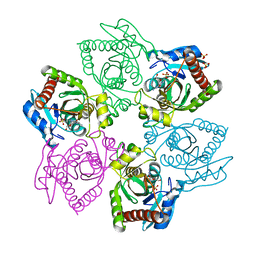 | | Crystal structure of E.coli purine nucleoside phosphorylase with acycloguanosine | | 分子名称: | 9-HYROXYETHOXYMETHYLGUANINE, Purine nucleoside phosphorylase DeoD-type, SULFATE ION | | 著者 | Timofeev, V.I, Abramchik, Y.A, Esipov, R.S, Kuranova, I.P. | | 登録日 | 2016-02-10 | | 公開日 | 2017-02-22 | | 最終更新日 | 2024-01-10 | | 実験手法 | X-RAY DIFFRACTION (2.32 Å) | | 主引用文献 | Crystal structure of Escherichia coli purine nucleoside phosphorylase complexed with acyclovir.
Acta Crystallogr F Struct Biol Commun, 74, 2018
|
|
6QDV
 | | Human post-catalytic P complex spliceosome | | 分子名称: | 116 kDa U5 small nuclear ribonucleoprotein component, ADENOSINE-5'-TRIPHOSPHATE, ATP-dependent RNA helicase DHX8, ... | | 著者 | Fica, S.M, Oubridge, C, Wilkinson, M.E, Newman, A.J, Nagai, K. | | 登録日 | 2019-01-03 | | 公開日 | 2019-02-20 | | 最終更新日 | 2024-11-13 | | 実験手法 | ELECTRON MICROSCOPY (3.3 Å) | | 主引用文献 | A human postcatalytic spliceosome structure reveals essential roles of metazoan factors for exon ligation.
Science, 363, 2019
|
|
5XJC
 | | Cryo-EM structure of the human spliceosome just prior to exon ligation at 3.6 angstrom | | 分子名称: | 116 kDa U5 small nuclear ribonucleoprotein component, ADENOSINE-5'-DIPHOSPHATE, ADENOSINE-5'-TRIPHOSPHATE, ... | | 著者 | Zhang, X, Yan, C, Hang, J, Finci, I.L, Lei, J, Shi, Y. | | 登録日 | 2017-04-30 | | 公開日 | 2017-07-05 | | 最終更新日 | 2024-11-20 | | 実験手法 | ELECTRON MICROSCOPY (3.6 Å) | | 主引用文献 | An Atomic Structure of the Human Spliceosome
Cell, 169, 2017
|
|
5IU6
 | | Crystal structure of E.coli purine nucleoside phosphorylase with 7-deazahypoxanthine | | 分子名称: | 7H-pyrrolo[2,3-d]pyrimidin-4-ol, Purine nucleoside phosphorylase DeoD-type, SULFATE ION | | 著者 | Timofeev, V.I, Abramchik, Y.A, Esipov, R.S, Kuranova, I.P. | | 登録日 | 2016-03-17 | | 公開日 | 2017-04-05 | | 最終更新日 | 2024-05-08 | | 実験手法 | X-RAY DIFFRACTION (2.51 Å) | | 主引用文献 | Crystal structure of Escherichia coli purine nucleoside phosphorylase in complex with 7-deazahypoxanthine.
Acta Crystallogr F Struct Biol Commun, 74, 2018
|
|
3MB8
 | | Crystal structure of purine nucleoside phosphorylase from toxoplasma gondii in complex with immucillin-H | | 分子名称: | 1,4-DIDEOXY-4-AZA-1-(S)-(9-DEAZAHYPOXANTHIN-9-YL)-D-RIBITOL, GLYCEROL, PHOSPHATE ION, ... | | 著者 | Ho, M, Almo, S.C, Schramm, V.L. | | 登録日 | 2010-03-25 | | 公開日 | 2011-04-06 | | 最終更新日 | 2024-05-22 | | 実験手法 | X-RAY DIFFRACTION (1.9 Å) | | 主引用文献 | Inhibition and Structure of Toxoplasma gondii Purine Nucleoside Phosphorylase.
Eukaryot Cell, 13, 2014
|
|
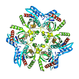
5IFK
 | | Purine nucleoside phosphorylase | | 分子名称: | HYPOXANTHINE, Purine nucleoside phosphorylase | | 著者 | Thakur, K.G, Priyanka, A. | | 登録日 | 2016-02-26 | | 公開日 | 2017-03-01 | | 最終更新日 | 2024-03-20 | | 実験手法 | X-RAY DIFFRACTION (1.967 Å) | | 主引用文献 | Functional and Structural Characterization of Purine Nucleoside Phosphorylase from Kluyveromyces lactis and Its Potential Applications in Reducing Purine Content in Food
PLoS ONE, 11, 2016
|
|
6NZO
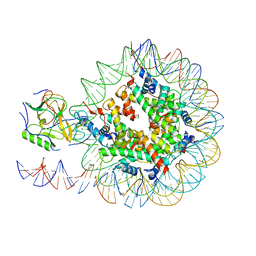 | | Set2 bound to nucleosome | | 分子名称: | DNA (149-MER), Histone H2B 1.1, Histone H3, ... | | 著者 | Halic, M, Bilokapic, S. | | 登録日 | 2019-02-14 | | 公開日 | 2019-08-28 | | 最終更新日 | 2025-06-04 | | 実験手法 | ELECTRON MICROSCOPY (3.8 Å) | | 主引用文献 | Nucleosome and ubiquitin position Set2 to methylate H3K36.
Nat Commun, 10, 2019
|
|
6PX3
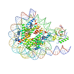 | | Set2 bound to nucleosome | | 分子名称: | DNA (145-MER), Histone H2B 1.1, Histone H3, ... | | 著者 | Halic, M, Bilokapic, S. | | 登録日 | 2019-07-24 | | 公開日 | 2019-08-28 | | 最終更新日 | 2025-05-28 | | 実験手法 | ELECTRON MICROSCOPY (4.1 Å) | | 主引用文献 | Nucleosome and ubiquitin position Set2 to methylate H3K36.
Nat Commun, 10, 2019
|
|
6PX1
 | | Set2 bound to nucleosome | | 分子名称: | DNA (149-MER), Histone H2B 1.1, Histone H3, ... | | 著者 | Halic, M, Bilokapic, S. | | 登録日 | 2019-07-24 | | 公開日 | 2019-08-28 | | 最終更新日 | 2025-05-21 | | 実験手法 | ELECTRON MICROSCOPY (3.3 Å) | | 主引用文献 | Nucleosome and ubiquitin position Set2 to methylate H3K36.
Nat Commun, 10, 2019
|
|
8W7H
 | | Purine Nucleoside Phosphorylase in complex with MMV000848 | | 分子名称: | (2R)-1-(9H-carbazol-9-yl)-3-(cyclopentylamino)propan-2-ol, 1,2-ETHANEDIOL, PHOSPHATE ION, ... | | 著者 | Chung, Z, Lin, J.Q, Lescar, J. | | 登録日 | 2023-08-30 | | 公開日 | 2024-01-17 | | 最終更新日 | 2024-01-31 | | 実験手法 | X-RAY DIFFRACTION (1.85 Å) | | 主引用文献 | Identification and structural validation of purine nucleoside phosphorylase from Plasmodium falciparum as a target of MMV000848.
J.Biol.Chem., 300, 2023
|
|
6GP1
 | | Structure of mEos4b in the red long-lived dark state | | 分子名称: | Green to red photoconvertible GFP-like protein EosFP | | 著者 | De Zitter, E, Adam, V, Byrdin, M, Van Meervelt, L, Dedecker, P, Bourgeois, D. | | 登録日 | 2018-06-04 | | 公開日 | 2019-05-22 | | 最終更新日 | 2024-01-17 | | 実験手法 | X-RAY DIFFRACTION (1.504 Å) | | 主引用文献 | Mechanistic investigation of mEos4b reveals a strategy to reduce track interruptions in sptPALM.
Nat.Methods, 16, 2019
|
|
6GOZ
 | | Structure of mEos4b in the green long-lived dark state | | 分子名称: | 1,2-ETHANEDIOL, DI(HYDROXYETHYL)ETHER, GLYCEROL, ... | | 著者 | De Zitter, E, Adam, V, Byrdin, M, Van Meervelt, L, Dedecker, P, Bourgeois, D. | | 登録日 | 2018-06-04 | | 公開日 | 2019-11-13 | | 最終更新日 | 2024-10-16 | | 実験手法 | X-RAY DIFFRACTION (2.406 Å) | | 主引用文献 | Mechanistic Investigations of Green mEos4b Reveal a Dynamic Long-Lived Dark State.
J.Am.Chem.Soc., 2020
|
|
7OOZ
 | | Purine nucleoside phosphorylase(DeoD-type) from H. pylori with 6-benzyloxo-2-chloropurine | | 分子名称: | 2-AMINO-2-HYDROXYMETHYL-PROPANE-1,3-DIOL, 6-benzyloxo-2-chloropurine, GLYCEROL, ... | | 著者 | Narczyk, M, Stefanic, Z. | | 登録日 | 2021-05-28 | | 公開日 | 2022-05-04 | | 最終更新日 | 2024-11-20 | | 実験手法 | X-RAY DIFFRACTION (1.7 Å) | | 主引用文献 | Interactions of 2,6-substituted purines with purine nucleoside phosphorylase from Helicobacter pylori in solution and in the crystal, and the effects of these compounds on cell cultures of this bacterium.
J Enzyme Inhib Med Chem, 37, 2022
|
|
7OP9
 | | Purine nucleoside phosphorylase(DeoD-type) from H. pylori with 2,6-dichloropurine | | 分子名称: | 2,6-bis(chloranyl)-7H-purine, IMIDAZOLE, MAGNESIUM ION, ... | | 著者 | Narczyk, M, Stefanic, Z. | | 登録日 | 2021-05-31 | | 公開日 | 2022-05-04 | | 最終更新日 | 2024-01-31 | | 実験手法 | X-RAY DIFFRACTION (1.5 Å) | | 主引用文献 | Interactions of 2,6-substituted purines with purine nucleoside phosphorylase from Helicobacter pylori in solution and in the crystal, and the effects of these compounds on cell cultures of this bacterium.
J Enzyme Inhib Med Chem, 37, 2022
|
|
7OPA
 | | Purine nucleoside phosphorylase(DeoD-type) from H. pylori with 6-benzylthiopurine | | 分子名称: | 2-AMINO-2-HYDROXYMETHYL-PROPANE-1,3-DIOL, 6-benzylthio-2-chloropurine, GLYCEROL, ... | | 著者 | Narczyk, M, Stefanic, Z. | | 登録日 | 2021-05-31 | | 公開日 | 2022-05-04 | | 最終更新日 | 2024-11-06 | | 実験手法 | X-RAY DIFFRACTION (2 Å) | | 主引用文献 | Interactions of 2,6-substituted purines with purine nucleoside phosphorylase from Helicobacter pylori in solution and in the crystal, and the effects of these compounds on cell cultures of this bacterium.
J Enzyme Inhib Med Chem, 37, 2022
|
|
7OOY
 | | Purine nucleoside phosphorylase(DeoD-type) from H. pylori with 6-benzylthio-2-chloropurine | | 分子名称: | 2-AMINO-2-HYDROXYMETHYL-PROPANE-1,3-DIOL, 6-benzylthio-2-chloropurine, GLYCEROL, ... | | 著者 | Narczyk, M, Stefanic, Z. | | 登録日 | 2021-05-28 | | 公開日 | 2022-05-04 | | 最終更新日 | 2024-11-20 | | 実験手法 | X-RAY DIFFRACTION (1.9 Å) | | 主引用文献 | Interactions of 2,6-substituted purines with purine nucleoside phosphorylase from Helicobacter pylori in solution and in the crystal, and the effects of these compounds on cell cultures of this bacterium.
J Enzyme Inhib Med Chem, 37, 2022
|
|
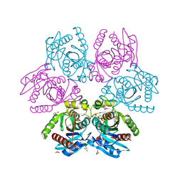
5TUS
 | | Potent competitive inhibition of human ribonucleotide reductase by a novel non-nucleoside small molecule | | 分子名称: | 2-hydroxy-N'-[(Z)-(2-hydroxynaphthalen-1-yl)methylidene]benzohydrazide, MAGNESIUM ION, Ribonucleoside-diphosphate reductase large subunit, ... | | 著者 | Mohammed, F.A, Alam, I, Dealwis, C.G. | | 登録日 | 2016-11-07 | | 公開日 | 2017-08-02 | | 最終更新日 | 2024-10-16 | | 実験手法 | X-RAY DIFFRACTION (2.66 Å) | | 主引用文献 | Potent competitive inhibition of human ribonucleotide reductase by a nonnucleoside small molecule.
Proc. Natl. Acad. Sci. U.S.A., 114, 2017
|
|
6N2G
 | |
6GP0
 | | Structure of mEos4b in the red fluorescent state | | 分子名称: | Green to red photoconvertible GFP-like protein EosFP | | 著者 | De Zitter, E, Adam, V, Byrdin, M, Van Meervelt, L, Dedecker, P, Bourgeois, D. | | 登録日 | 2018-06-04 | | 公開日 | 2019-05-22 | | 最終更新日 | 2025-07-16 | | 実験手法 | X-RAY DIFFRACTION (1.5 Å) | | 主引用文献 | Mechanistic investigation of mEos4b reveals a strategy to reduce track interruptions in sptPALM.
Nat.Methods, 16, 2019
|
|
6GOY
 | | Structure of mEos4b in the green fluorescent state | | 分子名称: | 1,2-ETHANEDIOL, DI(HYDROXYETHYL)ETHER, GLYCEROL, ... | | 著者 | De Zitter, E, Adam, V, Byrdin, M, Van Meervelt, L, Dedecker, P, Bourgeois, D. | | 登録日 | 2018-06-04 | | 公開日 | 2019-05-22 | | 最終更新日 | 2024-10-23 | | 実験手法 | X-RAY DIFFRACTION (1.65 Å) | | 主引用文献 | Mechanistic investigation of mEos4b reveals a strategy to reduce track interruptions in sptPALM.
Nat.Methods, 16, 2019
|
|
5TBU
 | | Crystal Structure of Isoform 2 of Purine Nucleoside Phosphorylase complexed with Hypoxanthine | | 分子名称: | DIMETHYL SULFOXIDE, HYPOXANTHINE, Purine nucleoside phosphorylase | | 著者 | Faheem, M, Torini, J.R, Romanello, L, Brandao-Neto, J, Pereira, H.M. | | 登録日 | 2016-09-13 | | 公開日 | 2017-10-11 | | 最終更新日 | 2023-10-04 | | 実験手法 | X-RAY DIFFRACTION (2.1 Å) | | 主引用文献 | The molecular structure of Schistosoma mansoni PNP isoform 2 provides insights into the nucleoside selectivity of PNPs.
PLoS ONE, 13, 2018
|
|
5TBT
 | | Crystal Structure of Isoform 2 of Purine Nucleoside Phosphorylase complexed with Cytidine | | 分子名称: | 4-AMINO-1-BETA-D-RIBOFURANOSYL-2(1H)-PYRIMIDINONE, Purine nucleoside phosphorylase, SULFATE ION | | 著者 | Faheem, M, Torini, J.R, Romanello, L, Brandao-Neto, J, Pereira, H.M. | | 登録日 | 2016-09-13 | | 公開日 | 2017-10-11 | | 最終更新日 | 2023-10-04 | | 実験手法 | X-RAY DIFFRACTION (2.101 Å) | | 主引用文献 | The molecular structure of Schistosoma mansoni PNP isoform 2 provides insights into the nucleoside selectivity of PNPs.
PLoS ONE, 13, 2018
|
|


