2KEW
 
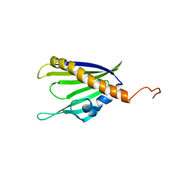
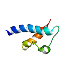
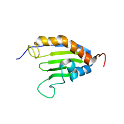 | | The solution structure of Bacillus subtilis SR211 START domain by NMR spectroscopy | | 分子名称: | Uncharacterized protein yndB | | 著者 | Mercier, K.A, Mueller, G.A, Powers, R, Acton, T.B, Ciano, M, Ho, C, Lui, J, Ma, L, Rost, B, Rossi, R, Xiao, R, Northeast Structural Genomics Consortium (NESG) | | 登録日 | 2009-02-05 | | 公開日 | 2009-03-17 | | 最終更新日 | 2024-05-01 | | 実験手法 | SOLUTION NMR | | 主引用文献 | (1)H, (13)C, and (15)N NMR assignments for the Bacillus subtilis yndB START domain.
Biomol.Nmr Assign., 3, 2009
|
|
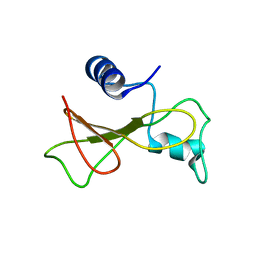
2KEY
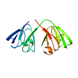
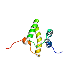 | | Solution NMR structure of a domain from a putative phage integrase protein BF2284 from Bacteroides fragilis, Northeast Structural Genomics Consortium Target BfR257C | | 分子名称: | Putative phage integrase | | 著者 | Mills, J.L, Sathyamoorthy, B, Sukumaran, D.K, Lee, D, Ciccosanti, C, Jiang, M, Xiao, R, Acton, T.B, Swapna, G.V.T, Rost, B, Nair, R, Everett, J.K, Montelione, G.T, Szyperski, T, Northeast Structural Genomics Consortium (NESG) | | 登録日 | 2009-02-06 | | 公開日 | 2009-04-14 | | 最終更新日 | 2024-05-01 | | 実験手法 | SOLUTION NMR | | 主引用文献 | Solution NMR Structure of a Domain from a Putative Phage Integrase Protein, BF2284, from Bacteroides fragilis, Northeast Structural Genomics Consortium Target BfR257C
To be Published
|
|
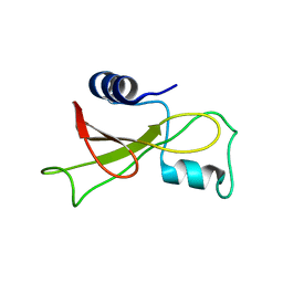
2KEZ
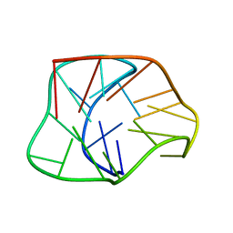
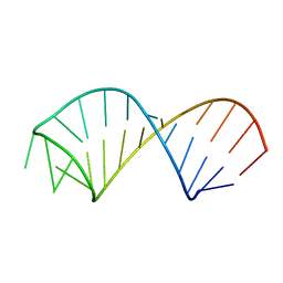 | | NMR structure of U6 ISL at pH 8.0 | | 分子名称: | RNA (5'-R(*GP*GP*UP*UP*CP*CP*CP*CP*UP*GP*CP*AP*UP*AP*AP*GP*GP*AP*UP*GP*AP*AP*CP*C)-3') | | 著者 | Venditti, V, Butcher, S.E. | | 登録日 | 2009-02-08 | | 公開日 | 2009-07-21 | | 最終更新日 | 2024-05-22 | | 実験手法 | SOLUTION NMR | | 主引用文献 | Minimum-energy path for a u6 RNA conformational change involving protonation, base-pair rearrangement and base flipping.
J.Mol.Biol., 391, 2009
|
|
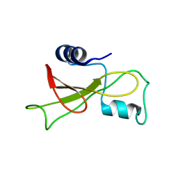
2KF0
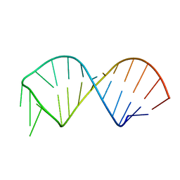 | | NMR structure of U6 ISL at pH 7.0 | | 分子名称: | RNA (5'-R(*GP*GP*UP*UP*CP*CP*CP*CP*UP*GP*CP*AP*UP*AP*AP*GP*GP*AP*UP*GP*AP*AP*CP*C)-3') | | 著者 | Venditti, V, Butcher, S.E. | | 登録日 | 2009-02-08 | | 公開日 | 2009-07-21 | | 最終更新日 | 2024-05-22 | | 実験手法 | SOLUTION NMR | | 主引用文献 | Minimum-energy path for a u6 RNA conformational change involving protonation, base-pair rearrangement and base flipping.
J.Mol.Biol., 391, 2009
|
|
2KF2
 | | Solution NMR structure of of Streptomyces coelicolor polyketide cyclase SCO5315. Northeast Structural Genomics Consortium target RR365 | | 分子名称: | Putative polyketide cyclase | | 著者 | Cort, J.R, Ramelot, T.A, Ding, K, Wang, H, Jiang, M, Maglaqui, M, Xiao, R, Nair, R, Baran, M.C, Everett, J.K, Swapna, G.V.T, Acton, T.B, Rost, B, Montelione, G.T, Kennedy, M.A, Northeast Structural Genomics Consortium (NESG) | | 登録日 | 2009-02-11 | | 公開日 | 2009-05-19 | | 最終更新日 | 2024-05-22 | | 実験手法 | SOLUTION NMR | | 主引用文献 | Solution NMR Structure of Streptomyces coelicolor polyketide cyclase SCO5315
To be Published
|
|
2KF3
 | |
2KF4
 | |
2KF5
 | |
2KF6
 | |
2KF7
 | | Structure of a two-G-tetrad basket-type intramolecular G-quadruplex formed by human telomeric repeats in K+ solution (with G7-to-BRG substitution) | | 分子名称: | HUMAN TELOMERE DNA | | 著者 | Lim, K.W, Amrane, S, Bouaziz, S, Xu, W, Mu, Y, Patel, D.J, Luu, K.N, Phan, A.T. | | 登録日 | 2009-02-12 | | 公開日 | 2009-03-24 | | 最終更新日 | 2024-05-01 | | 実験手法 | SOLUTION NMR | | 主引用文献 | Structure of the human telomere in K+ solution: a stable basket-type G-quadruplex with only two G-tetrad layers
J.Am.Chem.Soc., 131, 2009
|
|
2KF8
 | | Structure of a two-G-tetrad basket-type intramolecular G-quadruplex formed by human telomeric repeats in K+ solution | | 分子名称: | HUMAN TELOMERE DNA | | 著者 | Lim, K.W, Amrane, S, Bouaziz, S, Xu, W, Mu, Y, Patel, D.J, Luu, K.N, Phan, A.T. | | 登録日 | 2009-02-12 | | 公開日 | 2009-03-24 | | 最終更新日 | 2024-05-01 | | 実験手法 | SOLUTION NMR | | 主引用文献 | Structure of the human telomere in K+ solution: a stable basket-type G-quadruplex with only two G-tetrad layers
J.Am.Chem.Soc., 131, 2009
|
|
2KFB
 | | The structure of the cataract causing P23T mutant of human gamma-D crystallin | | 分子名称: | Gamma-crystallin D | | 著者 | Jung, J, Byeon, I.L, Wang, Y, King, J, Gronenborn, A.M. | | 登録日 | 2009-02-12 | | 公開日 | 2009-07-28 | | 最終更新日 | 2024-05-22 | | 実験手法 | SOLUTION NMR | | 主引用文献 | The structure of the cataract-causing P23T mutant of human gammaD-crystallin exhibits distinctive local conformational and dynamic changes.
Biochemistry, 48, 2009
|
|
2KFD
 | |
2KFE
 | |
2KFF
 | | Structure of the C-terminal domain of EHD1 with FNYESTNPFTAK | | 分子名称: | CALCIUM ION, EH domain-containing protein 1, Rab11-FIP2 NPF peptide FNYESTNPFTAK | | 著者 | Kieken, F, Jovic, M, Tonelli, M, Naslavsky, N, Caplan, S, Sorgen, P. | | 登録日 | 2009-02-20 | | 公開日 | 2009-12-22 | | 最終更新日 | 2024-05-01 | | 実験手法 | SOLUTION NMR | | 主引用文献 | Structural insight into the interaction of proteins containing NPF, DPF, and GPF motifs with the C-terminal EH-domain of EHD1.
Protein Sci., 18, 2009
|
|
2KFG
 | | Structure of the C-terminal domain of EHD1 in complex with FNYESTDPFTAK | | 分子名称: | CALCIUM ION, EH domain-containing protein 1, Rab11-FIP2 DPF peptide FNYESTDPFTAK | | 著者 | Kieken, F, Jovic, M, Tonelli, M, Naslavsky, N, Caplan, S, Sorgen, P. | | 登録日 | 2009-02-20 | | 公開日 | 2009-12-22 | | 最終更新日 | 2024-05-01 | | 実験手法 | SOLUTION NMR | | 主引用文献 | Structural insight into the interaction of proteins containing NPF, DPF, and GPF motifs with the C-terminal EH-domain of EHD1.
Protein Sci., 18, 2009
|
|
2KFH
 | | Structure of the C-terminal domain of EHD1 with FNYESTGPFTAK | | 分子名称: | CALCIUM ION, EH domain-containing protein 1, Rab11-FIP2 GPF peptide FNYESTGPFTAK | | 著者 | Kieken, F, Jovic, M, Tonelli, M, Naslavsky, N, Caplan, S, Sorgen, P. | | 登録日 | 2009-02-20 | | 公開日 | 2009-12-22 | | 最終更新日 | 2024-05-01 | | 実験手法 | SOLUTION NMR | | 主引用文献 | Structural insight into the interaction of proteins containing NPF, DPF, and GPF motifs with the C-terminal EH-domain of EHD1.
Protein Sci., 18, 2009
|
|
2KFJ
 | | Solution structure of the loop deletion mutant of PB1 domain of Cdc24p | | 分子名称: | Cell division control protein 24 | | 著者 | Ogura, K, Tandai, T, Yoshinaga, S, Kobashigawa, Y, Kumeta, H, Inagaki, F. | | 登録日 | 2009-02-22 | | 公開日 | 2009-10-06 | | 最終更新日 | 2024-05-29 | | 実験手法 | SOLUTION NMR | | 主引用文献 | NMR structure of the heterodimer of Bem1 and Cdc24 PB1 domains from Saccharomyces cerevisiae
J.Biochem., 146, 2009
|
|
2KFK
 | | Solution structure of Bem1p PB1 domain complexed with Cdc24p PB1 domain | | 分子名称: | Bud emergence protein 1, Cell division control protein 24 | | 著者 | Kobashigawa, Y, Yoshinaga, S, Tandai, T, Ogura, K, Inagaki, F. | | 登録日 | 2009-02-23 | | 公開日 | 2009-10-06 | | 最終更新日 | 2024-05-29 | | 実験手法 | SOLUTION NMR | | 主引用文献 | NMR structure of the heterodimer of Bem1 and Cdc24 PB1 domains from Saccharomyces cerevisiae
J.Biochem., 146, 2009
|
|
2KFL
 | |
2KFM
 | |
2KFN
 | | KLENOW FRAGMENT WITH BRIDGING-SULFUR SUBSTRATE AND MANGANESE | | 分子名称: | 5'-D(*GP*CP*TP*TP*AP*(US1)P*G)-3', KLENOW FRAGMENT OF DNA POLYMERASE I, MAGNESIUM ION, ... | | 著者 | Brautigam, C.A, Sun, S, Piccirilli, J.A, Steitz, T.A. | | 登録日 | 1998-07-01 | | 公開日 | 1998-11-11 | | 最終更新日 | 2024-02-21 | | 実験手法 | X-RAY DIFFRACTION (2.03 Å) | | 主引用文献 | Structures of normal single-stranded DNA and deoxyribo-3'-S-phosphorothiolates bound to the 3'-5' exonucleolytic active site of DNA polymerase I from Escherichia coli.
Biochemistry, 38, 1999
|
|
2KFO
 | |
2KFP
 | | Solution NMR structure of PSPTO_3016 from Pseudomonas syringae. Northeast Structural Genomics Consortium target PsR293. | | 分子名称: | PSPTO_3016 protein | | 著者 | Feldmann, E.A, Ramelot, T.A, Zhao, L, Hamilton, K, Ciccosanti, C, Xiao, R, Nair, R, Everett, J.K, Swapna, G, Acton, T.B, Rost, B, Montelione, G.T, Kennedy, M.A, Northeast Structural Genomics Consortium (NESG) | | 登録日 | 2009-02-24 | | 公開日 | 2009-03-24 | | 最終更新日 | 2024-05-08 | | 実験手法 | SOLUTION NMR | | 主引用文献 | Solution NMR and X-ray crystal structures of Pseudomonas syringae Pspto_3016 from protein domain family PF04237 (DUF419) adopt a "double wing" DNA binding motif.
J.Struct.Funct.Genom., 13, 2012
|
|
2KFQ
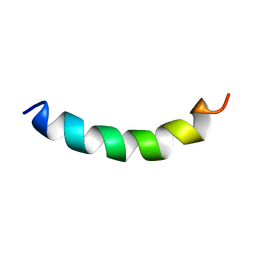
 | | NMR Structure of FP1 | | 分子名称: | FP1 | | 著者 | Araki, M, Tamura, A. | | 登録日 | 2009-02-26 | | 公開日 | 2009-03-31 | | 最終更新日 | 2024-05-29 | | 実験手法 | SOLUTION NMR | | 主引用文献 | Solubility-dependent structural formation of a 25-residue, natively unfolded protein, induced by addition of a seven-residue peptide fragment
Febs J., 276, 2009
|
|


