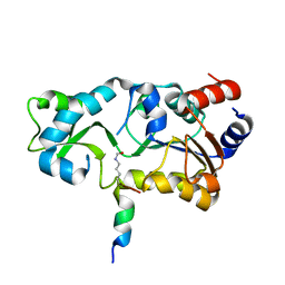
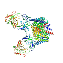
2H23
 
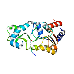
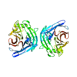
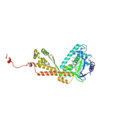 | | Structure of Rubisco LSMT bound to Trimethyllysine and AdoHcy | | 分子名称: | N-TRIMETHYLLYSINE, Ribulose-1,5 bisphosphate carboxylase/oxygenase large subunit N-methyltransferase, S-ADENOSYL-L-HOMOCYSTEINE | | 著者 | Couture, J.F, Hauk, G, Trievel, R.C. | | 登録日 | 2006-05-17 | | 公開日 | 2006-05-30 | | 最終更新日 | 2024-02-14 | | 実験手法 | X-RAY DIFFRACTION (2.45 Å) | | 主引用文献 | Catalytic Roles for Carbon-Oxygen Hydrogen Bonding in SET Domain Lysine Methyltransferases.
J.Biol.Chem., 281, 2006
|
|
2H24
 | | Crystal structure of human IL-10 | | 分子名称: | Interleukin-10 | | 著者 | Yoon, S.I, Walter, M.R. | | 登録日 | 2006-05-18 | | 公開日 | 2006-10-17 | | 最終更新日 | 2023-08-30 | | 実験手法 | X-RAY DIFFRACTION (2 Å) | | 主引用文献 | Conformational changes mediate interleukin-10 receptor 2 (IL-10R2) binding to IL-10 and assembly of the signaling complex.
J.Biol.Chem., 281, 2006
|
|
2H25
 | |
2H26
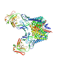
 | | human CD1b in complex with endogenous phosphatidylcholine and spacer | | 分子名称: | (4S,7R)-4-HYDROXY-N,N,N-TRIMETHYL-9-OXO-7-[(PALMITOYLOXY)METHYL]-3,5,8-TRIOXA-4-PHOSPHAHEXACOSAN-1-AMINIUM 4-OXIDE, Beta-2-microglobulin, GLYCEROL, ... | | 著者 | Garcia-Alles, L.F, Maveyraud, L, Vallina, A.T, Guillet, V, Mourey, L. | | 登録日 | 2006-05-18 | | 公開日 | 2006-07-04 | | 最終更新日 | 2024-10-09 | | 実験手法 | X-RAY DIFFRACTION (1.8 Å) | | 主引用文献 | Endogenous phosphatidylcholine and a long spacer ligand stabilize the lipid-binding groove of CD1b.
Embo J., 25, 2006
|
|
2H27
 | |
2H28
 | | Crystal structure of YeeU from E. coli. Northeast Structural Genomics target ER304 | | 分子名称: | CHLORIDE ION, GLYCEROL, Hypothetical protein yeeU, ... | | 著者 | Arbing, M, Su, M, Benach, J, Karpowich, N.K, Jiang, M, Xiao, R, Cunningham, K, Ma, L.-C, Chen, C.X, Acton, T.B, Montelione, G.T, Tong, L, Hunt, J.F, Northeast Structural Genomics Consortium (NESG) | | 登録日 | 2006-05-18 | | 公開日 | 2006-07-18 | | 最終更新日 | 2017-10-18 | | 実験手法 | X-RAY DIFFRACTION (2.1 Å) | | 主引用文献 | Crystal Structures of Phd-Doc, HigA, and YeeU Establish Multiple Evolutionary Links between Microbial Growth-Regulating Toxin-Antitoxin Systems.
Structure, 18, 2010
|
|
2H29
 | |
2H2A
 | |
2H2B
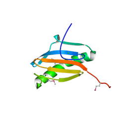
 | | Crystal Structure of ZO-1 PDZ1 Bound to a Phage-Derived Ligand (WRRTTYL) | | 分子名称: | ACETIC ACID, Tight junction protein ZO-1 | | 著者 | Appleton, B.A, Zhang, Y, Wu, P, Yin, J.P, Hunziker, W, Skelton, N.J, Sidhu, S.S, Wiesmann, C. | | 登録日 | 2006-05-18 | | 公開日 | 2006-06-13 | | 最終更新日 | 2023-08-30 | | 実験手法 | X-RAY DIFFRACTION (1.6 Å) | | 主引用文献 | Comparative structural analysis of the Erbin PDZ domain and the first PDZ domain of ZO-1. Insights into determinants of PDZ domain specificity.
J.Biol.Chem., 281, 2006
|
|
2H2C
 | | Crystal Structure of ZO-1 PDZ1 Bound to a Phage-Derived Ligand (WRRTTWV) | | 分子名称: | Tight junction protein ZO-1 | | 著者 | Appleton, B.A, Zhang, Y, Wu, P, Yin, J.P, Hunziker, W, Skelton, N.J, Sidhu, S.S, Wiesmann, C. | | 登録日 | 2006-05-18 | | 公開日 | 2006-06-13 | | 最終更新日 | 2023-08-30 | | 実験手法 | X-RAY DIFFRACTION (2 Å) | | 主引用文献 | Comparative structural analysis of the Erbin PDZ domain and the first PDZ domain of ZO-1. Insights into determinants of PDZ domain specificity.
J.Biol.Chem., 281, 2006
|
|
2H2D
 | |
2H2E
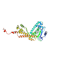 | | Structure of Rubisco LSMT bound to AzaAdoMet and Lysine | | 分子名称: | LYSINE, Ribulose-1,5 bisphosphate carboxylase/oxygenase large subunit N-methyltransferase, S-5'-AZAMETHIONINE-5'-DEOXYADENOSINE | | 著者 | Couture, J.F, Hauk, G, Trievel, R.C. | | 登録日 | 2006-05-18 | | 公開日 | 2006-05-30 | | 最終更新日 | 2024-02-14 | | 実験手法 | X-RAY DIFFRACTION (2.6 Å) | | 主引用文献 | Catalytic Roles for Carbon-Oxygen Hydrogen Bonding in SET Domain Lysine Methyltransferases.
J.Biol.Chem., 281, 2006
|
|
2H2F
 | |
2H2G
 | |
2H2H
 | |
2H2I
 | | The Structural basis of Sirtuin Substrate Affinity | | 分子名称: | (2S,5R,8R,11S,14S,17S,21R)-5,8,11,14,17-PENTAMETHYL-4,7,10,13,16,19-HEXAOXADOCOSANE-2,21-DIOL, NAD-dependent deacetylase, ZINC ION | | 著者 | Cosgrove, M.S, Wolberger, C. | | 登録日 | 2006-05-18 | | 公開日 | 2006-12-05 | | 最終更新日 | 2023-08-30 | | 実験手法 | X-RAY DIFFRACTION (1.8 Å) | | 主引用文献 | The structural basis of sirtuin substrate affinity
Biochemistry, 45, 2006
|
|
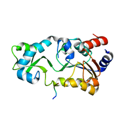
2H2J
 | | Structure of Rubisco LSMT bound to Sinefungin and Monomethyllysine | | 分子名称: | N-METHYL-LYSINE, Ribulose-1,5 bisphosphate carboxylase/oxygenase large subunit N-methyltransferase, SINEFUNGIN | | 著者 | Couture, J.F, Hauk, G, Trievel, R.C. | | 登録日 | 2006-05-18 | | 公開日 | 2006-05-30 | | 最終更新日 | 2024-02-14 | | 実験手法 | X-RAY DIFFRACTION (2.45 Å) | | 主引用文献 | Catalytic Roles for Carbon-Oxygen Hydrogen Bonding in SET Domain Lysine Methyltransferases.
J.Biol.Chem., 281, 2006
|
|
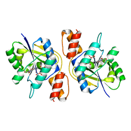
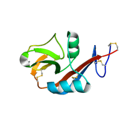
2H2K
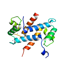 | | Crystal Structure Analysis of Human S100A13 | | 分子名称: | CALCIUM ION, Protein S100-A13 | | 著者 | Li, M, Zhang, P.F, Zhang, J.P, Chang, W.R. | | 登録日 | 2006-05-19 | | 公開日 | 2007-05-22 | | 最終更新日 | 2023-10-25 | | 実験手法 | X-RAY DIFFRACTION (2 Å) | | 主引用文献 | Crystal structure study on human S100A13 at 2.0 A resolution
Biochem.Biophys.Res.Commun., 356, 2007
|
|
2H2M
 | |
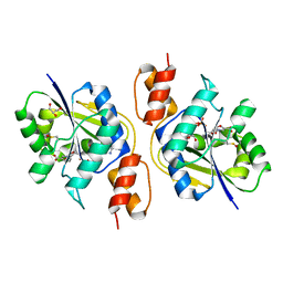
2H2N
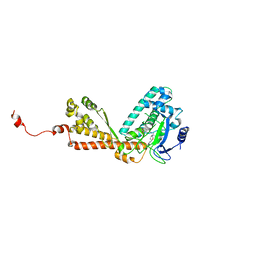
 | | Crystal structure of human soluble calcium-activated nucleotidase (SCAN) with calcium ion | | 分子名称: | ACETATE ION, CALCIUM ION, Soluble calcium-activated nucleotidase 1 | | 著者 | Yang, M, Horii, K, Herr, A.B, Kirley, T.L. | | 登録日 | 2006-05-19 | | 公開日 | 2006-07-18 | | 最終更新日 | 2024-02-14 | | 実験手法 | X-RAY DIFFRACTION (2.3 Å) | | 主引用文献 | Calcium-dependent dimerization of human soluble calcium activated nucleotidase: characterization of the dimer interface.
J.Biol.Chem., 281, 2006
|
|
2H2P
 | |
2H2Q
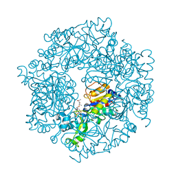
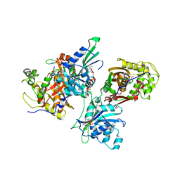 | | Crystal structure of Trypanosoma cruzi Dihydrofolate Reductase-Thymidylate synthase | | 分子名称: | 2'-DEOXYURIDINE-5'-MONOPHOSPHATE, Bifunctional dihydrofolate reductase-thymidylate synthase, NADP NICOTINAMIDE-ADENINE-DINUCLEOTIDE PHOSPHATE | | 著者 | Senkovich, O, Schormann, N, Chattopadhyay, D. | | 登録日 | 2006-05-19 | | 公開日 | 2008-04-08 | | 最終更新日 | 2023-08-30 | | 実験手法 | X-RAY DIFFRACTION (2.4 Å) | | 主引用文献 | Structure-based approach to pharmacophore identification, in silico screening, and three-dimensional quantitative structure-activity relationship studies for inhibitors of Trypanosoma cruzi dihydrofolate reductase function.
Proteins, 73, 2008
|
|
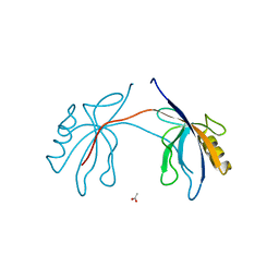
2H2R
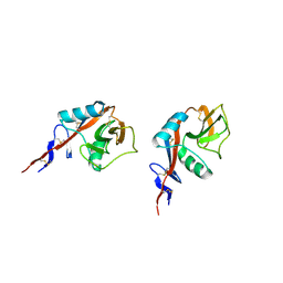 | | Crystal structure of the human CD23 Lectin domain, apo form | | 分子名称: | Low affinity immunoglobulin epsilon Fc receptor (Lymphocyte IgE receptor) (Fc-epsilon-RII)(Immunoglobulin E-binding factor) (CD23 antigen) | | 著者 | Wurzburg, B.A. | | 登録日 | 2006-05-19 | | 公開日 | 2006-06-20 | | 最終更新日 | 2023-08-30 | | 実験手法 | X-RAY DIFFRACTION (1.5 Å) | | 主引用文献 | Structural Changes in the Lectin Domain of CD23, the Low-Affinity IgE Receptor, upon Calcium Binding.
Structure, 14, 2006
|
|
2H2S
 | |
2H2T
 | | CD23 Lectin domain, Calcium 2+-bound | | 分子名称: | CALCIUM ION, Low affinity immunoglobulin epsilon Fc receptor (Lymphocyte IgE receptor) (Fc-epsilon-RII) (Immunoglobulin E-binding factor) (CD23 antigen) | | 著者 | Wurzburg, B.A. | | 登録日 | 2006-05-19 | | 公開日 | 2006-06-20 | | 最終更新日 | 2023-08-30 | | 実験手法 | X-RAY DIFFRACTION (1.3 Å) | | 主引用文献 | Structural Changes in the Lectin Domain of CD23, the Low-Affinity IgE Receptor, upon Calcium Binding.
Structure, 14, 2006
|
|


