2KNV
 
 | |
3KEG
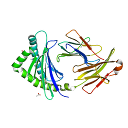
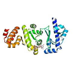
 | | X-ray Crystallographic Structure of a Y131F mutant of Pseudomonas Aeruginosa Azoreductase in complex with Methyl RED | | 分子名称: | 2-(4-DIMETHYLAMINOPHENYL)DIAZENYLBENZOIC ACID, FLAVIN MONONUCLEOTIDE, FMN-dependent NADH-azoreductase 1, ... | | 著者 | Wang, C.-J, Laurieri, N, Abuhammad, A, Lowe, E, Westwood, I, Ryan, A, Sim, E. | | 登録日 | 2009-10-26 | | 公開日 | 2010-01-12 | | 最終更新日 | 2023-11-01 | | 実験手法 | X-RAY DIFFRACTION (2.1 Å) | | 主引用文献 | Role of tyrosine 131 in the active site of paAzoR1, an azoreductase with specificity for the inflammatory bowel disease prodrug balsalazide
Acta Crystallogr.,Sect.F, 66, 2010
|
|
4ETL
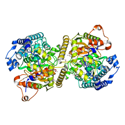
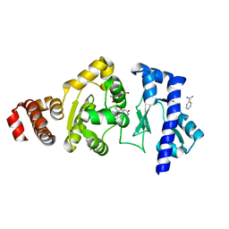
 | | Crystallographic structure of phenylalanine hydroxylase from Chromobacterium violaceum F258A mutation | | 分子名称: | COBALT (II) ION, Phenylalanine-4-hydroxylase | | 著者 | Ronau, J.A, Paul, L.P, Corn, I.R, Wagner, K.T, Abu-Omar, M.M, Das, C. | | 登録日 | 2012-04-24 | | 公開日 | 2013-05-08 | | 最終更新日 | 2023-09-13 | | 実験手法 | X-RAY DIFFRACTION (1.49 Å) | | 主引用文献 | An additional substrate binding site in a bacterial phenylalanine hydroxylase.
Eur.Biophys.J., 42, 2013
|
|
2KR2
 | |
4N1I
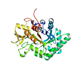
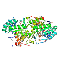
 | | Crystal Structure of the alpha-L-arabinofuranosidase UmAbf62A from Ustilago maidys | | 分子名称: | 2-AMINO-2-HYDROXYMETHYL-PROPANE-1,3-DIOL, CALCIUM ION, GLYCEROL, ... | | 著者 | Siguier, B, Dumon, C, Mourey, L, Tranier, S. | | 登録日 | 2013-10-04 | | 公開日 | 2014-01-15 | | 最終更新日 | 2024-11-27 | | 実験手法 | X-RAY DIFFRACTION (1 Å) | | 主引用文献 | First Structural Insights into alpha-L-Arabinofuranosidases from the Two GH62 Glycoside Hydrolase Subfamilies.
J.Biol.Chem., 289, 2014
|
|
4EXJ
 | | Crystal structure of glutathione s-transferase like protein lelg_03239 (target efi-501752) from lodderomyces elongisporus | | 分子名称: | CHLORIDE ION, SULFATE ION, uncharacterized protein | | 著者 | Patskovsky, Y, Toro, R, Bhosle, R, Zencheck, W.D, Hillerich, B, Seidel, R.D, Washington, E, Scott Glenn, A, Chowdhury, S, Evans, B, Hammonds, J, Imker, H.J, Armstrong, R.N, Gerlt, J.A, Almo, S.C, Enzyme Function Initiative (EFI) | | 登録日 | 2012-04-30 | | 公開日 | 2012-05-23 | | 最終更新日 | 2023-09-13 | | 実験手法 | X-RAY DIFFRACTION (1.64 Å) | | 主引用文献 | Crystal structure of glutathione s-transferase like protein lelg_03239 (target efi-501752) from lodderomyces elongisporus
To be Published
|
|
3P3O
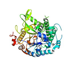
 | | Crystal Structure of the Cytochrome P450 Monooxygenase AurH (ntermII) from Streptomyces Thioluteus | | 分子名称: | CHLORIDE ION, Cytochrome P450, GLYCEROL, ... | | 著者 | Zocher, G, Richter, M.E.A, Mueller, U, Hertweck, C. | | 登録日 | 2010-10-05 | | 公開日 | 2011-02-16 | | 最終更新日 | 2024-02-21 | | 実験手法 | X-RAY DIFFRACTION (1.54 Å) | | 主引用文献 | Structural fine-tuning of a multifunctional cytochrome p450 monooxygenase.
J.Am.Chem.Soc., 133, 2011
|
|
4N57
 | | Crystal structure of aminoglycoside phosphotransferase APH(2'')-IVa ADP complex | | 分子名称: | ADENOSINE-5'-DIPHOSPHATE, APH(2'')-Id, MAGNESIUM ION | | 著者 | Kaplan, E, Leban, N, Chaloin, L, Guichou, J.-F, Lionne, C. | | 登録日 | 2013-10-09 | | 公開日 | 2014-12-17 | | 最終更新日 | 2023-09-20 | | 実験手法 | X-RAY DIFFRACTION (2.35 Å) | | 主引用文献 | Crystal structure of aminoglycoside phosphotransferase APH(2'')-IVa ADP complex
To be Published
|
|
3P73
 | | Crystal Structures of the Chicken YF1*7.1 molecule | | 分子名称: | ACETATE ION, Beta-2-microglobulin, CETYL-TRIMETHYL-AMMONIUM, ... | | 著者 | Hee, C.S, Gao, S, Loll, B, Miller, M.M, Uchanska-Ziegler, B, Daumke, O, Ziegler, A. | | 登録日 | 2010-10-12 | | 公開日 | 2010-11-24 | | 最終更新日 | 2024-11-06 | | 実験手法 | X-RAY DIFFRACTION (1.32 Å) | | 主引用文献 | Structure of a Classical MHC Class I Molecule That Binds "Non-Classical" Ligands.
Plos Biol., 8, 2010
|
|
2KLX
 | |
6KVR
 | | Fatty acid amide hydrolase | | 分子名称: | Fatty acid amide hydrolase | | 著者 | Min, C.A, Yun, J.S, Chang, J.H. | | 登録日 | 2019-09-05 | | 公開日 | 2021-09-15 | | 最終更新日 | 2024-10-16 | | 実験手法 | X-RAY DIFFRACTION (2.2 Å) | | 主引用文献 | Comparison of Candida Albicans Fatty Acid Amide Hydrolase Structure with Homologous Amidase Signature Family Enzymes
Crystals, 9, 2019
|
|
6LDU
 | |
4EDT
 | | The structure of the S. aureus DnaG RNA Polymerase Domain bound to ppGpp and Manganese | | 分子名称: | BENZAMIDINE, DNA primase, GUANOSINE-5',3'-TETRAPHOSPHATE, ... | | 著者 | Rymer, R.U, Solorio, F.A, Chu, C, Corn, J.E, Wang, J.D, Berger, J.M. | | 登録日 | 2012-03-27 | | 公開日 | 2012-07-25 | | 最終更新日 | 2024-02-28 | | 実験手法 | X-RAY DIFFRACTION (2.005 Å) | | 主引用文献 | Binding Mechanism of Metal-NTP Substrates and Stringent-Response Alarmones to Bacterial DnaG-Type Primases.
Structure, 20, 2012
|
|
4EF9
 | | Crystal structure of dihydroorotate dehydrogenase from Leishmania major in complex with 4-Nitrophenyl isothiocyanate | | 分子名称: | Dihydroorotate dehydrogenase, FLAVIN MONONUCLEOTIDE, GLYCEROL, ... | | 著者 | Pinheiro, M.P, Emery, F.S, Nonato, M.C. | | 登録日 | 2012-03-29 | | 公開日 | 2012-12-12 | | 最終更新日 | 2024-10-16 | | 実験手法 | X-RAY DIFFRACTION (1.6 Å) | | 主引用文献 | Target sites for the design of anti-trypanosomatid drugs based on the structure of dihydroorotate dehydrogenase.
Curr.Pharm.Des., 19, 2013
|
|
4EAN
 | | 1.75A resolution structure of indole bound beta-glycosidase (W33G) from sulfolobus solfataricus | | 分子名称: | (4S)-2-METHYL-2,4-PENTANEDIOL, Beta-galactosidase, CHLORIDE ION, ... | | 著者 | Lovell, S, Battaile, K.P, Deckert, K, Brunner, L.C, Budiardjo, S.J, Karanicolas, J. | | 登録日 | 2012-03-22 | | 公開日 | 2012-06-13 | | 最終更新日 | 2023-09-13 | | 実験手法 | X-RAY DIFFRACTION (1.75 Å) | | 主引用文献 | Designing allosteric control into enzymes by chemical rescue of structure.
J.Am.Chem.Soc., 134, 2012
|
|
4EDR
 | | The structure of the S. aureus DnaG RNA Polymerase Domain bound to UTP and Manganese | | 分子名称: | BENZAMIDINE, DNA primase, MANGANESE (II) ION, ... | | 著者 | Rymer, R.U, Solorio, F.A, Chu, C, Corn, J.E, Wang, J.D, Berger, J.M. | | 登録日 | 2012-03-27 | | 公開日 | 2012-07-25 | | 最終更新日 | 2024-02-28 | | 実験手法 | X-RAY DIFFRACTION (2.01 Å) | | 主引用文献 | Binding Mechanism of Metal-NTP Substrates and Stringent-Response Alarmones to Bacterial DnaG-Type Primases.
Structure, 20, 2012
|
|
4EF8
 | | Crystal structure of dihydroorotate dehydrogenase from Leishmania major in complex with Phenyl isothiocyanate | | 分子名称: | Dihydroorotate dehydrogenase, FLAVIN MONONUCLEOTIDE, GLYCEROL, ... | | 著者 | Pinheiro, M.P, Emery, F.S, Nonato, M.C. | | 登録日 | 2012-03-29 | | 公開日 | 2012-12-12 | | 最終更新日 | 2024-10-09 | | 実験手法 | X-RAY DIFFRACTION (1.56 Å) | | 主引用文献 | Target sites for the design of anti-trypanosomatid drugs based on the structure of dihydroorotate dehydrogenase.
Curr.Pharm.Des., 19, 2013
|
|
3P6V
 | |
6LLK
 | | Biphenyl-2,2',3-triol-soaked terminal oxygenase of carbazole 1,9a-dioxygenase | | 分子名称: | (4S)-2-METHYL-2,4-PENTANEDIOL, 3-(2-hydroxyphenyl)benzene-1,2-diol, DIMETHYL SULFOXIDE, ... | | 著者 | Wang, Y.X, Suzuki-Minakuchi, C, Nojiri, H. | | 登録日 | 2019-12-23 | | 公開日 | 2021-01-27 | | 最終更新日 | 2023-11-22 | | 実験手法 | X-RAY DIFFRACTION (2.3 Å) | | 主引用文献 | Biphenyl-2,2',3-triol-soaked terminal oxygenase of carbazole 1,9a-dioxygenase
To Be Published
|
|
3PA2
 | | Crystal Structure of P Domain from Norwalk Virus Strain Vietnam 026 in complex with HBGA type Ley | | 分子名称: | 1,2-ETHANEDIOL, Capsid protein, IMIDAZOLE, ... | | 著者 | Hansman, G.S, Biertumpfel, C, Chen, L, Georgiev, I, McLellan, J.S, Katayama, K, Kwong, P.D. | | 登録日 | 2010-10-18 | | 公開日 | 2011-05-11 | | 最終更新日 | 2023-09-06 | | 実験手法 | X-RAY DIFFRACTION (1.48 Å) | | 主引用文献 | Crystal Structures of GII.10 and GII.12 Norovirus Protruding Domains in Complex with Histo-Blood Group Antigens Reveal Details for a Potential Site of Vulnerability.
J.Virol., 85, 2011
|
|
4EP2
 | | Crystal Structure of inactive single chain wild-type HIV-1 Protease in Complex with the substrate RT-RH | | 分子名称: | GLYCEROL, PHOSPHATE ION, protease, ... | | 著者 | Schiffer, C.A, Mittal, S. | | 登録日 | 2012-04-16 | | 公開日 | 2012-06-06 | | 最終更新日 | 2024-02-28 | | 実験手法 | X-RAY DIFFRACTION (1.9 Å) | | 主引用文献 | Structural, kinetic, and thermodynamic studies of specificity designed HIV-1 protease.
Protein Sci., 21, 2012
|
|
3OUC
 | | MDR769 HIV-1 protease complexed with p2/NC hepta-peptide | | 分子名称: | MDR HIV-1 protease, p2/NC substrate peptide | | 著者 | Liu, Z, Wang, Y, Brunzelle, J, Kovari, I.A, Kovari, L.C. | | 登録日 | 2010-09-14 | | 公開日 | 2011-03-30 | | 最終更新日 | 2024-02-21 | | 実験手法 | X-RAY DIFFRACTION (2 Å) | | 主引用文献 | Nine Crystal Structures Determine the Substrate Envelope of the MDR HIV-1 Protease.
Protein J., 30, 2011
|
|
4EPJ
 | | Crystal Structure of inactive single chain wild-type HIV-1 Protease in Complex with the substrate p2-NC | | 分子名称: | 1,2-ETHANEDIOL, ACETATE ION, BETA-MERCAPTOETHANOL, ... | | 著者 | Schiffer, C.A, Mittal, S. | | 登録日 | 2012-04-17 | | 公開日 | 2012-06-06 | | 最終更新日 | 2024-02-28 | | 実験手法 | X-RAY DIFFRACTION (1.69 Å) | | 主引用文献 | Structural, kinetic, and thermodynamic studies of specificity designed HIV-1 protease.
Protein Sci., 21, 2012
|
|
4N8E
 | | DPP4 complexed with compound 12a | | 分子名称: | 1-[cis-4-(aminomethyl)-4-(3-chlorophenyl)cyclohexyl]piperidin-2-one, 2-acetamido-2-deoxy-beta-D-glucopyranose, Dipeptidyl peptidase 4, ... | | 著者 | Ostermann, N, Zink, F, Kroemer, M. | | 登録日 | 2013-10-17 | | 公開日 | 2014-02-12 | | 最終更新日 | 2024-11-20 | | 実験手法 | X-RAY DIFFRACTION (2.3 Å) | | 主引用文献 | Discovery of C-(1-aryl-cyclohexyl)-methylamines as selective, orally available inhibitors of dipeptidyl peptidase IV.
Bioorg.Med.Chem.Lett., 24, 2014
|
|
3OY4
 | | Crystal Structure of HIV-1 L76V Protease in Complex with the Protease Inhibitor Darunavir. | | 分子名称: | (3R,3AS,6AR)-HEXAHYDROFURO[2,3-B]FURAN-3-YL(1S,2R)-3-[[(4-AMINOPHENYL)SULFONYL](ISOBUTYL)AMINO]-1-BENZYL-2-HYDROXYPROPYLCARBAMATE, ACETATE ION, HIV-1 PROTEASE, ... | | 著者 | Schiffer, C.A, Nalivaika, E.A, Bandaranayake, R.M. | | 登録日 | 2010-09-22 | | 公開日 | 2011-02-09 | | 最終更新日 | 2024-02-21 | | 実験手法 | X-RAY DIFFRACTION (1.76 Å) | | 主引用文献 | Crystal Structure of HIV-1 L76V Protease in Complex with the Protease Inhibitor Darunavir.
To be Published
|
|


