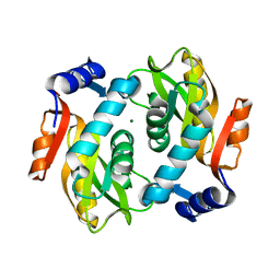
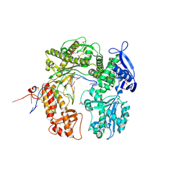
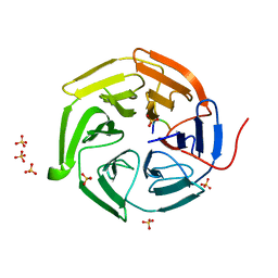
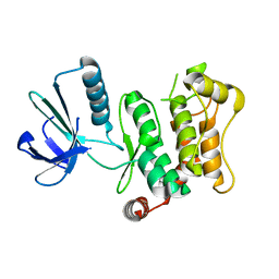
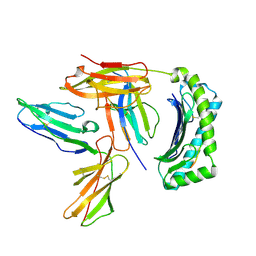
2DXY
 
 | | Structure of the complex of C-terminal lobe of bovine lactoferrin with trehalose at 2.0 A resolution | | 分子名称: | 2-acetamido-2-deoxy-beta-D-glucopyranose, CARBONATE ION, FE (III) ION, ... | | 著者 | Mir, R, Singh, N, Sinha, M, Sharma, S, Bhushan, A, Singh, T.P. | | 登録日 | 2006-09-03 | | 公開日 | 2006-09-19 | | 最終更新日 | 2023-10-25 | | 実験手法 | X-RAY DIFFRACTION (2.03 Å) | | 主引用文献 | Structure of the complex of C-terminal lobe of bovine lactoferrin with trehalose at 2.0 A resolution
To be Published
|
|
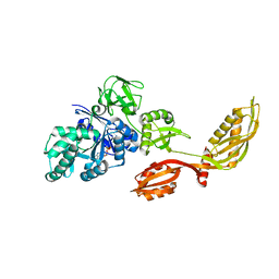
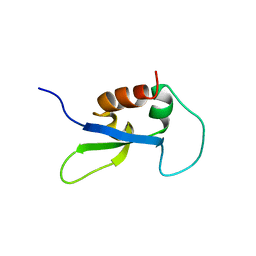
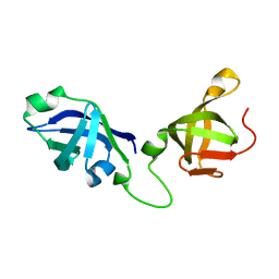
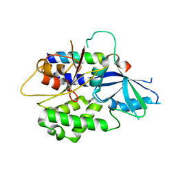
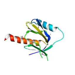
2DY0
 | |
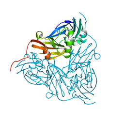
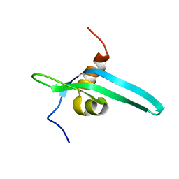
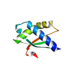
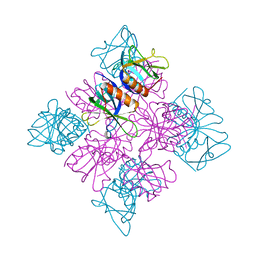
2DY1
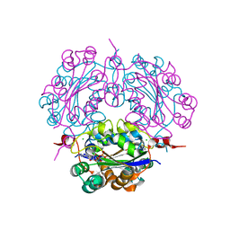
 | | Crystal structure of EF-G-2 from Thermus thermophilus | | 分子名称: | Elongation factor G, GUANOSINE-5'-TRIPHOSPHATE, MAGNESIUM ION | | 著者 | Wang, H, Takemoto, C, Murayama, K, Terada, T, Chen, L, Liu, Z.J, Wang, B.C, Shirouzu, M, Yokoyama, S, RIKEN Structural Genomics/Proteomics Initiative (RSGI) | | 登録日 | 2006-09-04 | | 公開日 | 2007-09-25 | | 最終更新日 | 2023-10-25 | | 実験手法 | X-RAY DIFFRACTION (1.6 Å) | | 主引用文献 | Crystal structure of EF-G-2 from Thermus thermophilus
To be Published
|
|
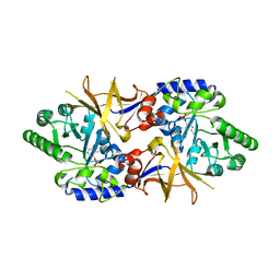
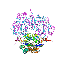
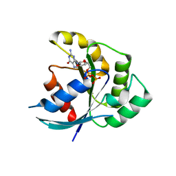
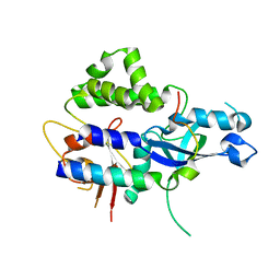
2DY2
 | | Nitrite reductase pH 6.0 | | 分子名称: | COPPER (II) ION, Copper-containing nitrite reductase | | 著者 | Jacobson, F. | | 登録日 | 2006-09-05 | | 公開日 | 2006-12-05 | | 最終更新日 | 2023-10-25 | | 実験手法 | X-RAY DIFFRACTION (2.26 Å) | | 主引用文献 | pH Dependence of Copper Geometry, Reduction Potential, and Nitrite Affinity in Nitrite Reductase
J.Biol.Chem., 282, 2007
|
|
2DY3
 | | Crystal Structure of alanine racemase from Corynebacterium glutamicum | | 分子名称: | Alanine racemase, PYRIDOXAL-5'-PHOSPHATE | | 著者 | Miyaguchi, I, Sasaki, C, Kato, R, Oikawa, T, Sugio, S. | | 登録日 | 2006-09-05 | | 公開日 | 2007-09-11 | | 最終更新日 | 2023-10-25 | | 実験手法 | X-RAY DIFFRACTION (2.1 Å) | | 主引用文献 | Crystal Structure of Alanine Racemase from Corynebacterium glutamicum at 2.1 A resolution
To be Published
|
|
2DY4
 | | Crystal structure of RB69 GP43 in complex with DNA containing Thymine Glycol | | 分子名称: | 5'-D(*CP*GP*(CTG)P*GP*GP*AP*AP*TP*GP*A*CP*AP*GP*CP*CP*GP*CP*G)-3', 5'-D(*GP*CP*GP*GP*CP*TP*GP*T*CP*AP*TP*TP*CP*CP*A)-3', DNA polymerase | | 著者 | Aller, P, Rould, M.A, Hogg, M, Wallace, S.S, Doublie, S. | | 登録日 | 2006-09-06 | | 公開日 | 2007-01-09 | | 最終更新日 | 2023-11-15 | | 実験手法 | X-RAY DIFFRACTION (2.65 Å) | | 主引用文献 | A structural rationale for stalling of a replicative DNA polymerase at the most common oxidative thymine lesion, thymine glycol.
Proc.Natl.Acad.Sci.USA, 104, 2007
|
|
2DY5
 | | Crystal structure of rat heme oxygenase-1 in complex with heme and 2-[2-(4-chlorophenyl)ethyl]-2-[(1H-imidazol-1-yl)methyl]-1,3-dioxolane | | 分子名称: | 1-({2-[2-(4-CHLOROPHENYL)ETHYL]-1,3-DIOXOLAN-2-YL}METHYL)-1H-IMIDAZOLE, CHLORIDE ION, Heme oxygenase 1, ... | | 著者 | Sugishima, M, Takahashi, H, Fukuyama, K. | | 登録日 | 2006-09-06 | | 公開日 | 2007-05-22 | | 最終更新日 | 2023-10-25 | | 実験手法 | X-RAY DIFFRACTION (2.7 Å) | | 主引用文献 | X-ray crystallographic and biochemical characterization of the inhibitory action of an imidazole-dioxolane compound on heme oxygenase
Biochemistry, 46, 2007
|
|
2DY7
 | |
2DY8
 | |
2DY9
 | | Crystal structure of nucleoside diphosphate kinase in complex with ADP | | 分子名称: | ADENOSINE-5'-DIPHOSPHATE, CHLORIDE ION, MAGNESIUM ION, ... | | 著者 | Kato-Murayama, M, Murayama, K, Terada, T, Shirouzu, M, Yokoyama, S, RIKEN Structural Genomics/Proteomics Initiative (RSGI) | | 登録日 | 2006-09-08 | | 公開日 | 2007-03-08 | | 最終更新日 | 2023-10-25 | | 実験手法 | X-RAY DIFFRACTION (2.01 Å) | | 主引用文献 | Crystal structure of nucleoside diphosphate kinase in complex with ADP
To be Published
|
|
2DYA
 | | Crystal structure of complex between Adenine nucleotide and nucleoside diphosphate kinase | | 分子名称: | ADENOSINE-5'-DIPHOSPHATE, CHLORIDE ION, MAGNESIUM ION, ... | | 著者 | Kato-Murayama, M, Murayama, K, Terada, T, Shirouzu, M, Yokoyama, S, RIKEN Structural Genomics/Proteomics Initiative (RSGI) | | 登録日 | 2006-09-08 | | 公開日 | 2007-03-08 | | 最終更新日 | 2023-10-25 | | 実験手法 | X-RAY DIFFRACTION (1.77 Å) | | 主引用文献 | Crystal structure of complex between Adenine nucleotide and nucleoside diphosphate kinase
To be Published
|
|
2DYB
 | | The crystal structure of human p40(phox) | | 分子名称: | Neutrophil cytosol factor 4 | | 著者 | Honbou, K. | | 登録日 | 2006-09-08 | | 公開日 | 2007-01-23 | | 最終更新日 | 2023-10-25 | | 実験手法 | X-RAY DIFFRACTION (3.15 Å) | | 主引用文献 | Full-length p40phox structure suggests a basis for regulation mechanism of its membrane binding.
Embo J., 26, 2007
|
|
2DYC
 | | Crystal structure of the N-terminal domain of mouse galectin-4 | | 分子名称: | Galectin-4 | | 著者 | Kato-Murayama, M, Murayama, K, Terada, T, Shirouzu, M, Yokoyama, S, RIKEN Structural Genomics/Proteomics Initiative (RSGI) | | 登録日 | 2006-09-08 | | 公開日 | 2007-10-16 | | 最終更新日 | 2024-10-09 | | 実験手法 | X-RAY DIFFRACTION (2.4 Å) | | 主引用文献 | Crystal structure of the N-terminal domain of mouse galectin-4
To be Published
|
|
2DYD
 | |
2DYF
 | |
2DYH
 | |
2DYI
 | | Crystal structure of 16S ribosomal RNA processing protein RimM from Thermus thermophilus HB8 | | 分子名称: | Probable 16S rRNA-processing protein rimM | | 著者 | Kawazoe, M, Takemoto, C, Kaminishi, T, Tatsuguchi, A, Shirouzu, M, Yokoyama, S, RIKEN Structural Genomics/Proteomics Initiative (RSGI) | | 登録日 | 2006-09-14 | | 公開日 | 2007-03-14 | | 最終更新日 | 2024-04-03 | | 実験手法 | X-RAY DIFFRACTION (2 Å) | | 主引用文献 | Crystal structure of 16S ribosomal RNA processing protein RimM from Thermus thermophilus HB8
To be published
|
|
2DYJ
 | | Crystal structure of ribosome-binding factor A from Thermus thermophilus HB8 | | 分子名称: | Ribosome-binding factor A | | 著者 | Kawazoe, M, Takemoto, C, Nakayama-Ushikoshi, R, Terada, T, Shirouzu, M, Yokoyama, S, RIKEN Structural Genomics/Proteomics Initiative (RSGI) | | 登録日 | 2006-09-14 | | 公開日 | 2007-03-14 | | 最終更新日 | 2023-10-25 | | 実験手法 | X-RAY DIFFRACTION (1.84 Å) | | 主引用文献 | Structural aspects of RbfA action during small ribosomal subunit assembly.
Mol.Cell, 28, 2007
|
|
2DYK
 | | Crystal structure of N-terminal GTP-binding domain of EngA from Thermus thermophilus HB8 | | 分子名称: | GTP-binding protein, GUANOSINE-5'-DIPHOSPHATE | | 著者 | Kawazoe, M, Takemoto, C, Fusatomi, E, Terada, T, Shirouzu, M, Yokoyama, S, RIKEN Structural Genomics/Proteomics Initiative (RSGI) | | 登録日 | 2006-09-14 | | 公開日 | 2007-03-14 | | 最終更新日 | 2023-10-25 | | 実験手法 | X-RAY DIFFRACTION (1.96 Å) | | 主引用文献 | Crystal structure of N-terminal GTP-binding domain of EngA from Thermus thermophilus HB8
to be published
|
|
2DYL
 | | Crystal structure of human mitogen-activated protein kinase kinase 7 activated mutant (S287D, T291D) | | 分子名称: | Dual specificity mitogen-activated protein kinase kinase 7 | | 著者 | Kukimoto-Niino, M, Takagi, T, Kaminishi, T, Uchikubo-Kamo, T, Terada, T, Matsuzaki, O, Shirouzu, M, Yokoyama, S, RIKEN Structural Genomics/Proteomics Initiative (RSGI) | | 登録日 | 2006-09-15 | | 公開日 | 2007-08-28 | | 最終更新日 | 2023-10-25 | | 実験手法 | X-RAY DIFFRACTION (2.45 Å) | | 主引用文献 | Crystal structure of human mitogen-activated protein kinase kinase 7 activated mutant (S287D, T291D)
To be Published
|
|
2DYM
 | |
2DYN
 | |
2DYO
 | |
2DYP
 | | Crystal Structure of LILRB2(LIR2/ILT4/CD85d) complexed with HLA-G | | 分子名称: | 9 Mer Peptide From Histone H2A.x, Beta-2-microglobulin, HLA class I histocompatibility antigen, ... | | 著者 | Shiroishi, M, Kuroki, K, Rasubala, L, Kohda, D, Maenaka, K. | | 登録日 | 2006-09-15 | | 公開日 | 2006-11-07 | | 最終更新日 | 2023-10-25 | | 実験手法 | X-RAY DIFFRACTION (2.5 Å) | | 主引用文献 | Structural basis for recognition of the nonclassical MHC molecule HLA-G by the leukocyte Ig-like receptor B2 (LILRB2/LIR2/ILT4/CD85d)
Proc.Natl.Acad.Sci.Usa, 103, 2006
|
|
2DYQ
 | | Crystal Structure of the C-terminal Phophotyrosine Interaction Domain of Human APBB3 | | 分子名称: | Amyloid beta A4 precursor protein-binding family B member 3 | | 著者 | Nishino, A, Saijo, S, Kishishita, S, Shirouzu, M, Yokoyama, S, RIKEN Structural Genomics/Proteomics Initiative (RSGI) | | 登録日 | 2006-09-16 | | 公開日 | 2007-09-18 | | 最終更新日 | 2011-07-13 | | 実験手法 | X-RAY DIFFRACTION (3.1 Å) | | 主引用文献 | Crystal Structure of the C-terminal Phophotyrosine Interaction Domain of Human APBB3
To be Published
|
|


