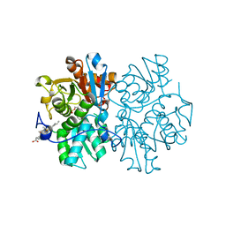
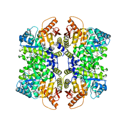
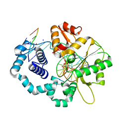
7QU5
 
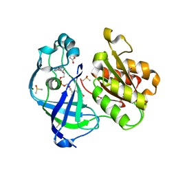 | | X-ray structure of FAD domain of NqrF of Pseudomonas aeruginosa | | 分子名称: | DIMETHYL SULFOXIDE, FLAVIN-ADENINE DINUCLEOTIDE, MAGNESIUM ION, ... | | 著者 | Stegmann, D, Steuber, J, Fritz, G. | | 登録日 | 2022-01-17 | | 公開日 | 2022-02-09 | | 最終更新日 | 2024-01-31 | | 実験手法 | X-RAY DIFFRACTION (1.25 Å) | | 主引用文献 | Fast fragment- and compound-screening pipeline at the Swiss Light Source.
Acta Crystallogr D Struct Biol, 78, 2022
|
|
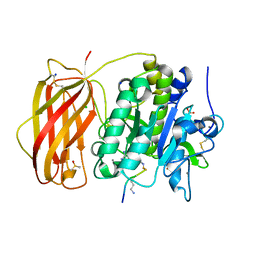
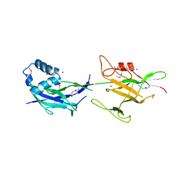
7QTY
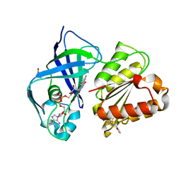 | | X-ray structure of FAD domain of NqrF of Klebsiella pneumoniae | | 分子名称: | 1-(furan-2-ylmethyl)-3-(2-methylphenyl)thiourea, DIMETHYL SULFOXIDE, FLAVIN-ADENINE DINUCLEOTIDE, ... | | 著者 | Stegmann, D, Steuber, J, Fritz, G. | | 登録日 | 2022-01-17 | | 公開日 | 2022-02-09 | | 最終更新日 | 2024-01-31 | | 実験手法 | X-RAY DIFFRACTION (1.69 Å) | | 主引用文献 | Fast fragment- and compound-screening pipeline at the Swiss Light Source.
Acta Crystallogr D Struct Biol, 78, 2022
|
|
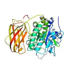
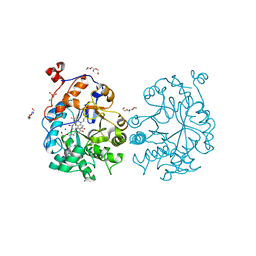
7QU3
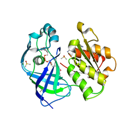 | | X-ray structure of FAD domain of NqrF of Pseudomonas aeruginosa | | 分子名称: | 4-(benzimidazol-1-ylmethyl)benzenecarbonitrile, DIMETHYL SULFOXIDE, FLAVIN-ADENINE DINUCLEOTIDE, ... | | 著者 | Stegmann, D, Steuber, J, Fritz, G. | | 登録日 | 2022-01-17 | | 公開日 | 2022-02-09 | | 最終更新日 | 2024-01-31 | | 実験手法 | X-RAY DIFFRACTION (1.6 Å) | | 主引用文献 | Fast fragment- and compound-screening pipeline at the Swiss Light Source.
Acta Crystallogr D Struct Biol, 78, 2022
|
|
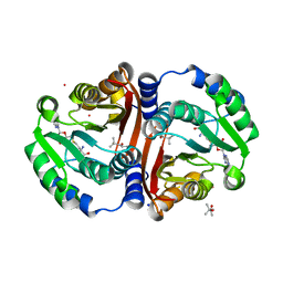
7QU0
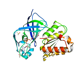 | | X-ray structure of FAD domain of NqrF of Klebsiella pneumoniae | | 分子名称: | DIMETHYL SULFOXIDE, FLAVIN-ADENINE DINUCLEOTIDE, Na(+)-translocating NADH-quinone reductase subunit F, ... | | 著者 | Stegmann, D, Steuber, J, Fritz, G. | | 登録日 | 2022-01-17 | | 公開日 | 2022-02-09 | | 最終更新日 | 2024-01-31 | | 実験手法 | X-RAY DIFFRACTION (1.62 Å) | | 主引用文献 | Fast fragment- and compound-screening pipeline at the Swiss Light Source.
Acta Crystallogr D Struct Biol, 78, 2022
|
|
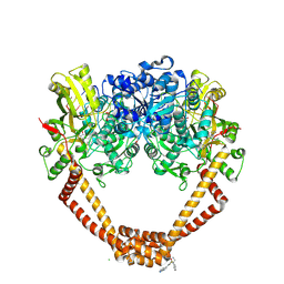
5MRE
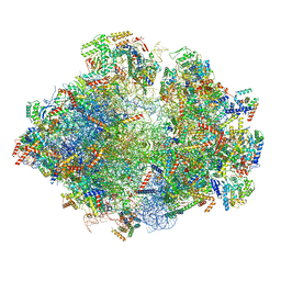 | | Structure of the yeast mitochondrial ribosome - Class B | | 分子名称: | 15S ribosomal RNA, 21S ribosomal RNA, GUANOSINE-5'-DIPHOSPHATE, ... | | 著者 | Desai, N, Brown, A, Amunts, A, Ramakrishnan, V. | | 登録日 | 2016-12-22 | | 公開日 | 2017-02-15 | | 最終更新日 | 2024-05-15 | | 実験手法 | ELECTRON MICROSCOPY (3.75 Å) | | 主引用文献 | The structure of the yeast mitochondrial ribosome.
Science, 355, 2017
|
|
5MRT
 | | Crystal structure of L5 protease Lysobacter sp. XL1 | | 分子名称: | CHLORIDE ION, FORMIC ACID, GLYCEROL, ... | | 著者 | Gabdulkhakov, A, Tishchenko, S, Lisov, A, Leontievsky, A. | | 登録日 | 2016-12-26 | | 公開日 | 2018-01-17 | | 最終更新日 | 2024-01-17 | | 実験手法 | X-RAY DIFFRACTION (1.6 Å) | | 主引用文献 | Crystal structure of L5 protease Lysobacter sp. XL1
To Be Published
|
|
4L3G
 | |
3ZVQ
 | |
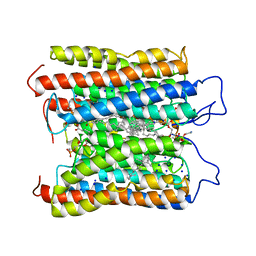
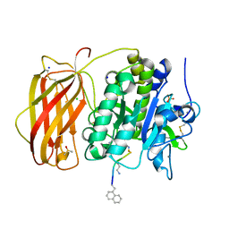
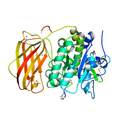
5MMS
 | | Human cystathionine beta-synthase (CBS) p.P49L delta409-551 variant | | 分子名称: | Cystathionine beta-synthase, PROTOPORPHYRIN IX CONTAINING FE, PYRIDOXAL-5'-PHOSPHATE, ... | | 著者 | Vicente, J.B, Colaco, H.G, Malagrino, F, Santo, P.E, Gutierres, A, Bandeiras, T.M, Leandro, P, Brito, J.A, Giuffre, A. | | 登録日 | 2016-12-12 | | 公開日 | 2017-05-03 | | 最終更新日 | 2024-01-17 | | 実験手法 | X-RAY DIFFRACTION (2.8 Å) | | 主引用文献 | A Clinically Relevant Variant of the Human Hydrogen Sulfide-Synthesizing Enzyme Cystathionine beta-Synthase: Increased CO Reactivity as a Novel Molecular Mechanism of Pathogenicity?
Oxid Med Cell Longev, 2017, 2017
|
|
4L3H
 | |
6HLB
 | |
6HLE
 | |
7QDN
 | |
8IFU
 | | HcCCR in NaCl | | 分子名称: | (1S)-2-{[(S)-(2-aminoethoxy)(hydroxy)phosphoryl]oxy}-1-[(octadecanoyloxy)methyl]ethyl (9Z)-octadec-9-enoate, HcCCR, RETINAL, ... | | 著者 | Zhang, M.F. | | 登録日 | 2023-02-19 | | 公開日 | 2024-02-28 | | 実験手法 | ELECTRON MICROSCOPY (2.37 Å) | | 主引用文献 | Structure of HcCCR
To Be Published
|
|
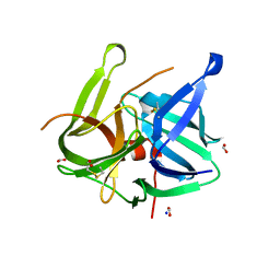
4LMF
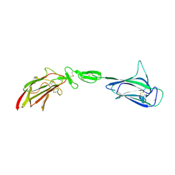 | | C1s CUB1-EGF-CUB2 | | 分子名称: | CALCIUM ION, Complement C1s subcomponent heavy chain, SODIUM ION | | 著者 | Wallis, R, Venkatraman Girija, U, Moody, P.C.E, Marshall, J.E. | | 登録日 | 2013-07-10 | | 公開日 | 2013-08-07 | | 最終更新日 | 2018-01-24 | | 実験手法 | X-RAY DIFFRACTION (2.921 Å) | | 主引用文献 | Structural basis of the C1q/C1s interaction and its central role in assembly of the C1 complex of complement activation.
Proc.Natl.Acad.Sci.USA, 110, 2013
|
|
5MDQ
 | |
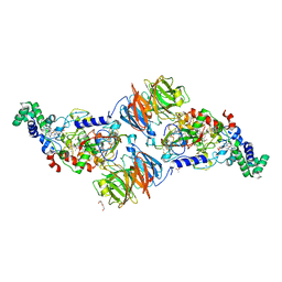
5NPK
 | | 1.98A STRUCTURE OF THIOPHENE1 WITH S.AUREUS DNA GYRASE AND DNA | | 分子名称: | CHLORIDE ION, DNA (5'-D(*AP*GP*CP*CP*GP*TP*AP*GP*GP*TP*AP*CP*CP*TP*AP*CP*GP*GP*CP*T)-3'), DNA gyrase subunit B,DNA gyrase subunit B,DNA gyrase subunit A, ... | | 著者 | Bax, B.D, Chan, P.F, Stavenger, R.A. | | 登録日 | 2017-04-17 | | 公開日 | 2017-07-12 | | 最終更新日 | 2018-10-24 | | 実験手法 | X-RAY DIFFRACTION (1.98 Å) | | 主引用文献 | Thiophene antibacterials that allosterically stabilize DNA-cleavage complexes with DNA gyrase.
Proc. Natl. Acad. Sci. U.S.A., 114, 2017
|
|
6HLD
 | |
6HRL
 | | Crystal structure of the Kelch domain of human KLHL17 | | 分子名称: | 1,2-ETHANEDIOL, CHLORIDE ION, Kelch-like protein 17, ... | | 著者 | Chen, Z, Williams, E, Sorrell, F.J, Newman, J.A, Shrestha, L, Burgess-Brown, N, von Delft, F, Arrowsmith, F, Edwards, C.H, Bountra, C, Bullock, A.N. | | 登録日 | 2018-09-27 | | 公開日 | 2018-10-17 | | 最終更新日 | 2024-01-24 | | 実験手法 | X-RAY DIFFRACTION (2.6 Å) | | 主引用文献 | Crystal structure of the Kelch domain of human KLHL17
To Be Published
|
|
6HZD
 | |
4KHS
 | |
4KLL
 | | DNA polymerase beta matched product complex with Mg2+, 45 min | | 分子名称: | 5'-D(*CP*CP*GP*AP*CP*GP*GP*CP*GP*CP*AP*TP*CP*AP*GP*C)-3', 5'-D(*GP*CP*TP*GP*AP*TP*GP*CP*GP*CP*C)-3', 5'-D(P*GP*TP*CP*GP*G)-3', ... | | 著者 | Freudenthal, B.D, Beard, W.A, Shock, D.D, Wilson, S.H. | | 登録日 | 2013-05-07 | | 公開日 | 2013-07-17 | | 最終更新日 | 2023-09-20 | | 実験手法 | X-RAY DIFFRACTION (1.841 Å) | | 主引用文献 | Observing a DNA polymerase choose right from wrong.
Cell(Cambridge,Mass.), 154, 2013
|
|
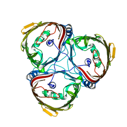
6JBV
 | | Crystal structure of SpaE basal pilin from Lactobacillus rhamnosus GG - Selenium derivative | | 分子名称: | Pilus assembly protein, SODIUM ION | | 著者 | Megta, A.K, Mishra, A.K, Palva, A, von Ossowski, I, Krishnan, V. | | 登録日 | 2019-01-26 | | 公開日 | 2019-06-26 | | 最終更新日 | 2021-09-15 | | 実験手法 | X-RAY DIFFRACTION (1.712 Å) | | 主引用文献 | Crystal structure of basal pilin SpaE reveals the molecular basis of its incorporation in the lactobacillar SpaFED pilus.
J.Struct.Biol., 207, 2019
|
|
4K8E
 | | OYE1-W116V complexed with the aromatic product of (R)-carvone dismutation | | 分子名称: | 2-methyl-5-(prop-1-en-2-yl)phenol, CHLORIDE ION, DI(HYDROXYETHYL)ETHER, ... | | 著者 | Sullivan, B, Pompeu, Y.A, Stewart, J.D. | | 登録日 | 2013-04-18 | | 公開日 | 2013-10-09 | | 最終更新日 | 2024-02-28 | | 実験手法 | X-RAY DIFFRACTION (1.269 Å) | | 主引用文献 | X‑ray Crystallography Reveals How Subtle Changes Control the
Orientation of Substrate Binding in an Alkene Reductase
ACS CATALYSIS, 3, 2013
|
|
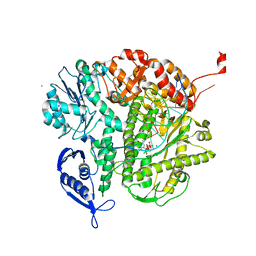
6JCL
 | | Crystal structure of cofactor-bound Rv0187 from MTB | | 分子名称: | (4S)-2-METHYL-2,4-PENTANEDIOL, Probable O-methyltransferase, S-ADENOSYL-L-HOMOCYSTEINE, ... | | 著者 | Kim, J, Lee, S. | | 登録日 | 2019-01-29 | | 公開日 | 2019-12-11 | | 最終更新日 | 2023-11-22 | | 実験手法 | X-RAY DIFFRACTION (1.644 Å) | | 主引用文献 | Structural and biochemical characterization of Rv0187, an O-methyltransferase from Mycobacterium tuberculosis.
Sci Rep, 9, 2019
|
|


