Loading
PDBj
メニューPDBj@FacebookPDBj@TwitterPDBj@YouTubewwPDB FoundationwwPDB
RCSB PDBPDBeBMRBAdv. SearchSearch help

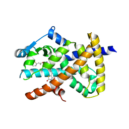
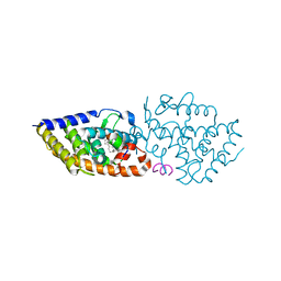
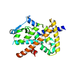
6LOA
DownloadVisualize
BU of 6loa by Molmil
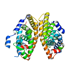
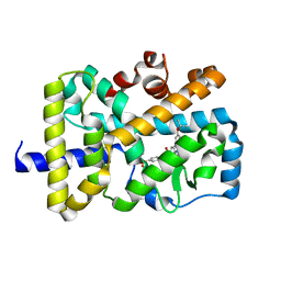
Crystal structure of RORgammat with ligand C46D bound
分子名称: 9-ethyl-~{N}-[(4-ethylsulfonylphenyl)methyl]-6-propan-2-yloxy-carbazole-3-carboxamide, Nuclear receptor ROR-gamma
著者Feng, Y, Shijie, C.
登録日2020-01-04
公開日2021-01-06
最終更新日2023-11-29
実験手法X-RAY DIFFRACTION (2.50003314 Å)
主引用文献Crystal structure of RORgammat with ligand C46D bound
To Be Published
5UBP
DownloadVisualize
BU of 5ubp by Molmil
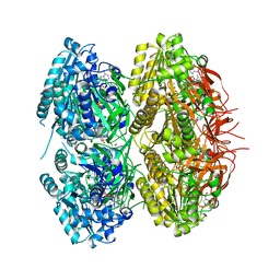
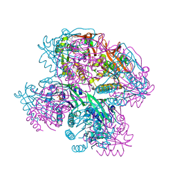
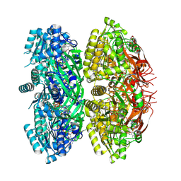
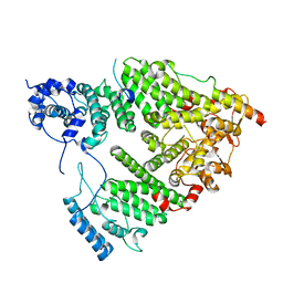
TREX2 M-region
分子名称: 26S proteasome complex subunit SEM1, Leucine permease transcriptional regulator, Nuclear mRNA export protein THP1
著者Stewart, M, Gordon, J.
登録日2016-12-21
公開日2017-04-05
最終更新日2024-01-17
実験手法X-RAY DIFFRACTION (2.3 Å)
主引用文献Structure of the Sac3 RNA-binding M-region in the Saccharomyces cerevisiae TREX-2 complex.
Nucleic Acids Res., 45, 2017
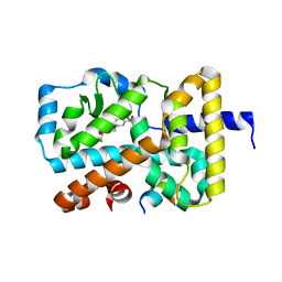
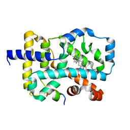
6DGQ
DownloadVisualize
BU of 6dgq by Molmil
Crystal Structure of Human PPARgamma Ligand Binding Domain in Complex with CAY10506
分子名称: N-(2-{4-[(2,4-dioxo-3,4-dihydro-2H-1lambda~4~,3-thiazol-5-yl)methyl]phenoxy}ethyl)-5-[(3R)-1,2-dithiolan-3-yl]pentanamide, Peroxisome proliferator-activated receptor gamma
著者Shang, J, Kojetin, D.J.
登録日2018-05-17
公開日2019-05-22
最終更新日2023-10-11
実験手法X-RAY DIFFRACTION (2.45 Å)
主引用文献Quantitative structural assessment of graded receptor agonism.
Proc.Natl.Acad.Sci.USA, 116, 2019
6NIT
DownloadVisualize
BU of 6nit by Molmil
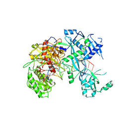
Human Argonaute2-miR-122 bound to a target RNA with four central mismatches (bu4)
分子名称: Protein argonaute-2, RNA (5'-R(P*AP*AP*CP*AP*CP*CP*AP*UP*CP*CP*AP*AP*CP*AP*CP*UP*CP*CP*AP*AP*A)-3'), RNA (5'-R(P*UP*GP*GP*AP*GP*UP*GP*UP*GP*AP*CP*AP*AP*UP*GP*GP*UP*GP*UP*UP*U)-3')
著者Sheu-Gruttadauria, J, MacRae, I.J.
登録日2018-12-31
公開日2019-08-07
最終更新日2024-03-13
実験手法X-RAY DIFFRACTION (3.8 Å)
主引用文献Structural Basis for Target-Directed MicroRNA Degradation.
Mol.Cell, 75, 2019
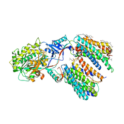
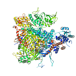
5VHM
DownloadVisualize
BU of 5vhm by Molmil
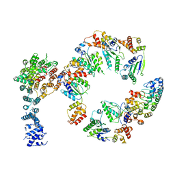
Conformational Landscape of the p28-Bound Human Proteasome Regulatory Particle
分子名称: 26S proteasome non-ATPase regulatory subunit 10, 26S proteasome non-ATPase regulatory subunit 2, 26S proteasome regulatory subunit 10B, ...
著者Lu, Y, Wu, J, Dong, Y, Chen, S, Sun, S, Ma, Y.B, Ouyang, Q, Finley, D, Kirschner, M.W, Mao, Y.
登録日2017-04-13
公開日2017-08-23
最終更新日2024-03-13
実験手法ELECTRON MICROSCOPY (8.3 Å)
主引用文献Conformational Landscape of the p28-Bound Human Proteasome Regulatory Particle.
Mol. Cell, 67, 2017
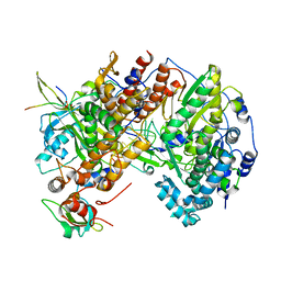
5VHP
DownloadVisualize
BU of 5vhp by Molmil
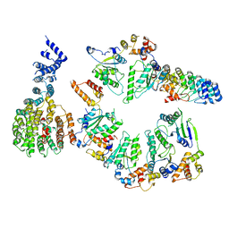
Conformational Landscape of the p28-Bound Human Proteasome Regulatory Particle
分子名称: 26S proteasome non-ATPase regulatory subunit 10, 26S proteasome non-ATPase regulatory subunit 2, 26S proteasome regulatory subunit 10B, ...
著者Lu, Y, Wu, J, Dong, Y, Chen, S, Sun, S, Ma, Y.B, Ouyang, Q, Finley, D, Kirschner, M.W, Mao, Y.
登録日2017-04-13
公開日2017-08-23
最終更新日2024-03-13
実験手法ELECTRON MICROSCOPY (7.9 Å)
主引用文献Conformational Landscape of the p28-Bound Human Proteasome Regulatory Particle.
Mol. Cell, 67, 2017
6G72
DownloadVisualize
BU of 6g72 by Molmil
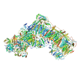
Mouse mitochondrial complex I in the deactive state
分子名称: ACETYL GROUP, ADENOSINE-5'-DIPHOSPHATE, Acyl carrier protein, ...
著者Agip, A.N.A, Blaza, J.N, Bridges, H.R, Viscomi, C, Rawson, S, Muench, S.P, Hirst, J.
登録日2018-04-04
公開日2018-06-06
最終更新日2024-05-29
実験手法ELECTRON MICROSCOPY (3.9 Å)
主引用文献Cryo-EM structures of complex I from mouse heart mitochondria in two biochemically defined states.
Nat. Struct. Mol. Biol., 25, 2018
7B9C
DownloadVisualize
BU of 7b9c by Molmil
Structure of a minimal SF3B core in complex with spliceostatin A (form I)
分子名称: PHD finger-like domain-containing protein 5A, Splicing factor 3B subunit 1, Splicing factor 3B subunit 3, ...
著者Cretu, C, Pena, V.
登録日2020-12-14
公開日2021-08-04
最終更新日2024-10-16
実験手法X-RAY DIFFRACTION (2.4 Å)
主引用文献Structural basis of intron selection by U2 snRNP in the presence of covalent inhibitors.
Nat Commun, 12, 2021
7B91
DownloadVisualize
BU of 7b91 by Molmil
Structure of a minimal SF3B core in complex with pladienolide D (form I)
分子名称: PHD finger-like domain-containing protein 5A, Splicing factor 3B subunit 1, Splicing factor 3B subunit 3, ...
著者Cretu, C, Pena, V.
登録日2020-12-13
公開日2021-08-04
最終更新日2024-01-31
実験手法X-RAY DIFFRACTION (3 Å)
主引用文献Structural basis of intron selection by U2 snRNP in the presence of covalent inhibitors.
Nat Commun, 12, 2021
7BK4
DownloadVisualize
BU of 7bk4 by Molmil
Crystal structure of RXRalpha ligand binding domain in complex with a fragment of the TIF2 coactivator
分子名称: 6-[1-(3,5,5,8,8-PENTAMETHYL-5,6,7,8-TETRAHYDRONAPHTHALEN-2-YL)CYCLOPROPYL]PYRIDINE-3-CARBOXYLIC ACID, Nuclear receptor coactivator 2, Retinoic acid receptor RXR-alpha
著者le Maire, A, Bourguet, W.
登録日2021-01-15
公開日2021-08-04
最終更新日2024-01-31
実験手法X-RAY DIFFRACTION (2.8 Å)
主引用文献Structural Insights into the Interaction of the Intrinsically Disordered Co-activator TIF2 with Retinoic Acid Receptor Heterodimer (RXR/RAR).
J.Mol.Biol., 433, 2021
7BO6
DownloadVisualize
BU of 7bo6 by Molmil
VDR complex with LCA derivative
分子名称: (4R)-4-[(3R,5R,8R,9S,10S,13R,14S,17R)-10,13-dimethyl-3-(2-methyl-2-oxidanyl-propyl)-2,3,4,5,6,7,8,9,11,12,14,15,16,17-tetradecahydro-1H-cyclopenta[a]phenanthren-17-yl]pentanoic acid, Nuclear receptor coactivator 1, Vitamin D3 receptor A
著者Rochel, N.
登録日2021-01-24
公開日2021-08-11
最終更新日2024-01-31
実験手法X-RAY DIFFRACTION (2.86 Å)
主引用文献Lithocholic acid-based design of noncalcemic vitamin D receptor agonists.
Bioorg.Chem., 111, 2021
7Y5T
DownloadVisualize
BU of 7y5t by Molmil
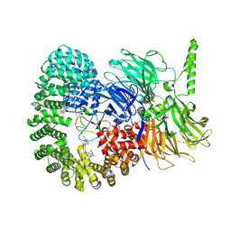
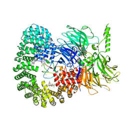
CryoEM structure of PS1-containing gamma-secretase in complex with MRK-560
分子名称: 1,2-DIACYL-SN-GLYCERO-3-PHOSPHOCHOLINE, 2-acetamido-2-deoxy-beta-D-glucopyranose, 2-acetamido-2-deoxy-beta-D-glucopyranose-(1-4)-2-acetamido-2-deoxy-beta-D-glucopyranose, ...
著者Guo, X, Wang, Y, Zhou, J, Jin, C, Wang, J, Jia, B, Jing, D, Yan, C, Lei, J, Zhou, R, Shi, Y.
登録日2022-06-17
公開日2022-11-02
実験手法ELECTRON MICROSCOPY (2.9 Å)
主引用文献Molecular basis for isoform-selective inhibition of presenilin-1 by MRK-560.
Nat Commun, 13, 2022
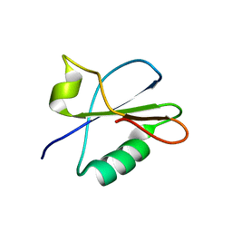
7BB5
DownloadVisualize
BU of 7bb5 by Molmil
Crystal structure of anti-CRISPR protein AcrIF9
分子名称: AcrIF9
著者Tamulaitiene, G, Sinkunas, T, Kupcinskaite, E.
登録日2020-12-17
公開日2021-11-24
最終更新日2024-05-01
実験手法X-RAY DIFFRACTION (2.3 Å)
主引用文献Disarming of type I-F CRISPR-Cas surveillance complex by anti-CRISPR proteins AcrIF6 and AcrIF9.
Sci Rep, 12, 2022
6G07
DownloadVisualize
BU of 6g07 by Molmil
RORGT (264-518;C455S) IN COMPLEX WITH INVERSE AGONIST "CPD-9" AND RIP140 PEPTIDE AT 1.66A
分子名称: Nuclear receptor ROR-gamma, Nuclear receptor-interacting protein 1, ~{N}-[5-chloranyl-6-[(1~{S})-1-phenylethoxy]pyridin-3-yl]-2-(4-ethylsulfonylphenyl)ethanamide
著者Kallen, J.
登録日2018-03-16
公開日2018-07-18
最終更新日2024-01-17
実験手法X-RAY DIFFRACTION (1.66 Å)
主引用文献Optimizing a Weakly Binding Fragment into a Potent ROR gamma t Inverse Agonist with Efficacy in an in Vivo Inflammation Model.
J. Med. Chem., 61, 2018
7E2O
DownloadVisualize
BU of 7e2o by Molmil
X-ray Crystal structure of PPARgamma R288H mutant.
分子名称: Peroxisome proliferator-activated receptor gamma
著者Egawa, D, Itoh, T.
登録日2021-02-06
公開日2022-01-19
最終更新日2023-11-29
実験手法X-RAY DIFFRACTION (3.2 Å)
主引用文献Structural Insights into the Loss-of-Function R288H Mutant of Human PPAR gamma.
Biol.Pharm.Bull., 44, 2021
7Z43
DownloadVisualize
BU of 7z43 by Molmil
Influenza B polymerase with Pol II pSer5 CTD peptide mimic bound in site 1B and 2B
分子名称: PHOSPHATE ION, Polymerase acidic protein, Polymerase basic protein 2, ...
著者Cusack, S, Drncova, P.
登録日2022-03-03
公開日2022-05-11
最終更新日2024-01-31
実験手法X-RAY DIFFRACTION (3.123 Å)
主引用文献Type B and type A influenza polymerases have evolved distinct binding interfaces to recruit the RNA polymerase II CTD.
Plos Pathog., 18, 2022
7Z4O
DownloadVisualize
BU of 7z4o by Molmil
Influenza A/H7N9 polymerase core dimer with Pol II pSer5 CTD peptide mimic bound in site 2A
分子名称: MAGNESIUM ION, Polymerase acidic protein, Polymerase basic protein 2, ...
著者Cusack, S, Pflug, A.
登録日2022-03-04
公開日2022-05-11
最終更新日2024-10-09
実験手法X-RAY DIFFRACTION (3.412 Å)
主引用文献Type B and type A influenza polymerases have evolved distinct binding interfaces to recruit the RNA polymerase II CTD.
Plos Pathog., 18, 2022
7E3M
DownloadVisualize
BU of 7e3m by Molmil
RORgamma LBD complexed with Panaxatriol and SRC2.2
分子名称: (3R,5R,6S,8R,9R,10R,12R,13R,14R,17S)-4,4,8,10,14-pentamethyl-17-[(2R)-2,6,6-trimethyloxan-2-yl]-2,3,5,6,7,9,11,12,13,15,16,17-dodecahydro-1H-cyclopenta[a]phenanthrene-3,6,12-triol, LYS-ILE-LEU-HIS-ARG-LEU-LEU-GLN, Nuclear receptor ROR-gamma
著者Liu, Z.H, Huang, J, Lu, W.Q, Tang, Y, Wu, Z.R.
登録日2021-02-09
公開日2022-03-23
最終更新日2023-11-29
実験手法X-RAY DIFFRACTION (2.8 Å)
主引用文献Structure of human RORgammat LBD with SRC2.2 at 2.80 Angstroms resolution
To Be Published
7DYK
DownloadVisualize
BU of 7dyk by Molmil
Crystal Structure of Cyanobacterial Circadian Clock Protein KaiC
分子名称: ADENOSINE-5'-TRIPHOSPHATE, Circadian clock protein kinase KaiC, MAGNESIUM ION
著者Furuike, Y, Akiyama, S.
登録日2021-01-22
公開日2022-04-27
最終更新日2023-11-29
実験手法X-RAY DIFFRACTION (2.99 Å)
主引用文献Elucidation of master allostery essential for circadian clock oscillation in cyanobacteria.
Sci Adv, 8, 2022
7DYI
DownloadVisualize
BU of 7dyi by Molmil
Crystal Structure of Cyanobacterial Circadian Clock Protein KaiC
分子名称: ADENOSINE-5'-TRIPHOSPHATE, Circadian clock protein kinase KaiC, MAGNESIUM ION
著者Furuike, Y, Akiyama, S.
登録日2021-01-21
公開日2022-04-27
最終更新日2023-11-29
実験手法X-RAY DIFFRACTION (2.64 Å)
主引用文献Elucidation of master allostery essential for circadian clock oscillation in cyanobacteria.
Sci Adv, 8, 2022
7DYJ
DownloadVisualize
BU of 7dyj by Molmil
Crystal Structure of Cyanobacterial Circadian Clock Protein KaiC
分子名称: ADENOSINE-5'-TRIPHOSPHATE, Circadian clock protein kinase KaiC, MAGNESIUM ION
著者Furuike, Y, Akiyama, S.
登録日2021-01-22
公開日2022-04-27
最終更新日2023-11-29
実験手法X-RAY DIFFRACTION (2.4 Å)
主引用文献Elucidation of master allostery essential for circadian clock oscillation in cyanobacteria.
Sci Adv, 8, 2022
7DXQ
DownloadVisualize
BU of 7dxq by Molmil
Crystal Structure of Cyanobacterial Circadian Clock Protein KaiC
分子名称: ADENOSINE-5'-TRIPHOSPHATE, Circadian clock protein kinase KaiC, MAGNESIUM ION
著者Furuike, Y, Akiyama, S.
登録日2021-01-20
公開日2022-04-27
最終更新日2023-11-29
実験手法X-RAY DIFFRACTION (2.8 Å)
主引用文献Elucidation of master allostery essential for circadian clock oscillation in cyanobacteria.
Sci Adv, 8, 2022
7DY2
DownloadVisualize
BU of 7dy2 by Molmil
Crystal Structure of Cyanobacterial Circadian Clock Protein KaiC
分子名称: ADENOSINE-5'-DIPHOSPHATE, ADENOSINE-5'-TRIPHOSPHATE, Circadian clock protein kinase KaiC, ...
著者Furuike, Y, Akiyama, S.
登録日2021-01-20
公開日2022-04-27
最終更新日2023-11-29
実験手法X-RAY DIFFRACTION (3.04 Å)
主引用文献Elucidation of master allostery essential for circadian clock oscillation in cyanobacteria.
Sci Adv, 8, 2022
7DYE
DownloadVisualize
BU of 7dye by Molmil
Crystal Structure of Cyanobacterial Circadian Clock Protein KaiC
分子名称: ADENOSINE-5'-TRIPHOSPHATE, Circadian clock protein kinase KaiC, MAGNESIUM ION
著者Furuike, Y, Akiyama, S.
登録日2021-01-21
公開日2022-05-18
最終更新日2023-11-29
実験手法X-RAY DIFFRACTION (2.6 Å)
主引用文献Regulation mechanisms of the dual ATPase in KaiC.
Proc.Natl.Acad.Sci.USA, 119, 2022
7DY1
DownloadVisualize
BU of 7dy1 by Molmil
Crystal Structure of Cyanobacterial Circadian Clock Protein KaiC
分子名称: ADENOSINE-5'-TRIPHOSPHATE, Circadian clock protein kinase KaiC, MAGNESIUM ION
著者Furuike, Y, Akiyama, S.
登録日2021-01-20
公開日2022-05-18
最終更新日2023-11-29
実験手法X-RAY DIFFRACTION (2.2 Å)
主引用文献Regulation mechanisms of the dual ATPase in KaiC.
Proc.Natl.Acad.Sci.USA, 119, 2022

226262

件を2024-10-16に公開中

PDB statisticsPDBj update infoContact PDBjnumon