3EG0
 
 | |
4GHA
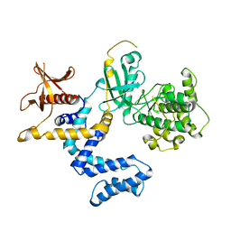
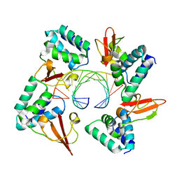 | | Crystal structure of Marburg virus VP35 RNA binding domain bound to 12-bp dsRNA | | 分子名称: | Polymerase cofactor VP35, RNA (5'-R(*CP*UP*AP*GP*AP*CP*GP*UP*CP*UP*AP*G)-3') | | 著者 | Bale, S, Jean-Philippe, J, Bornholdt, Z.A, Kimberlin, C.K, Halfmann, P, Zandonatti, M.A, Kunert, J, Kroon, G.J.A, Kawaoka, Y, MacRae, I.J, Wilson, I.A, Saphire, E.O. | | 登録日 | 2012-08-07 | | 公開日 | 2012-08-22 | | 最終更新日 | 2023-09-13 | | 実験手法 | X-RAY DIFFRACTION (2.5 Å) | | 主引用文献 | Marburg Virus VP35 Can Both Fully Coat the Backbone and Cap the Ends of dsRNA for Interferon Antagonism.
Plos Pathog., 8, 2012
|
|
2JU2
 | |
2RK9
 | | The crystal structure of a glyoxalase/bleomycin resistance protein/dioxygenase superfamily member from Vibrio splendidus 12B01 | | 分子名称: | Glyoxalase/bleomycin resistance protein/dioxygenase | | 著者 | Tyagi, R, Eswaramoorthy, S, Sauder, J.M, Burley, S.K, Swaminathan, S, New York SGX Research Center for Structural Genomics (NYSGXRC) | | 登録日 | 2007-10-16 | | 公開日 | 2007-10-30 | | 最終更新日 | 2021-10-20 | | 実験手法 | X-RAY DIFFRACTION (1.6 Å) | | 主引用文献 | The crystal structure of a glyoxalase/bleomycin resistance protein/dioxygenase superfamily member from Vibrio splendidus 12B01.
To be Published
|
|
3A70
 | | Crystal structure of Pseudomonas sp. MIS38 lipase in complex with diethyl phosphate | | 分子名称: | ACETATE ION, CALCIUM ION, DIETHYL PHOSPHONATE, ... | | 著者 | Angkawidjaja, C, Matsumura, H, Koga, Y, Takano, K, Kanaya, S. | | 登録日 | 2009-09-10 | | 公開日 | 2010-05-26 | | 最終更新日 | 2023-11-01 | | 実験手法 | X-RAY DIFFRACTION (2.15 Å) | | 主引用文献 | X-ray Crystallographic and MD Simulation Studies on the Mechanism of Interfacial Activation of a Family I.3 Lipase with Two Lids
J.Mol.Biol., 2010
|
|
3QT9
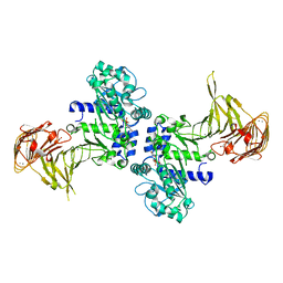
 | | Analysis of a new family of widely distributed metal-independent alpha mannosidases provides unique insight into the processing of N-linked glycans, Clostridium perfringens CPE0426 complexed with alpha-1,6-linked 1-thio-alpha-mannobiose | | 分子名称: | 1,2-ETHANEDIOL, Putative uncharacterized protein CPE0426, alpha-D-mannopyranose-(1-6)-6-thio-alpha-D-mannopyranose | | 著者 | Gregg, K.J, Zandberg, W.F, Hehemann, J.-H, Whitworth, G.E, Deng, L.E, Vocadlo, D.J, Boraston, A.B. | | 登録日 | 2011-02-22 | | 公開日 | 2011-03-09 | | 最終更新日 | 2024-02-21 | | 実験手法 | X-RAY DIFFRACTION (2.05 Å) | | 主引用文献 | Analysis of a New Family of Widely Distributed Metal-independent {alpha}-Mannosidases Provides Unique Insight into the Processing of N-Linked Glycans.
J.Biol.Chem., 286, 2011
|
|
1SI2
 | |
3A6Z
 | | Crystal structure of Pseudomonas sp. MIS38 lipase (PML) in the open conformation following dialysis against Ca-free buffer | | 分子名称: | CALCIUM ION, Lipase | | 著者 | Angkawidjaja, C, Matsumura, H, Koga, Y, Takano, K, Kanaya, S. | | 登録日 | 2009-09-10 | | 公開日 | 2010-05-26 | | 最終更新日 | 2023-11-01 | | 実験手法 | X-RAY DIFFRACTION (2.15 Å) | | 主引用文献 | X-ray Crystallographic and MD Simulation Studies on the Mechanism of Interfacial Activation of a Family I.3 Lipase with Two Lids
J.Mol.Biol., 2010
|
|
2Y91
 | | Crystal structure of class A beta-lactamase from Bacillus licheniformis BS3 with clavulanic acid | | 分子名称: | 5-HYDROXY-3-OXOPENTANOIC ACID, BETA-LACTAMASE, CITRIC ACID, ... | | 著者 | Power, P, Sauvage, E, Herman, R, Kerff, F, Charlier, P. | | 登録日 | 2011-02-11 | | 公開日 | 2012-02-22 | | 最終更新日 | 2023-12-20 | | 実験手法 | X-RAY DIFFRACTION (2 Å) | | 主引用文献 | Novel Fragments of Clavulanate Observed in the Structure of the Class a Beta-Lactamase from Bacillus Licheniformis Bs3.
J.Antimicrob.Chemother., 67, 2012
|
|
4I06
 | | Crystal structure of human Arginase-2 complexed with inhibitor 14 | | 分子名称: | Arginase-2, mitochondrial, BENZAMIDINE, ... | | 著者 | Cousido-Siah, A, Mitschler, A, Ruiz, F.X, Whitehouse, D.L, Golebiowski, A, Ji, M, Zhang, M, Beckett, P, Sheeler, R, Andreoli, M, Conway, B, Mahboubi, K, Schroeter, H, Van Zandt, M.C, Podjarny, A. | | 登録日 | 2012-11-16 | | 公開日 | 2013-03-20 | | 最終更新日 | 2023-09-20 | | 実験手法 | X-RAY DIFFRACTION (1.8 Å) | | 主引用文献 | Discovery of (R)-2-Amino-6-borono-2-(2-(piperidin-1-yl)ethyl)hexanoic Acid and Congeners As Highly Potent Inhibitors of Human Arginases I and II for Treatment of Myocardial Reperfusion Injury.
J.Med.Chem., 56, 2013
|
|
4MKM
 | | Repeat domains 1 & 2 of Clostridium perfringens Cpe0147 | | 分子名称: | CALCIUM ION, Putative surface anchored protein | | 著者 | Kwon, H, Squire, C.J, Young, P.G, Baker, E.N. | | 登録日 | 2013-09-05 | | 公開日 | 2013-12-04 | | 最終更新日 | 2014-02-12 | | 実験手法 | X-RAY DIFFRACTION (1.75 Å) | | 主引用文献 | Autocatalytically generated Thr-Gln ester bond cross-links stabilize the repetitive Ig-domain shaft of a bacterial cell surface adhesin.
Proc.Natl.Acad.Sci.USA, 111, 2014
|
|
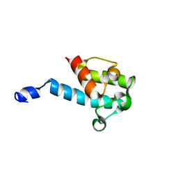
4GH9
 | | Crystal structure of Marburg virus VP35 RNA binding domain | | 分子名称: | ACETATE ION, Polymerase cofactor VP35 | | 著者 | Bale, S, Jean-Philippe, J, Bornholdt, Z.A, Kimberlin, C.K, Halfmann, P, Zandonatti, M.A, Kunert, J, Kroon, G.J.A, Kawaoka, Y, MacRae, I.J, Wilson, I.A, Saphire, E.O. | | 登録日 | 2012-08-07 | | 公開日 | 2012-08-15 | | 最終更新日 | 2023-09-13 | | 実験手法 | X-RAY DIFFRACTION (1.65 Å) | | 主引用文献 | Marburg Virus VP35 Can Both Fully Coat the Backbone and Cap the Ends of dsRNA for Interferon Antagonism.
Plos Pathog., 8, 2012
|
|
1K64
 | | NMR Structue of alpha-conotoxin EI | | 分子名称: | alpha-conotoxin EI | | 著者 | Park, K.H, Suk, J.E, Jacobsen, R, Gray, W.R, McIntosh, J.M, Han, K.H. | | 登録日 | 2001-10-15 | | 公開日 | 2003-09-09 | | 最終更新日 | 2022-02-23 | | 実験手法 | SOLUTION NMR | | 主引用文献 | Solution conformation of alpha-conotoxin EI, a neuromuscular toxin specific for the alpha 1/delta subunit interface of torpedo nicotinic acetylcholine receptor
J.BIOL.CHEM., 276, 2001
|
|
1YDP
 | | 1.9A crystal structure of HLA-G | | 分子名称: | Beta-2-microglobulin, CHLORIDE ION, COBALT (II) ION, ... | | 著者 | Clements, C.S, Kjer-nielsen, L, Kostenko, L, Hoare, H.L, Dunstone, M.A, Moses, E, Freed, K, Brooks, A.G, Rossjohn, J, Mccluskey, J. | | 登録日 | 2004-12-25 | | 公開日 | 2005-03-08 | | 最終更新日 | 2021-11-10 | | 実験手法 | X-RAY DIFFRACTION (1.9 Å) | | 主引用文献 | Crystal structure of HLA-G: A nonclassical MHC class I molecule expressed at the fetal-maternal interface
PROC.NATL.ACAD.SCI.USA, 102, 2005
|
|
1UWN
 | |
1RWE
 | | Enhancing the activity of insulin at receptor edge: crystal structure and photo-cross-linking of A8 analogues | | 分子名称: | CHLORIDE ION, Insulin, PHENOL, ... | | 著者 | Wan, Z, Xu, B, Chu, Y.C, Li, B, Nakagawa, S.H, Qu, Y, Hu, S.Q, Katsoyannis, P.G, Weiss, M.A. | | 登録日 | 2003-12-16 | | 公開日 | 2005-02-15 | | 最終更新日 | 2023-08-23 | | 実験手法 | X-RAY DIFFRACTION (1.8 Å) | | 主引用文献 | Enhancing the activity of insulin at the receptor interface: crystal structure and photo-cross-linking of A8 analogues.
Biochemistry, 43, 2004
|
|
4HZE
 | | Crystal structure of human Arginase-2 complexed with inhibitor 9 | | 分子名称: | Arginase-2, mitochondrial, BENZAMIDINE, ... | | 著者 | Cousido-Siah, A, Mitschler, A, Ruiz, F.X, Whitehouse, D.L, Golebiowski, A, Ji, M, Zhang, M, Beckett, P, Sheeler, R, Andreoli, M, Conway, B, Mahboubi, K, Schroeter, H, Van Zandt, M.C, Podjarny, A. | | 登録日 | 2012-11-15 | | 公開日 | 2013-03-20 | | 最終更新日 | 2023-09-20 | | 実験手法 | X-RAY DIFFRACTION (1.602 Å) | | 主引用文献 | Discovery of (R)-2-Amino-6-borono-2-(2-(piperidin-1-yl)ethyl)hexanoic Acid and Congeners As Highly Potent Inhibitors of Human Arginases I and II for Treatment of Myocardial Reperfusion Injury.
J.Med.Chem., 56, 2013
|
|
3FVJ
 | |
3EG1
 | |
3EGU
 | |
3EG3
 | |
1YM7
 | | G Protein-Coupled Receptor Kinase 2 (GRK2) | | 分子名称: | Beta-adrenergic receptor kinase 1 | | 著者 | Lodowski, D.T, Barnhill, J.F, Pyskadlo, R.M, Ghirlando, R, Sterne-Marr, R, Tesmer, J.J.G. | | 登録日 | 2005-01-20 | | 公開日 | 2005-07-05 | | 最終更新日 | 2023-08-23 | | 実験手法 | X-RAY DIFFRACTION (4.5 Å) | | 主引用文献 | The role of Gbetagamma and domain interfaces in the activation of G protein-coupled receptor kinase 2
Biochemistry, 44, 2005
|
|
1LJ7
 | | Crystal structure of calcium-depleted human C-reactive protein from perfectly twinned data | | 分子名称: | C-reactive protein | | 著者 | Ramadan, M.A, Shrive, A.K, Holden, D, Myles, D.A, Volanakis, J.E, DeLucas, L.J, Greenhough, T.J. | | 登録日 | 2002-04-19 | | 公開日 | 2002-06-05 | | 最終更新日 | 2023-08-16 | | 実験手法 | X-RAY DIFFRACTION (3.15 Å) | | 主引用文献 | The three-dimensional structure of calcium-depleted human C-reactive protein from perfectly twinned crystals.
Acta Crystallogr.,Sect.D, 58, 2002
|
|
3FVI
 | |
3BDL
 | | Crystal structure of a truncated human Tudor-SN | | 分子名称: | CITRIC ACID, Staphylococcal nuclease domain-containing protein 1 | | 著者 | Li, C.L. | | 登録日 | 2007-11-15 | | 公開日 | 2008-08-26 | | 最終更新日 | 2024-03-13 | | 実験手法 | X-RAY DIFFRACTION (1.9 Å) | | 主引用文献 | Structural and functional insights into human Tudor-SN, a key component linking RNA interference and editing.
Nucleic Acids Res., 36, 2008
|
|


