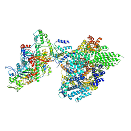
3JBN
 
 | | Cryo-electron microscopy reconstruction of the Plasmodium falciparum 80S ribosome bound to P-tRNA | | 分子名称: | 18S ribosomal RNA, 28S ribosomal RNA, 40S ribosomal protein eS1, ... | | 著者 | Sun, M, Li, W, Blomqvist, K, Das, S, Hashem, Y, Dvorin, J.D, Frank, J. | | 登録日 | 2015-09-16 | | 公開日 | 2015-10-14 | | 最終更新日 | 2024-02-21 | | 実験手法 | ELECTRON MICROSCOPY (4.7 Å) | | 主引用文献 | Dynamical features of the Plasmodium falciparum ribosome during translation.
Nucleic Acids Res., 43, 2015
|
|
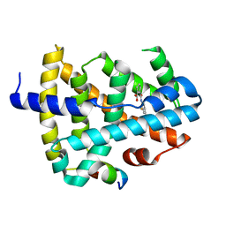
5YCP
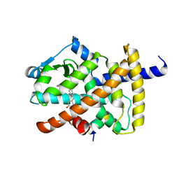 | |
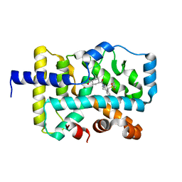
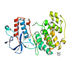
2ZDY
 | | Inhibitor-bound structures of human pyruvate dehydrogenase kinase 4 | | 分子名称: | 4-(2-HYDROXYETHYL)-1-PIPERAZINE ETHANESULFONIC ACID, ADENOSINE-5'-DIPHOSPHATE, MAGNESIUM ION, ... | | 著者 | Kawamoto, M, Shiromizu, I, Kukimoto-Niino, M, Tokmakov, A, Terada, T, Shirouzu, M, Matsusue, T, Yokoyama, S. | | 登録日 | 2007-12-01 | | 公開日 | 2008-12-09 | | 最終更新日 | 2023-11-01 | | 実験手法 | X-RAY DIFFRACTION (2.4 Å) | | 主引用文献 | Inhibitor-bound structures of human pyruvate dehydrogenase kinase 4.
Acta Crystallogr.,Sect.D, 67, 2011
|
|
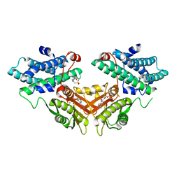
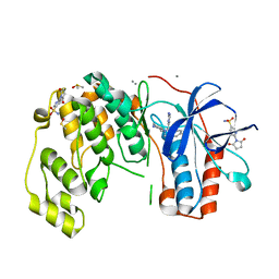
3JV0
 | |
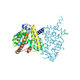
2ZG2
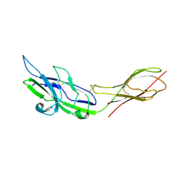 | |
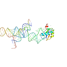
2ZHB
 | | Complex structure of AFCCA with tRNAminiDUC | | 分子名称: | CCA-adding enzyme, SULFATE ION, tRNA (34-MER) | | 著者 | Toh, Y, Tomita, K. | | 登録日 | 2008-02-01 | | 公開日 | 2008-08-05 | | 最終更新日 | 2024-03-13 | | 実験手法 | X-RAY DIFFRACTION (3.05 Å) | | 主引用文献 | Molecular basis for maintenance of fidelity during the CCA-adding reaction by a CCA-adding enzyme
Embo J., 27, 2008
|
|
6XAE
 | | SUBSTITUTED BENZYLOXYTRICYCLIC COMPOUNDS AS RETINOIC ACID-RELATED ORPHAN RECEPTOR GAMMA T AGONISTS | | 分子名称: | Nuclear receptor ROR-gamma, trans-4-{(3aR,9bR)-7-[(2-chloro-6-fluorophenyl)methoxy]-9b-[(4-fluorophenyl)sulfonyl]-1,2,3a,4,5,9b-hexahydro-3H-benzo[e]indole-3-carbonyl}cyclohexane-1-carboxylic acid | | 著者 | Sack, J.S. | | 登録日 | 2020-06-04 | | 公開日 | 2020-06-17 | | 最終更新日 | 2023-10-18 | | 実験手法 | X-RAY DIFFRACTION (2.257 Å) | | 主引用文献 | Substituted benzyloxytricyclic compounds as retinoic acid-related orphan receptor gamma t (ROR gamma t) agonists.
Bioorg.Med.Chem.Lett., 30, 2020
|
|
1M6Y
 | | Crystal Structure Analysis of TM0872, a Putative SAM-dependent Methyltransferase, Complexed with SAH | | 分子名称: | S-ADENOSYL-L-HOMOCYSTEINE, S-adenosyl-methyltransferase mraW, SULFATE ION | | 著者 | Miller, D.J, Anderson, W.F, Midwest Center for Structural Genomics (MCSG) | | 登録日 | 2002-07-17 | | 公開日 | 2003-01-28 | | 最終更新日 | 2016-03-30 | | 実験手法 | X-RAY DIFFRACTION (1.9 Å) | | 主引用文献 | Crystal complexes of a predicted S-adenosylmethionine-dependent methyltransferase reveal a typical AdoMet binding domain and a substrate recognition domain
Protein Sci., 12, 2003
|
|
1M5V
 | | Transition State Stabilization by a Catalytic RNA | | 分子名称: | (4S)-2-METHYL-2,4-PENTANEDIOL, CALCIUM ION, RNA HAIRPIN RIBOZYME, ... | | 著者 | Rupert, P.B, Massey, A.P, Sigurdsson, S.T, Ferre-D'Amare, A.R. | | 登録日 | 2002-07-09 | | 公開日 | 2002-10-12 | | 最終更新日 | 2024-02-14 | | 実験手法 | X-RAY DIFFRACTION (2.4 Å) | | 主引用文献 | Transition state stabilization by a catalytic RNA
Science, 298, 2002
|
|
3JPV
 | | Crystal structure of human proto-oncogene serine threonine kinase (PIM1) in complex with a consensus peptide and a pyrrolo[2,3-a]carbazole ligand | | 分子名称: | 1,10-dihydropyrrolo[2,3-a]carbazole-3-carbaldehyde, Peptide (PIMTIDE) ARKRRRHPSGPPTA, Proto-oncogene serine/threonine-protein kinase Pim-1 | | 著者 | Filippakopoulos, P, Bullock, A.N, Fedorov, O, Akue-Gedu, R, Rossignol, E, Azzaro, S, Bain, J, Cohen, P, Prudhomme, M, Moreau, P, Amizon, F, von Delft, F, Arrowsmith, C.H, Weigelt, J, Edwards, A, Bountra, C, Knapp, S, Structural Genomics Consortium (SGC) | | 登録日 | 2009-09-04 | | 公開日 | 2009-10-27 | | 最終更新日 | 2023-09-06 | | 実験手法 | X-RAY DIFFRACTION (2.35 Å) | | 主引用文献 | Synthesis, kinase inhibitory potencies, and in vitro antiproliferative evaluation of new pim kinase inhibitors.
J.Med.Chem., 52, 2009
|
|
1M90
 | | Co-crystal structure of CCA-Phe-caproic acid-biotin and sparsomycin bound to the 50S ribosomal subunit | | 分子名称: | 23S RRNA, 5S RRNA, 6-AMINOHEXANOIC ACID, ... | | 著者 | Hansen, J.L, Schmeing, T.M, Moore, P.B, Steitz, T.A. | | 登録日 | 2002-07-26 | | 公開日 | 2002-09-06 | | 最終更新日 | 2023-11-15 | | 実験手法 | X-RAY DIFFRACTION (2.8 Å) | | 主引用文献 | Structural insights into peptide bond formation.
Proc.Natl.Acad.Sci.USA, 99, 2002
|
|
6SFO
 | | MAPK14 with bound inhibitor SR-318 | | 分子名称: | 5-azanyl-~{N}-[[4-(3-cyclohexylpropylcarbamoyl)phenyl]methyl]-1-phenyl-pyrazole-4-carboxamide, GLYCEROL, Mitogen-activated protein kinase 14, ... | | 著者 | Schroeder, M, Edwards, A.M, Arrowsmith, C.H, Bountra, C, Knapp, S, Structural Genomics Consortium (SGC) | | 登録日 | 2019-08-01 | | 公開日 | 2019-09-11 | | 最終更新日 | 2024-01-24 | | 実験手法 | X-RAY DIFFRACTION (1.75 Å) | | 主引用文献 | MAPK14 with bound inhibitor SR-318
To Be Published
|
|
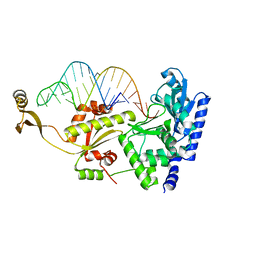
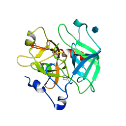
5Y1J
 | |
6SP9
 | | Fragment KCL802 in complex with MAP kinase p38-alpha | | 分子名称: | 4-(4-FLUOROPHENYL)-1-(4-PIPERIDINYL)-5-(2-AMINO-4-PYRIMIDINYL)-IMIDAZOLE, 6-[2,5-bis(oxidanylidene)pyrrolidin-1-yl]pyridine-3-sulfonamide, CALCIUM ION, ... | | 著者 | Nichols, C.E, De Nicola, G.F. | | 登録日 | 2019-08-31 | | 公開日 | 2019-10-02 | | 最終更新日 | 2024-01-24 | | 実験手法 | X-RAY DIFFRACTION (1.22 Å) | | 主引用文献 | Mining the PDB for Tractable Cases Where X-ray Crystallography Combined with Fragment Screens Can Be Used to Systematically Design Protein-Protein Inhibitors: Two Test Cases Illustrated by IL1 beta-IL1R and p38 alpha-TAB1 Complexes.
J.Med.Chem., 63, 2020
|
|
5Y2O
 | | Structure of PPARgamma ligand binding domain-pioglitazone complex | | 分子名称: | (5S)-5-[[4-[2-(5-ethylpyridin-2-yl)ethoxy]phenyl]methyl]-1,3-thiazolidine-2,4-dione, Peroxisome proliferator-activated receptor gamma | | 著者 | Im, Y.J, Lee, M. | | 登録日 | 2017-07-26 | | 公開日 | 2017-12-20 | | 最終更新日 | 2023-11-22 | | 実験手法 | X-RAY DIFFRACTION (1.801 Å) | | 主引用文献 | Structures of PPAR gamma complexed with lobeglitazone and pioglitazone reveal key determinants for the recognition of antidiabetic drugs
Sci Rep, 7, 2017
|
|
1MH0
 | | Crystal structure of the anticoagulant slow form of thrombin | | 分子名称: | 2-acetamido-2-deoxy-beta-D-glucopyranose, Prothrombin | | 著者 | Pineda, A.O, Savvides, S, Waksman, G, Di Cera, E. | | 登録日 | 2002-08-18 | | 公開日 | 2002-11-08 | | 最終更新日 | 2021-10-27 | | 実験手法 | X-RAY DIFFRACTION (2.8 Å) | | 主引用文献 | Crystal structure of the anticoagulant slow form of thrombin
J.Biol.Chem., 277, 2002
|
|
6SOU
 | | Fragment N13565a in complex with MAP kinase p38-alpha | | 分子名称: | 2-(4-methylphenoxy)-1-(4-methylpiperazin-4-ium-1-yl)ethanone, CHLORIDE ION, Mitogen-activated protein kinase 14, ... | | 著者 | Nichols, C.E, De Nicola, G.F. | | 登録日 | 2019-08-29 | | 公開日 | 2019-10-02 | | 最終更新日 | 2024-01-24 | | 実験手法 | X-RAY DIFFRACTION (1.5 Å) | | 主引用文献 | Mining the PDB for Tractable Cases Where X-ray Crystallography Combined with Fragment Screens Can Be Used to Systematically Design Protein-Protein Inhibitors: Two Test Cases Illustrated by IL1 beta-IL1R and p38 alpha-TAB1 Complexes.
J.Med.Chem., 63, 2020
|
|
3JUZ
 | |
5VB3
 | |
3JY0
 | |
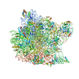
7YFN
 | | Core module of the NuA4 complex in S. cerevisiae | | 分子名称: | ADENOSINE-5'-TRIPHOSPHATE, ARP4 isoform 1, Actin, ... | | 著者 | Ji, L.T, Zhao, L.X, Xu, K, Gao, H.H, Zhou, Y, Kornberg, R.D, Zhang, H.Q. | | 登録日 | 2022-07-08 | | 公開日 | 2023-03-08 | | 実験手法 | ELECTRON MICROSCOPY (3.8 Å) | | 主引用文献 | Structure of the NuA4 histone acetyltransferase complex.
Proc.Natl.Acad.Sci.USA, 119, 2022
|
|
8RTT
 | | Structure of the formin Cdc12 bound to the barbed end of phalloidin-stabilized F-actin. | | 分子名称: | ADENOSINE-5'-DIPHOSPHATE, Actin, cytoplasmic 1, ... | | 著者 | Oosterheert, W, Boiero Sanders, M, Funk, J, Prumbaum, D, Raunser, S, Bieling, P. | | 登録日 | 2024-01-29 | | 公開日 | 2024-04-10 | | 最終更新日 | 2024-06-05 | | 実験手法 | ELECTRON MICROSCOPY (3.56 Å) | | 主引用文献 | Molecular mechanism of actin filament elongation by formins.
Science, 384, 2024
|
|
6XZV
 | | Structure of zVDR LBD-Calcitriol in complex with chimera 18 | | 分子名称: | 5-{2-[1-(5-HYDROXY-1,5-DIMETHYL-HEXYL)-7A-METHYL-OCTAHYDRO-INDEN-4-YLIDENE]-ETHYLIDENE}-4-METHYLENE-CYCLOHEXANE-1,3-DIOL, URA-UIA-URL-URY-URV-UZN-LYS, Vitamin D3 receptor A | | 著者 | Buratto, J, Belorusova, A.Y, Rochel, N, Guichard, G. | | 登録日 | 2020-02-05 | | 公開日 | 2021-02-17 | | 最終更新日 | 2024-01-24 | | 実験手法 | X-RAY DIFFRACTION (2.3 Å) | | 主引用文献 | Structural Basis for alpha-Helix Mimicry and Inhibition of Protein-Protein Interactions with Oligourea Foldamers.
Angew.Chem.Int.Ed.Engl., 60, 2021
|
|
8GXP
 | | Complex structure of RORgama with betulinic acid | | 分子名称: | Betulinic acid, Nuclear receptor ROR-gamma | | 著者 | Zhang, X.L, Xu, C, Bai, F. | | 登録日 | 2022-09-20 | | 公開日 | 2023-06-07 | | 最終更新日 | 2023-11-08 | | 実験手法 | X-RAY DIFFRACTION (2.45 Å) | | 主引用文献 | Discovery, structural optimization, and anti-tumor bioactivity evaluations of betulinic acid derivatives as a new type of ROR gamma antagonists.
Eur.J.Med.Chem., 257, 2023
|
|
5SYB
 | | Crystal structure of human PHF5A | | 分子名称: | 1,2-ETHANEDIOL, PHD finger-like domain-containing protein 5A, ZINC ION | | 著者 | Tsai, J.H.C, Teng, T, Zhu, P, Fekkes, P, Larsen, N.A. | | 登録日 | 2016-08-10 | | 公開日 | 2016-09-07 | | 最終更新日 | 2024-03-06 | | 実験手法 | X-RAY DIFFRACTION (1.82 Å) | | 主引用文献 | Splicing modulators act at the branch point adenosine binding pocket defined by the PHF5A-SF3b complex.
Nat Commun, 8, 2017
|
|


