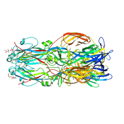
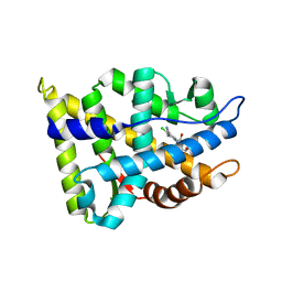
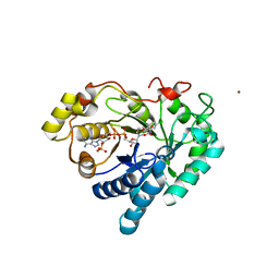
3P3L
 
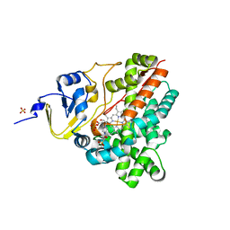 | | Crystal Structure of the Cytochrome P450 monooxygenase AurH (wildtype) from Streptomyces Thioluteus | | 分子名称: | 4-(2-HYDROXYETHYL)-1-PIPERAZINE ETHANESULFONIC ACID, CHLORIDE ION, Cytochrome P450, ... | | 著者 | Zocher, G, Richter, M.E.A, Mueller, U, Hertweck, C. | | 登録日 | 2010-10-05 | | 公開日 | 2011-02-16 | | 最終更新日 | 2024-02-21 | | 実験手法 | X-RAY DIFFRACTION (2.1 Å) | | 主引用文献 | Structural fine-tuning of a multifunctional cytochrome p450 monooxygenase.
J.Am.Chem.Soc., 133, 2011
|
|
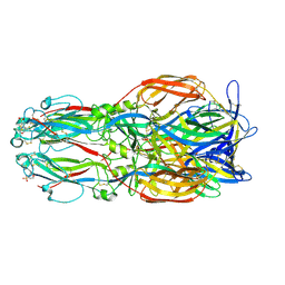
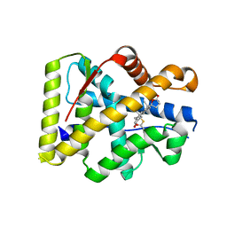
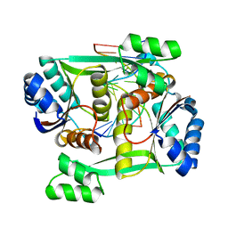
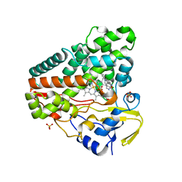
3G5F
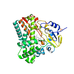 | | Crystallographic analysis of cytochrome P450 cyp121 | | 分子名称: | Cytochrome P450 121, PROTOPORPHYRIN IX CONTAINING FE, SULFATE ION | | 著者 | Belin, P, Le Du, M.H, Gondry, M. | | 登録日 | 2009-02-05 | | 公開日 | 2009-04-21 | | 最終更新日 | 2023-11-01 | | 実験手法 | X-RAY DIFFRACTION (1.4 Å) | | 主引用文献 | Identification and structural basis of the reaction catalyzed by CYP121, an essential cytochrome P450 in Mycobacterium tuberculosis.
Proc.Natl.Acad.Sci.USA, 106, 2009
|
|
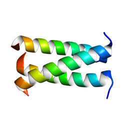
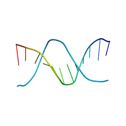
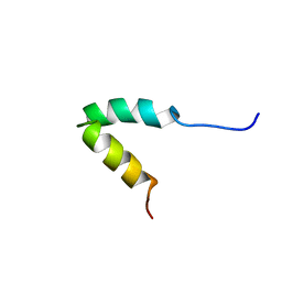
1ZTR
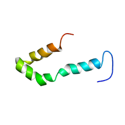 | | Solution structure of Engrailed homeodomain L16A mutant | | 分子名称: | Segmentation polarity homeobox protein engrailed | | 著者 | Religa, T.L, Markson, J.S, Mayor, U, Freund, S.M.V, Fersht, A.R. | | 登録日 | 2005-05-27 | | 公開日 | 2005-10-18 | | 最終更新日 | 2024-05-29 | | 実験手法 | SOLUTION NMR | | 主引用文献 | Solution structure of a protein denatured state and folding intermediate.
Nature, 437, 2005
|
|
3GUG
 | |
2KY8
 | |
3FL6
 | |
2HOS
 | | Phage-Selected Homeodomain Bound to Unmodified DNA | | 分子名称: | 3-METHYL-1,3-OXAZOLIDIN-2-ONE, 5'-D(*AP*TP*CP*CP*GP*GP*GP*GP*AP*TP*TP*AP*CP*AP*TP*GP*GP*CP*AP*AP*A)-3', 5'-D(*TP*TP*TP*TP*GP*CP*CP*AP*TP*GP*TP*AP*AP*TP*CP*CP*CP*CP*GP*GP*A)-3', ... | | 著者 | Shokat, K.M, Feldman, M.E, Simon, M.D. | | 登録日 | 2006-07-16 | | 公開日 | 2006-12-12 | | 最終更新日 | 2023-08-30 | | 実験手法 | X-RAY DIFFRACTION (1.9 Å) | | 主引用文献 | Structure and properties of a re-engineered homeodomain protein-DNA interface.
Acs Chem.Biol., 1, 2006
|
|
2HOT
 | | Phage selected homeodomain bound to modified DNA | | 分子名称: | 3-PROP-2-YN-1-YL-1,3-OXAZOLIDIN-2-ONE, 5'-D(*AP*TP*CP*CP*GP*GP*GP*GP*AP*TP*TP*AP*CP*AP*TP*GP*GP*CP*AP*AP*A)-3', 5'-D(*TP*TP*TP*TP*GP*CP*CP*AP*TP*GP*TP*AP*AP*TP*CP*CP*CP*CP*GP*GP*A)-3', ... | | 著者 | Feldman, M.E, Simon, M.D, Shokat, K.M. | | 登録日 | 2006-07-16 | | 公開日 | 2006-12-12 | | 最終更新日 | 2024-02-14 | | 実験手法 | X-RAY DIFFRACTION (2.19 Å) | | 主引用文献 | Structure and properties of a re-engineered homeodomain protein-DNA interface.
Acs Chem.Biol., 1, 2006
|
|
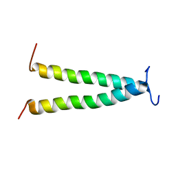
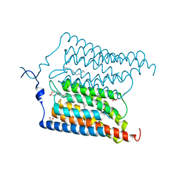
3HFE
 | | A trimeric form of the Kv7.1 A domain Tail | | 分子名称: | Potassium voltage-gated channel subfamily KQT member 1 | | 著者 | Xu, Q, Minor, D.L. | | 登録日 | 2009-05-11 | | 公開日 | 2009-09-01 | | 最終更新日 | 2024-02-21 | | 実験手法 | X-RAY DIFFRACTION (1.7 Å) | | 主引用文献 | Crystal structure of a trimeric form of the K(V)7.1 (KCNQ1) A-domain tail coiled-coil reveals structural plasticity and context dependent changes in a putative coiled-coil trimerization motif.
Protein Sci., 18, 2009
|
|
2JWT
 | |
2L6W
 | | PDGFR beta-TM | | 分子名称: | Beta-type platelet-derived growth factor receptor | | 著者 | Muhle-Goll, C, Hoffmann, S, Ulrich, A.S. | | 登録日 | 2010-11-29 | | 公開日 | 2012-05-30 | | 最終更新日 | 2024-05-01 | | 実験手法 | SOLUTION NMR | | 主引用文献 | Hydrophobic matching controls the tilt and stability of the dimeric platelet-derived growth factor receptor (PDGFR) beta transmembrane segment.
J.Biol.Chem., 287, 2012
|
|
2LDI
 | |
6EGU
 | |
6EIG
 | | Crystal structure of N24Q/C128T mutant of Channelrhodopsin 2 | | 分子名称: | (2R)-2,3-dihydroxypropyl (9Z)-octadec-9-enoate, Archaeal-type opsin 2, EICOSANE, ... | | 著者 | Kovalev, K, Borshchevskiy, V, Volkov, O, Polovinkin, V, Marin, E, Balandin, T, Astashkin, R, Bamann, C, Bueldt, G, Willlbold, D, Popov, A, Bamberg, E, Gordeliy, V. | | 登録日 | 2017-09-19 | | 公開日 | 2017-12-06 | | 最終更新日 | 2024-01-17 | | 実験手法 | X-RAY DIFFRACTION (2.7 Å) | | 主引用文献 | Structural insights into ion conduction by channelrhodopsin 2.
Science, 358, 2017
|
|
6EGT
 | |
6EDQ
 | | Crystal Structure of the Light-Gated Anion Channelrhodopsin GtACR1 | | 分子名称: | (2R)-2,3-dihydroxypropyl (9Z)-octadec-9-enoate, Anion channelrhodopsin 1, GLYCEROL | | 著者 | Li, H, Huang, C.Y, Wang, M, Zheng, L, Spudich, J.L. | | 登録日 | 2018-08-10 | | 公開日 | 2019-01-16 | | 最終更新日 | 2023-10-11 | | 実験手法 | X-RAY DIFFRACTION (2.9 Å) | | 主引用文献 | Crystal structure of a natural light-gated anion channelrhodopsin.
Elife, 8, 2019
|
|
2NW4
 | | Crystal Structure of the Rat Androgen Receptor Ligand Binding Domain Complex with BMS-564929 | | 分子名称: | 2-CHLORO-4-[(7R,7AS)-7-HYDROXY-1,3-DIOXOTETRAHYDRO-1H-PYRROLO[1,2-C]IMIDAZOL-2(3H)-YL]-3-METHYLBENZONITRILE, Androgen receptor | | 著者 | Ostrowski, J, Kuhns, J.E, Lupisella, J.A, Manfredi, M.C, Beehler, B.C, Krystek, S.R, Bi, Y, Sun, C, Seethala, R, Golla, R, Sleph, P.G, Fura, A, An, Y, Kish, K.F, Sack, J.S, Mookhtiar, K.A, Grover, G.J, Hamann, L.G. | | 登録日 | 2006-11-14 | | 公開日 | 2006-12-12 | | 最終更新日 | 2023-08-30 | | 実験手法 | X-RAY DIFFRACTION (3 Å) | | 主引用文献 | Pharmacological and x-ray structural characterization of a novel selective androgen receptor modulator: potent hyperanabolic stimulation of skeletal muscle with hypostimulation of prostate in rats.
Endocrinology, 148, 2007
|
|
2OAX
 | | Crystal structure of the S810L mutant mineralocorticoid receptor associated with SC9420 | | 分子名称: | Mineralocorticoid receptor, SPIRONOLACTONE | | 著者 | Huyet, J, Pinon, G.M, Fay, M.R, Rafestin-Oblin, M.E, Fagart, J. | | 登録日 | 2006-12-18 | | 公開日 | 2007-08-14 | | 最終更新日 | 2023-08-30 | | 実験手法 | X-RAY DIFFRACTION (2.29 Å) | | 主引用文献 | Structural basis of spirolactone recognition by the mineralocorticoid receptor
Mol.Pharmacol., 72, 2007
|
|
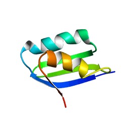
2O8N
 | | Crystal Structure of Mouse Apolipoprotein A-I Binding Protein | | 分子名称: | ApoA-I binding protein, CHLORIDE ION, SULFATE ION | | 著者 | Shumilin, I.A, Jha, K.N, Zheng, H, Chruszcz, M, Cymborowski, M, Herr, J.C, Minor, W. | | 登録日 | 2006-12-12 | | 公開日 | 2007-12-25 | | 最終更新日 | 2023-12-27 | | 実験手法 | X-RAY DIFFRACTION (2 Å) | | 主引用文献 | Biochemical and Structural Characterization of Apolipoprotein A-I Binding Protein, a Novel Phosphoprotein with a Potential Role in Sperm Capacitation.
Endocrinology, 149, 2008
|
|
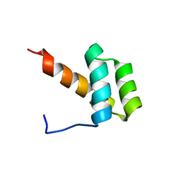
2P8D
 | |
2P81
 | | Engrailed homeodomain helix-turn-helix motif | | 分子名称: | Segmentation polarity homeobox protein engrailed | | 著者 | Religa, T.L. | | 登録日 | 2007-03-21 | | 公開日 | 2007-06-12 | | 最終更新日 | 2024-05-22 | | 実験手法 | SOLUTION NMR | | 主引用文献 | The helix-turn-helix motif as an ultrafast independently folding domain: The pathway of folding of Engrailed homeodomain.
Proc.Natl.Acad.Sci.Usa, 104, 2007
|
|
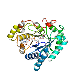
3C3U
 | | Crystal structure of AKR1C1 in complex with NADP and 3,5-dichlorosalicylic acid | | 分子名称: | 3,5-dichloro-2-hydroxybenzoic acid, Aldo-keto reductase family 1 member C1, NADP NICOTINAMIDE-ADENINE-DINUCLEOTIDE PHOSPHATE, ... | | 著者 | Dhagat, U, El-Kabbani, O. | | 登録日 | 2008-01-28 | | 公開日 | 2008-08-26 | | 最終更新日 | 2023-11-01 | | 実験手法 | X-RAY DIFFRACTION (1.8 Å) | | 主引用文献 | Selectivity determinants of inhibitor binding to human 20alpha-hydroxysteroid dehydrogenase: crystal structure of the enzyme in ternary complex with coenzyme and the potent inhibitor 3,5-dichlorosalicylic acid
J.Med.Chem., 51, 2008
|
|
2P0J
 | | Structure of restriction endonuclease BstYI bound to non-cognate DNA | | 分子名称: | 5'-D(*AP*TP*GP*AP*AP*TP*CP*CP*AP*TP*A)-3', 5'-D(*TP*AP*TP*GP*GP*AP*TP*TP*CP*AP*T)-3', BstYI | | 著者 | Townson, S.A, Samuelson, J.C, Bao, Y, Xu, S.Y, Aggarwal, A.K. | | 登録日 | 2007-02-28 | | 公開日 | 2007-05-01 | | 最終更新日 | 2024-02-21 | | 実験手法 | X-RAY DIFFRACTION (2.1 Å) | | 主引用文献 | BstYI Bound to Noncognate DNA Reveals a "Hemispecific" Complex: Implications for DNA Scanning.
Structure, 15, 2007
|
|
6FYJ
 | | Cytochrome P450 peroxygenase CYP152K6 in complex with Myristic Acid | | 分子名称: | DI(HYDROXYETHYL)ETHER, Fatty-acid peroxygenase, MYRISTIC ACID, ... | | 著者 | Girvan, H.M, Poddar, H, Mclean, K.J, Leys, D, Munro, A.W. | | 登録日 | 2018-03-12 | | 公開日 | 2018-08-22 | | 最終更新日 | 2024-01-17 | | 実験手法 | X-RAY DIFFRACTION (1.3 Å) | | 主引用文献 | Structural and catalytic properties of the peroxygenase P450 enzyme CYP152K6 from Bacillus methanolicus.
J. Inorg. Biochem., 188, 2018
|
|
6GEO
 | |


