7P8D
 
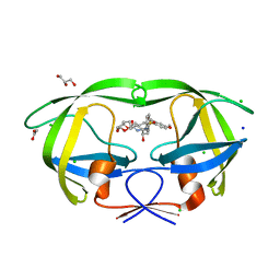
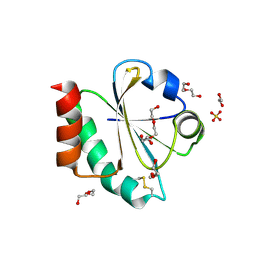
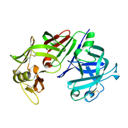
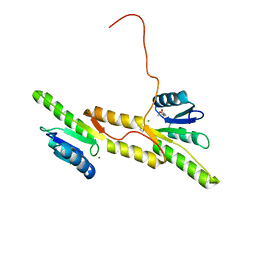 | | Crystal structure of the Receiver domain of A. thaliana cytokinin receptor AtCRE1 in complex with Mg2+ | | 分子名称: | (4S)-2-METHYL-2,4-PENTANEDIOL, 1,2-ETHANEDIOL, MAGNESIUM ION, ... | | 著者 | Tran, L.H, Urbanowicz, A, Jasinski, M, Jaskolski, M, Ruszkowski, M. | | 登録日 | 2021-07-21 | | 公開日 | 2021-10-20 | | 最終更新日 | 2024-05-01 | | 実験手法 | X-RAY DIFFRACTION (1.7 Å) | | 主引用文献 | 3D Domain Swapping Dimerization of the Receiver Domain of Cytokinin Receptor CRE1 From Arabidopsis thaliana and Medicago truncatula .
Front Plant Sci, 12, 2021
|
|
5WIW
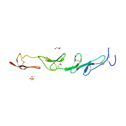 | |
5WJK
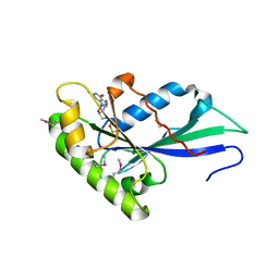
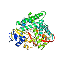
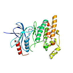
 | | 2.0-Angstrom In situ Mylar structure of sperm whale myoglobin (SWMb) at 293 K | | 分子名称: | CHLORIDE ION, Myoglobin, PROTOPORPHYRIN IX CONTAINING FE, ... | | 著者 | Broecker, J, Ou, W.-L, Ernst, O.P. | | 登録日 | 2017-07-23 | | 公開日 | 2017-12-13 | | 最終更新日 | 2023-10-04 | | 実験手法 | X-RAY DIFFRACTION (2 Å) | | 主引用文献 | High-throughput in situ X-ray screening of and data collection from protein crystals at room temperature and under cryogenic conditions.
Nat Protoc, 13, 2018
|
|
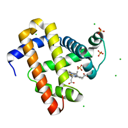
3TDA
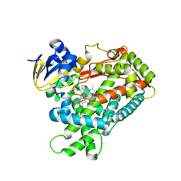
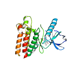
 | | Competitive replacement of thioridazine by prinomastat in crystals of cytochrome P450 2D6 | | 分子名称: | Cytochrome P450 2D6, PROTOPORPHYRIN IX CONTAINING FE, Prinomastat, ... | | 著者 | Wang, A, Stout, C.D, Johnson, E.F. | | 登録日 | 2011-08-10 | | 公開日 | 2012-08-15 | | 最終更新日 | 2023-09-13 | | 実験手法 | X-RAY DIFFRACTION (2.67 Å) | | 主引用文献 | Contributions of Ionic Interactions and Protein Dynamics to Cytochrome P450 2D6 (CYP2D6) Substrate and Inhibitor Binding.
J.Biol.Chem., 290, 2015
|
|
5WLO
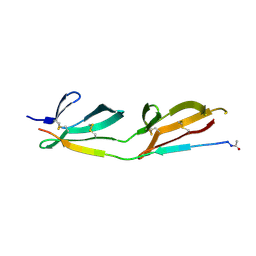
 | | a novel 13-ring macrocyclic HIV-1 protease inhibitors involving the P1'-P2' ligands | | 分子名称: | (3R,3aS,6aR)-hexahydrofuro[2,3-b]furan-3-yl {(2S,3R)-3-hydroxy-4-[(7E)-13-methoxy-1,1-dioxo-1,4,5,6,9,11-hexahydro-10,1lambda~6~,2-benzoxathiazacyclotridecin-2(3H)-yl]-1-phenylbutan-2-yl}carbamate, ACETATE ION, CHLORIDE ION, ... | | 著者 | Wang, Y.-F, Agniswamy, J, Weber, I.T. | | 登録日 | 2017-07-27 | | 公開日 | 2017-10-11 | | 最終更新日 | 2023-10-04 | | 実験手法 | X-RAY DIFFRACTION (1.27 Å) | | 主引用文献 | Design, synthesis, X-ray studies, and biological evaluation of novel macrocyclic HIV-1 protease inhibitors involving the P1'-P2' ligands.
Bioorg. Med. Chem. Lett., 27, 2017
|
|
3T1V
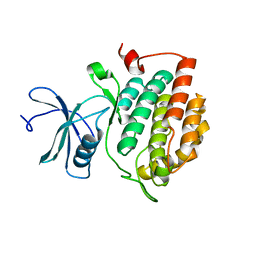
 | | MglA bound to GDP in P2(1) tetrameric arrangement | | 分子名称: | GUANOSINE-5'-DIPHOSPHATE, Gliding protein mglA | | 著者 | Miertzschke, M, Vetter, I.R, Koerner, C, Wittinghofer, A. | | 登録日 | 2011-07-22 | | 公開日 | 2011-08-31 | | 最終更新日 | 2011-11-02 | | 実験手法 | X-RAY DIFFRACTION (2.4 Å) | | 主引用文献 | Structural analysis of the Ras-like G protein MglA and its cognate GAP MglB and implications for bacterial polarity.
Embo J., 30, 2011
|
|
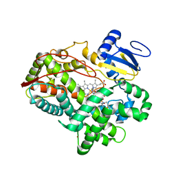
3T3Q
 | | Human Cytochrome P450 2A6 I208S/I300F/G301A/S369G in complex with Pilocarpine | | 分子名称: | (3S,4R)-3-ethyl-4-[(1-methyl-1H-imidazol-5-yl)methyl]dihydrofuran-2(3H)-one, Cytochrome P450 2A6, PROTOPORPHYRIN IX CONTAINING FE | | 著者 | DeVore, N.M, Scott, E.E. | | 登録日 | 2011-07-25 | | 公開日 | 2011-12-07 | | 最終更新日 | 2023-09-13 | | 実験手法 | X-RAY DIFFRACTION (2.1 Å) | | 主引用文献 | Structural comparison of cytochromes P450 2A6, 2A13, and 2E1 with pilocarpine.
Febs J., 279, 2012
|
|
3CAL
 | |
7OOL
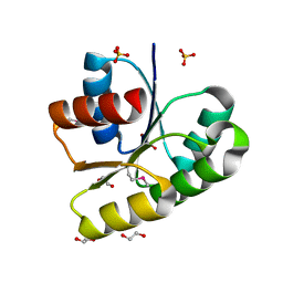
 | | Crystal structure of a Candidatus photodesmus katoptron thioredoxin chimera containing an ancestral loop | | 分子名称: | 1,2-ETHANEDIOL, DI(HYDROXYETHYL)ETHER, GLYCEROL, ... | | 著者 | Gavira, J.A, Ibarra-Molero, B, Gamiz-Arco, G, Risso, V, Sanchez-Ruiz, J.M. | | 登録日 | 2021-05-28 | | 公開日 | 2021-11-10 | | 最終更新日 | 2024-01-31 | | 実験手法 | X-RAY DIFFRACTION (2.85 Å) | | 主引用文献 | Combining Ancestral Reconstruction with Folding-Landscape Simulations to Engineer Heterologous Protein Expression.
J.Mol.Biol., 433, 2021
|
|
3CBD
 | |
5WNJ
 | |
3SV0
 | |
3CAD
 | | Crystal structure of Natural Killer Cell Receptor, Ly49G | | 分子名称: | Lectin-related NK cell receptor LY49G1 | | 著者 | Cho, S. | | 登録日 | 2008-02-19 | | 公開日 | 2008-04-08 | | 最終更新日 | 2011-07-13 | | 実験手法 | X-RAY DIFFRACTION (2.6 Å) | | 主引用文献 | Molecular Architecture of the Major Histocompatibility Complex Class I-binding Site of Ly49 Natural Killer Cell Receptors.
J.Biol.Chem., 283, 2008
|
|
3T04
 | | Crystal structure of monobody 7c12/abl1 sh2 domain complex | | 分子名称: | GLYCEROL, MONOBODY 7C12, SULFATE ION, ... | | 著者 | Wojcik, J.B, Wyrzucki, A.M, Koide, S. | | 登録日 | 2011-07-19 | | 公開日 | 2011-11-23 | | 最終更新日 | 2023-09-13 | | 実験手法 | X-RAY DIFFRACTION (2.1 Å) | | 主引用文献 | Targeting the SH2-Kinase Interface in Bcr-Abl Inhibits Leukemogenesis.
Cell(Cambridge,Mass.), 147, 2011
|
|
3T1P
 | |
7OUB
 | |
3CE1
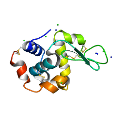
 | | Crystal Structure of the Cu/Zn Superoxide Dismutase from Cryptococcus liquefaciens Strain N6 | | 分子名称: | ACETATE ION, COPPER (II) ION, Superoxide dismutase [Cu-Zn], ... | | 著者 | Teh, A.H, Kanamasa, S, Kajiwara, S, Kumasaka, T. | | 登録日 | 2008-02-27 | | 公開日 | 2008-08-26 | | 最終更新日 | 2023-11-01 | | 実験手法 | X-RAY DIFFRACTION (1.2 Å) | | 主引用文献 | Structure of Cu/Zn superoxide dismutase from the heavy-metal-tolerant yeast Cryptococcus liquefaciens strain N6.
Biochem.Biophys.Res.Commun., 374, 2008
|
|
3T27
 | | Orthorhombic trypsin (bovine) in the presence of betaine | | 分子名称: | BENZAMIDINE, CALCIUM ION, Cationic trypsin, ... | | 著者 | Venkat, M, Saxby, C, Cahn, J, Juers, D. | | 登録日 | 2011-07-22 | | 公開日 | 2011-12-28 | | 最終更新日 | 2023-11-15 | | 実験手法 | X-RAY DIFFRACTION (1.95 Å) | | 主引用文献 | The use of trimethylamine N-oxide as a primary precipitating agent and related methylamine osmolytes as cryoprotective agents for macromolecular crystallography.
Acta Crystallogr.,Sect.D, 68, 2012
|
|
3CAA
 | | CLEAVED ANTICHYMOTRYPSIN A347R | | 分子名称: | ANTICHYMOTRYPSIN | | 著者 | Lukacs, C.M, Christianson, D.W. | | 登録日 | 1997-08-18 | | 公開日 | 1998-02-25 | | 最終更新日 | 2024-05-22 | | 実験手法 | X-RAY DIFFRACTION (2.4 Å) | | 主引用文献 | Engineering an anion-binding cavity in antichymotrypsin modulates the "spring-loaded" serpin-protease interaction.
Biochemistry, 37, 1998
|
|
3SVA
 | | Crystal structure of V57D mutant of human cystatin C | | 分子名称: | ACETATE ION, Cystatin-C, DI(HYDROXYETHYL)ETHER | | 著者 | Orlikowska, M, Szymanska, A, Borek, D, Otwinowski, Z, Skowron, P, Jankowska, E. | | 登録日 | 2011-07-12 | | 公開日 | 2012-08-01 | | 最終更新日 | 2024-10-09 | | 実験手法 | X-RAY DIFFRACTION (3.02 Å) | | 主引用文献 | Structural characterization of V57D and V57P mutants of human cystatin C, an amyloidogenic protein.
Acta Crystallogr.,Sect.D, 69, 2013
|
|
3CMS
 | | ENGINEERING ENZYME SUB-SITE SPECIFICITY: PREPARATION, KINETIC CHARACTERIZATION AND X-RAY ANALYSIS AT 2.0-ANGSTROMS RESOLUTION OF VAL111PHE SITE-MUTATED CALF CHYMOSIN | | 分子名称: | CHYMOSIN B | | 著者 | Newman, M, Frazao, C, Shearer, A, Tickle, I.J, Blundell, T.L. | | 登録日 | 1990-02-26 | | 公開日 | 1992-10-15 | | 最終更新日 | 2017-11-29 | | 実験手法 | X-RAY DIFFRACTION (2 Å) | | 主引用文献 | Engineering enzyme subsite specificity: preparation, kinetic characterization, and X-ray analysis at 2.0-A resolution of Val111Phe site-mutated calf chymosin.
Biochemistry, 29, 1990
|
|
3CGO
 | | IRAK-4 Inhibitors (Part II)- A structure based assessment of imidazo[1,2 a]pyridine binding | | 分子名称: | 2-{4-[(4-imidazo[1,2-a]pyridin-3-ylpyrimidin-2-yl)amino]piperidin-1-yl}-N-methylacetamide, Mitogen-activated protein kinase 10 | | 著者 | Ceska, T.A, Platt, A, Fortunato, M, Dickson, K.M, Beevers, R. | | 登録日 | 2008-03-06 | | 公開日 | 2008-06-03 | | 最終更新日 | 2023-11-01 | | 実験手法 | X-RAY DIFFRACTION (3 Å) | | 主引用文献 | IRAK-4 inhibitors. Part II: A structure-based assessment of imidazo[1,2-a]pyridine binding
Bioorg.Med.Chem.Lett., 18, 2008
|
|
3T6K
 | |
3T6U
 | |
3CQT
 | | N53I V55L MUTANT of FYN SH3 DOMAIN | | 分子名称: | Proto-oncogene tyrosine-protein kinase Fyn | | 著者 | Neculai, A.M, Zarrine-Afsar, A, Howell, P.L, Davidson, A, Chan, H.S. | | 登録日 | 2008-04-03 | | 公開日 | 2008-07-01 | | 最終更新日 | 2023-08-30 | | 実験手法 | X-RAY DIFFRACTION (1.6 Å) | | 主引用文献 | Theoretical and experimental demonstration of the importance of specific nonnative interactions in protein folding.
Proc.Natl.Acad.Sci.Usa, 105, 2008
|
|


