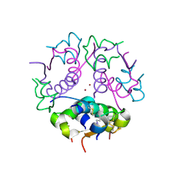
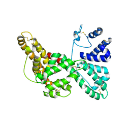
1J6Z
 
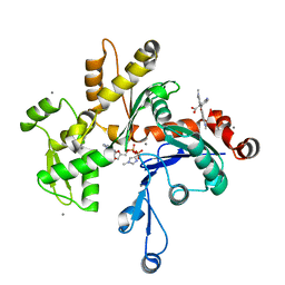 | | UNCOMPLEXED ACTIN | | 分子名称: | ACTIN ALPHA 1, ADENOSINE-5'-DIPHOSPHATE, CALCIUM ION, ... | | 著者 | Otterbein, L.R, Graceffa, P, Dominguez, R. | | 登録日 | 2001-05-15 | | 公開日 | 2001-08-15 | | 最終更新日 | 2023-08-16 | | 実験手法 | X-RAY DIFFRACTION (1.54 Å) | | 主引用文献 | The crystal structure of uncomplexed actin in the ADP state.
Science, 293, 2001
|
|
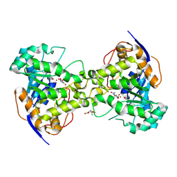
1J70
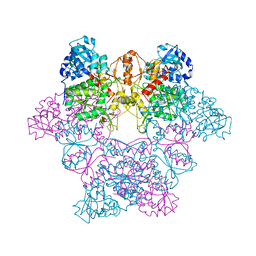 | | CRYSTAL STRUCTURE OF YEAST ATP SULFURYLASE | | 分子名称: | ATP SULPHURYLASE, PHOSPHATE ION, SODIUM ION | | 著者 | Lalor, D.J, Schnyder, T, Saridakis, V, Pilloff, D.E, Dong, A, Tang, H, Leyh, T.S, Pai, E.F. | | 登録日 | 2001-05-15 | | 公開日 | 2003-06-17 | | 最終更新日 | 2024-02-07 | | 実験手法 | X-RAY DIFFRACTION (2.3 Å) | | 主引用文献 | Structural and functional analysis of a truncated form of Saccharomyces cerevisiae ATP sulfurylase: C-terminal domain essential for oligomer formation but not for activity.
Protein Eng., 16, 2003
|
|
1J71
 | |
1J72
 | |
1J73
 | | Crystal structure of an unstable insulin analog with native activity. | | 分子名称: | ZINC ION, insulin a, insulin b | | 著者 | Wan, Z, Zhao, M, Nakagawa, S, Jia, W, Weiss, M.A. | | 登録日 | 2001-05-15 | | 公開日 | 2001-05-30 | | 最終更新日 | 2021-10-27 | | 実験手法 | X-RAY DIFFRACTION (2 Å) | | 主引用文献 | Non-standard insulin design: structure-activity relationships at the periphery of the insulin receptor.
J.Mol.Biol., 315, 2002
|
|
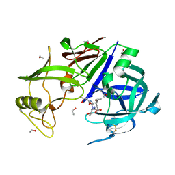
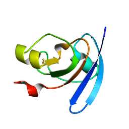
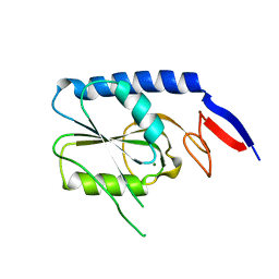
1J74
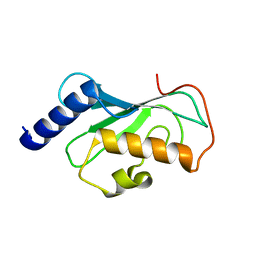 | | Crystal Structure of Mms2 | | 分子名称: | MMS2 | | 著者 | Moraes, T.F, Edwards, R.A, McKenna, S, Pastushok, L, Xiao, W, Glover, J.N.M, Ellison, M.J. | | 登録日 | 2001-05-15 | | 公開日 | 2001-08-08 | | 最終更新日 | 2023-08-16 | | 実験手法 | X-RAY DIFFRACTION (1.9 Å) | | 主引用文献 | Crystal structure of the human ubiquitin conjugating enzyme complex, hMms2-hUbc13.
Nat.Struct.Biol., 8, 2001
|
|
1J75
 | | Crystal Structure of the DNA-Binding Domain Zalpha of DLM-1 Bound to Z-DNA | | 分子名称: | 5'-D(*TP*CP*GP*CP*GP*CP*G)-3', Tumor Stroma and Activated Macrophage Protein DLM-1 | | 著者 | Schwartz, T, Behlke, J, Lowenhaupt, K, Heinemann, U, Rich, A. | | 登録日 | 2001-05-15 | | 公開日 | 2001-09-01 | | 最終更新日 | 2023-08-16 | | 実験手法 | X-RAY DIFFRACTION (1.85 Å) | | 主引用文献 | Structure of the DLM-1-Z-DNA complex reveals a conserved family of Z-DNA-binding proteins.
Nat.Struct.Biol., 8, 2001
|
|
1J77
 | | Crystal Structure of Gram-negative Bacterial Heme Oxygenase Complexed with Heme | | 分子名称: | HemO, PROTOPORPHYRIN IX CONTAINING FE | | 著者 | Schuller, D.J, Zhu, W, Stojiljkovic, I, Wilks, A, Poulos, T.L. | | 登録日 | 2001-05-15 | | 公開日 | 2001-05-30 | | 最終更新日 | 2024-02-07 | | 実験手法 | X-RAY DIFFRACTION (1.5 Å) | | 主引用文献 | Crystal structure of heme oxygenase from the gram-negative pathogen Neisseria meningitidis and a comparison with mammalian heme oxygenase-1.
Biochemistry, 40, 2001
|
|
1J78
 | | Crystallographic analysis of the human vitamin D binding protein | | 分子名称: | 3-{2-[1-(5-HYDROXY-1,5-DIMETHYL-HEXYL)-7A-METHYL-OCTAHYDRO-INDEN-4-YLIDENE]-ETHYLIDENE}-4-METHYLENE-CYCLOHEXANOL, OLEIC ACID, vitamin D binding protein | | 著者 | Verboven, C, Rabijns, A, De Maeyer, M, Van Baelen, H, Bouillon, R, De Ranter, C. | | 登録日 | 2001-05-16 | | 公開日 | 2002-02-06 | | 最終更新日 | 2018-01-31 | | 実験手法 | X-RAY DIFFRACTION (2.31 Å) | | 主引用文献 | A structural basis for the unique binding features of the human vitamin D-binding protein.
Nat.Struct.Biol., 9, 2002
|
|
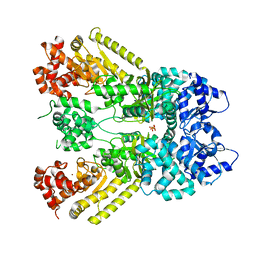
1J79
 | | Molecular Structure of Dihydroorotase: A Paradigm for Catalysis Through the Use of a Binuclear Metal Center | | 分子名称: | N-CARBAMOYL-L-ASPARTATE, OROTIC ACID, ZINC ION, ... | | 著者 | Thoden, J.B, Phillips Jr, G.N, Neal, T.M, Raushel, F.M, Holden, H.M. | | 登録日 | 2001-05-16 | | 公開日 | 2001-06-20 | | 最終更新日 | 2011-07-13 | | 実験手法 | X-RAY DIFFRACTION (1.7 Å) | | 主引用文献 | Molecular structure of dihydroorotase: a paradigm for catalysis through the use of a binuclear metal center.
Biochemistry, 40, 2001
|
|
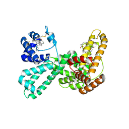
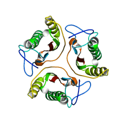
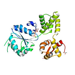
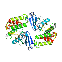
1J7A
 | | STRUCTURE OF THE ANABAENA FERREDOXIN D68K MUTANT | | 分子名称: | FE2/S2 (INORGANIC) CLUSTER, FERREDOXIN I | | 著者 | Hurley, J.K, Weber-Main, A.M, Stankovich, M.T, Benning, M.M, Thoden, J.B, VanHooke, J.L, Holden, H.M, Chae, Y.K, Xia, B, Cheng, H, Markley, J.L, Martinez-Julvez, M, Gomez-Moreno, C, Schmeits, J.L, Tollen, G. | | 登録日 | 2001-05-16 | | 公開日 | 2001-05-23 | | 最終更新日 | 2024-02-07 | | 実験手法 | X-RAY DIFFRACTION (1.8 Å) | | 主引用文献 | Structure-function relationships in Anabaena ferredoxin: correlations between X-ray crystal structures, reduction potentials, and rate constants of electron transfer to ferredoxin:NADP+ reductase for site-specific ferredoxin mutants.
Biochemistry, 36, 1997
|
|
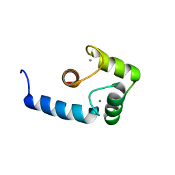
1J7B
 | | STRUCTURE OF THE ANABAENA FERREDOXIN MUTANT E94K | | 分子名称: | FE2/S2 (INORGANIC) CLUSTER, FERREDOXIN I | | 著者 | Hurley, J.K, Weber-Main, A.M, Stankovich, M.T, Benning, M.M, Thoden, J.B, Vanhooke, J.L, Holden, H.M, Chae, Y.K, Xia, B, Cheng, H, Markley, J.L, Martinez-Julvez, M, Gomez-Moreno, C, Schmeits, J.L, Tollin, G. | | 登録日 | 2001-05-16 | | 公開日 | 2001-05-23 | | 最終更新日 | 2024-02-07 | | 実験手法 | X-RAY DIFFRACTION (1.8 Å) | | 主引用文献 | Structure-function relationships in Anabaena ferredoxin: correlations between X-ray crystal structures, reduction potentials, and rate constants of electron transfer to ferredoxin:NADP+ reductase for site-specific ferredoxin mutants.
Biochemistry, 36, 1997
|
|
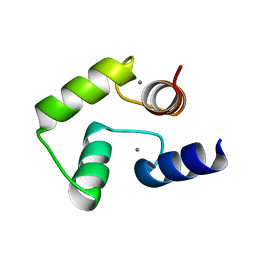
1J7C
 | | STRUCTURE OF THE ANABAENA FERREDOXIN MUTANT E95K | | 分子名称: | FE2/S2 (INORGANIC) CLUSTER, FERREDOXIN I | | 著者 | Hurley, J.K, Weber-Main, A.M, Stankovich, M.T, Benning, M.M, Thoden, J.B, Vanhooke, J.L, Holden, H.M, Chae, Y.K, Xia, B, Cheng, H, Markley, J.L, Martinez-Julvez, M, Gomez-Moreno, C, Schmeits, J.L, Tollin, G. | | 登録日 | 2001-05-16 | | 公開日 | 2001-05-23 | | 最終更新日 | 2024-02-07 | | 実験手法 | X-RAY DIFFRACTION (1.8 Å) | | 主引用文献 | Structure-function relationships in Anabaena ferredoxin: correlations between X-ray crystal structures, reduction potentials, and rate constants of electron transfer to ferredoxin:NADP+ reductase for site-specific ferredoxin mutants.
Biochemistry, 36, 1997
|
|
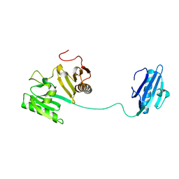
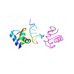
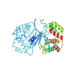
1J7D
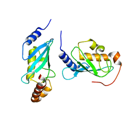 | | Crystal Structure of hMms2-hUbc13 | | 分子名称: | MMS2, UBIQUITIN-CONJUGATING ENZYME E2-17 KDA | | 著者 | Moraes, T.F, Edwards, R.A, McKenna, S, Pashushok, L, Xiao, W, Glover, J.N.M, Ellison, M.J. | | 登録日 | 2001-05-16 | | 公開日 | 2001-08-08 | | 最終更新日 | 2024-02-07 | | 実験手法 | X-RAY DIFFRACTION (1.85 Å) | | 主引用文献 | Crystal structure of the human ubiquitin conjugating enzyme complex, hMms2-hUbc13.
Nat.Struct.Biol., 8, 2001
|
|
1J7E
 | | A Structural Basis for the Unique Binding Features of the Human Vitamin D-binding Protein | | 分子名称: | 3-(2-{4-[2-(5-HYDROXY-2-METHYLENE-CYCLOHEXYLIDENE)-ETHYLIDENE]-7A-METHYL-OCTAHYDRO-INDEN-1-YL}-PROPYL)-PHENOL, OLEIC ACID, vitamin D binding protein | | 著者 | Verboven, C, Rabijns, A, De Maeyer, M, Van Baelen, H, Bouillon, R, De Ranter, C. | | 登録日 | 2001-05-16 | | 公開日 | 2002-02-06 | | 最終更新日 | 2023-08-16 | | 実験手法 | X-RAY DIFFRACTION (2.55 Å) | | 主引用文献 | A structural basis for the unique binding features of the human vitamin D-binding protein.
Nat.Struct.Biol., 9, 2002
|
|
1J7G
 | |
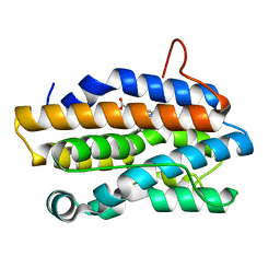
1J7H
 | | Solution Structure of HI0719, a Hypothetical Protein From Haemophilus Influenzae | | 分子名称: | HYPOTHETICAL PROTEIN HI0719 | | 著者 | Parsons, L, Bonander, N, Eisenstein, E, Gilson, M, Kairys, V, Orban, J, Structure 2 Function Project (S2F) | | 登録日 | 2001-05-16 | | 公開日 | 2003-02-11 | | 最終更新日 | 2024-05-22 | | 実験手法 | SOLUTION NMR | | 主引用文献 | Solution Structure and Functional Ligand Screening of HI0719, a Highly Conserved Protein from Bacteria to Humans in the YjgF/YER057c/UK114 Family
Biochemistry, 42, 2003
|
|
1J7I
 | | Crystal Structure of 3',5"-Aminoglycoside Phosphotransferase Type IIIa Apoenzyme | | 分子名称: | AMINOGLYCOSIDE 3'-PHOSPHOTRANSFERASE | | 著者 | Burk, D.L, Hon, W.C, Leung, A.K.-W, Berghuis, A.M. | | 登録日 | 2001-05-16 | | 公開日 | 2001-08-08 | | 最終更新日 | 2024-02-07 | | 実験手法 | X-RAY DIFFRACTION (3.2 Å) | | 主引用文献 | Structural analyses of nucleotide binding to an aminoglycoside phosphotransferase.
Biochemistry, 40, 2001
|
|
1J7J
 | |
1J7K
 | | THERMOTOGA MARITIMA RUVB P216G MUTANT | | 分子名称: | ACETATE ION, ADENOSINE-5'-TRIPHOSPHATE, COBALT (II) ION, ... | | 著者 | Putnam, C.D, Clancy, S.B, Tsuruta, H, Wetmur, J.G, Tainer, J.A. | | 登録日 | 2001-05-16 | | 公開日 | 2001-08-08 | | 最終更新日 | 2023-08-16 | | 実験手法 | X-RAY DIFFRACTION (1.8 Å) | | 主引用文献 | Structure and mechanism of the RuvB Holliday junction branch migration motor.
J.Mol.Biol., 311, 2001
|
|
1J7L
 | | Crystal Structure of 3',5"-Aminoglycoside Phosphotransferase Type IIIa ADP Complex | | 分子名称: | ADENOSINE-5'-DIPHOSPHATE, AMINOGLYCOSIDE 3'-PHOSPHOTRANSFERASE, MAGNESIUM ION | | 著者 | Burk, D.L, Hon, W.C, Leung, A.K.-W, Berghuis, A.M. | | 登録日 | 2001-05-17 | | 公開日 | 2001-08-08 | | 最終更新日 | 2011-07-13 | | 実験手法 | X-RAY DIFFRACTION (2.2 Å) | | 主引用文献 | Structural analyses of nucleotide binding to an aminoglycoside phosphotransferase.
Biochemistry, 40, 2001
|
|
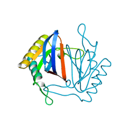
1J7M
 | | The Third Fibronectin Type II Module from Human Matrix Metalloproteinase 2 | | 分子名称: | MATRIX METALLOPROTEINASE 2 | | 著者 | Briknarova, K, Gehrmann, M, Banyai, L, Tordai, H, Patthy, L, Llinas, M. | | 登録日 | 2001-05-17 | | 公開日 | 2001-05-30 | | 最終更新日 | 2022-02-23 | | 実験手法 | SOLUTION NMR | | 主引用文献 | Gelatin-binding region of human matrix metalloproteinase-2: solution structure, dynamics, and function of the COL-23 two-domain construct.
J.Biol.Chem., 276, 2001
|
|
1J7N
 | | Anthrax Toxin Lethal factor | | 分子名称: | Lethal Factor precursor, SULFATE ION, ZINC ION | | 著者 | Pannifer, A.D, Wong, T.Y, Schwarzenbacher, R, Renatus, M, Petosa, C, Collier, R.J, Bienkowska, J, Lacy, D.B, Park, S, Leppla, S.H, Hanna, P, Liddington, R.C. | | 登録日 | 2001-05-17 | | 公開日 | 2001-11-07 | | 最終更新日 | 2024-02-07 | | 実験手法 | X-RAY DIFFRACTION (2.3 Å) | | 主引用文献 | Crystal structure of the anthrax lethal factor.
Nature, 414, 2001
|
|
1J7O
 | |
1J7P
 | |


