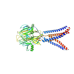
2XSP
 
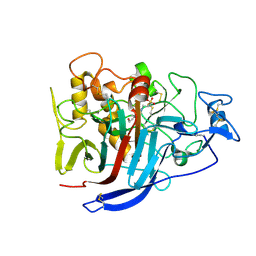 | | Structure of Cellobiohydrolase 1 (Cel7A) from Heterobasidion annosum | | 分子名称: | 2-acetamido-2-deoxy-beta-D-glucopyranose-(1-4)-2-acetamido-2-deoxy-beta-D-glucopyranose, 4-(2-HYDROXYETHYL)-1-PIPERAZINE ETHANESULFONIC ACID, CELLULOSE 1,4-BETA-CELLOBIOSIDASE, ... | | 著者 | Haddad-momeni, M, Hansson, H, Mikkelsen, N.E, Wang, X, Svedberg, J, Sandgren, M, Stahlberg, J. | | 登録日 | 2010-09-29 | | 公開日 | 2011-10-12 | | 最終更新日 | 2023-12-20 | | 実験手法 | X-RAY DIFFRACTION (1.7 Å) | | 主引用文献 | Structural, Biochemical, and Computational Characterization of the Glycoside Hydrolase Family 7 Cellobiohydrolase of the Tree-Killing Fungus Heterobasidion Irregulare.
J.Biol.Chem., 288, 2013
|
|
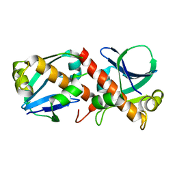
2JXL
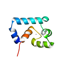 | | Solution structure of cardiac N-domain troponin C mutant F77W-V82A | | 分子名称: | CALCIUM ION, Troponin C, slow skeletal and cardiac muscles | | 著者 | Julien, O, Sun, Y, Wang, X, Lindhout, D.A, Thiessen, A, Irving, M, Sykes, B.D. | | 登録日 | 2007-11-20 | | 公開日 | 2007-12-04 | | 最終更新日 | 2024-05-29 | | 実験手法 | SOLUTION NMR | | 主引用文献 | Tryptophan Mutants of Cardiac Troponin C: 3D Structure, Troponin I Affinity, and in Situ Activity.
Biochemistry, 47, 2008
|
|
8H8F
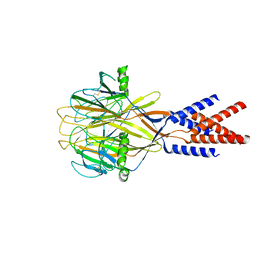 | | Structure of Xenopus tropicalis acid-sensitive outwardly rectifying channel ASOR (resting state) | | 分子名称: | Proton-activated chloride channel | | 著者 | Chi, P, Wang, X, Li, J, Li, K, Zhang, Y, Geng, J, Wu, J, Deng, D. | | 登録日 | 2022-10-22 | | 公開日 | 2024-05-01 | | 実験手法 | ELECTRON MICROSCOPY (3.48 Å) | | 主引用文献 | Structure of Xenopus tropicalis acid-sensitive outwardly rectifying channel ASOR (resting state)
To Be Published
|
|
8H8E
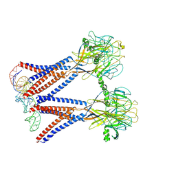 | | Structure of the dimeric Xenopus tropical acid-sensitive outwardly rectifying channel ASOR trimer bound with tRNA (closed state) | | 分子名称: | Proton-activated chloride channel, tRNA (75-MER)of Spodoptera frugiperda | | 著者 | Chi, P, Wang, X, Li, J, Li, K, Zhang, Y, Geng, J, Wu, J, Deng, D. | | 登録日 | 2022-10-22 | | 公開日 | 2024-05-01 | | 実験手法 | ELECTRON MICROSCOPY (3.81 Å) | | 主引用文献 | Structure of the dimeric Xenopus tropical acid-sensitive outwardly rectifying channel ASOR trimer bound with tRNA (closed state)
To Be Published
|
|
8H8D
 | | Structure of Xenopus tropicalis acid-sensitive outwardly rectifying channel ASOR trimer bound with tRNA (intermediate state) | | 分子名称: | Proton-activated chloride channel | | 著者 | Chi, P, Wang, X, Li, J, Li, K, Zhang, Y, Geng, J, Wu, J, Deng, D. | | 登録日 | 2022-10-22 | | 公開日 | 2024-05-01 | | 実験手法 | ELECTRON MICROSCOPY (4.26 Å) | | 主引用文献 | Structure of Xenopus tropicalis acid-sensitive outwardly rectifying channel ASOR trimer bound with tRNA (intermediate state)
To Be Published
|
|
8A2F
 | | Crystal Structure of Ljunganvirus 1 2A protein | | 分子名称: | 2A protein | | 著者 | von Castelmur, E, Zhu, L, Wang, X, Fry, E, Ren, J, Perrakis, A, Stuart, D.I. | | 登録日 | 2022-06-03 | | 公開日 | 2023-06-14 | | 最終更新日 | 2024-02-07 | | 実験手法 | X-RAY DIFFRACTION (1.73 Å) | | 主引用文献 | Structural plasticity of 2A proteins in the Parechovirus family
To Be Published
|
|
7VYV
 | | Cryo-EM structure of Depo32, a Klebsiella phage depolymerase targets the K2 serotype K. pneumoniae | | 分子名称: | Depolymerase | | 著者 | Cai, R, Ren, Z, Zhao, R, Wang, X, Guo, Z, Du, R, Han, W, Ru, H, Gu, J. | | 登録日 | 2021-11-15 | | 公開日 | 2023-08-30 | | 実験手法 | ELECTRON MICROSCOPY (2.32 Å) | | 主引用文献 | Depo32, a depolymerase of Klebsiella phage, efficiently controls infection caused by Klebsiella pneumoniae with K2 serotype CPS
To Be Published
|
|
7VZ3
 | | Cryo-EM structure of Depo32, a Klebsiella phage depolymerase targets the K2 serotype K. pneumoniae | | 分子名称: | Depolymerase | | 著者 | Cai, R, Ren, Z, Zhao, R, Wang, X, Guo, Z, Du, R, Han, W, Ru, H, Gu, J. | | 登録日 | 2021-11-15 | | 公開日 | 2023-08-30 | | 実験手法 | ELECTRON MICROSCOPY (2.46 Å) | | 主引用文献 | Depo32, a depolymerase of Klebsiella phage, efficiently controls infection caused by Klebsiella pneumoniae with K2 serotype CPS
To Be Published
|
|
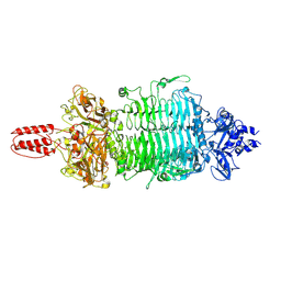
2YG1
 | | APO STRUCTURE OF CELLOBIOHYDROLASE 1 (CEL7A) FROM HETEROBASIDION ANNOSUM | | 分子名称: | 2-acetamido-2-deoxy-beta-D-glucopyranose-(1-4)-2-acetamido-2-deoxy-beta-D-glucopyranose, CELLULOSE 1,4-BETA-CELLOBIOSIDASE, MAGNESIUM ION | | 著者 | Haddad-Momeni, M, Hansson, H, Mikkelsen, N.E, Wang, X, Svedberg, J, Sandgren, M, Stahlberg, J. | | 登録日 | 2011-04-11 | | 公開日 | 2012-04-25 | | 最終更新日 | 2023-12-20 | | 実験手法 | X-RAY DIFFRACTION (1.9 Å) | | 主引用文献 | Structural, Biochemical, and Computational Characterization of the Glycoside Hydrolase Family 7 Cellobiohydrolase of the Tree-Killing Fungus Heterobasidion Irregulare.
J.Biol.Chem., 288, 2013
|
|
2ZNV
 | | Crystal structure of human AMSH-LP DUB domain in complex with Lys63-linked ubiquitin dimer | | 分子名称: | 1,2-ETHANEDIOL, AMSH-like protease, Ubiquitin, ... | | 著者 | Sato, Y, Azusa, Y, Yamagata, A, Mimura, H, Wang, X, Yamashita, M, Ookata, K, Nureki, O, Iwai, K, Komada, M, Fukai, S. | | 登録日 | 2008-05-01 | | 公開日 | 2008-09-02 | | 最終更新日 | 2023-11-01 | | 実験手法 | X-RAY DIFFRACTION (1.6 Å) | | 主引用文献 | Structural basis for specific cleavage of Lys 63-linked polyubiquitin chains
Nature, 455, 2008
|
|
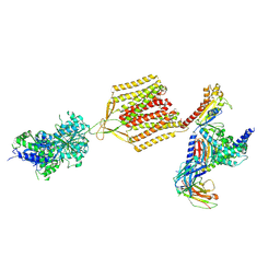
7D5Z
 | | Crystal structure of EBV gH/gL bound with neutralizing antibody 1D8 | | 分子名称: | Envelope glycoprotein H, Envelope glycoprotein L, heavy chain of 1D8, ... | | 著者 | Zhu, Q, Shan, S, Yu, J, Wang, X, Zhang, L, Zeng, M. | | 登録日 | 2020-09-28 | | 公開日 | 2021-10-20 | | 最終更新日 | 2023-11-29 | | 実験手法 | X-RAY DIFFRACTION (4.2 Å) | | 主引用文献 | A Neutralizing Antibody Targeting a New Site of Vulnerability on Epstein-Barr Virus gH/gL Protects against Dual-Tropic Infection
To Be Published
|
|
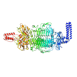
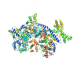
7E3N
 | | Crystal structure of Isocitrate dehydrogenase D252N mutant from Trypanosoma brucei in complexed with NADP+, alpha-ketoglutarate, and ca2+ | | 分子名称: | CALCIUM ION, ISOCITRIC ACID, Isocitrate dehydrogenase [NADP], ... | | 著者 | Arai, N, Shiba, T, Inaoka, D.K, Kita, K, Wang, X, Otani, M, Matsushiro, S, Kojima, C. | | 登録日 | 2021-02-09 | | 公開日 | 2022-02-16 | | 最終更新日 | 2023-11-29 | | 実験手法 | X-RAY DIFFRACTION (1.9 Å) | | 主引用文献 | Crystal structure of Isocitrate dehydrogenase from Trypanosoma brucei.
To Be Published
|
|
2ZNR
 | | Crystal structure of the DUB domain of human AMSH-LP | | 分子名称: | 1,2-ETHANEDIOL, AMSH-like protease, PRASEODYMIUM ION, ... | | 著者 | Sato, Y, Azusa, Y, Yamagata, A, Mimura, H, Wang, X, Yamashita, M, Ookata, K, Nureki, O, Iwai, K, Komada, M, Fukai, S. | | 登録日 | 2008-05-01 | | 公開日 | 2008-09-02 | | 最終更新日 | 2024-03-13 | | 実験手法 | X-RAY DIFFRACTION (1.2 Å) | | 主引用文献 | Structural basis for specific cleavage of Lys 63-linked polyubiquitin chains
Nature, 455, 2008
|
|
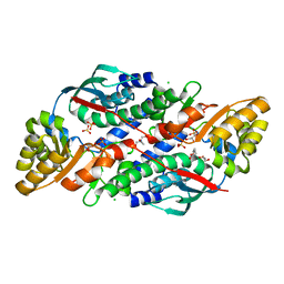
7DHD
 | | Vibrio vulnificus Wzb | | 分子名称: | CHLORIDE ION, Protein-tyrosine-phosphatase | | 著者 | Ma, Q, Wang, X. | | 登録日 | 2020-11-14 | | 公開日 | 2021-01-20 | | 最終更新日 | 2023-11-29 | | 実験手法 | X-RAY DIFFRACTION (1.71 Å) | | 主引用文献 | Wzb of Vibrio vulnificus represents a new group of low-molecular-weight protein tyrosine phosphatases with a unique insertion in the W-loop.
J.Biol.Chem., 296, 2021
|
|
7DHE
 | | Vibrio vulnificus Wzb in complex with benzylphosphonate | | 分子名称: | Protein-tyrosine-phosphatase, benzylphosphonic acid | | 著者 | Ma, Q, Wang, X. | | 登録日 | 2020-11-14 | | 公開日 | 2021-01-20 | | 最終更新日 | 2023-11-29 | | 実験手法 | X-RAY DIFFRACTION (2.79 Å) | | 主引用文献 | Wzb of Vibrio vulnificus represents a new group of low-molecular-weight protein tyrosine phosphatases with a unique insertion in the W-loop.
J.Biol.Chem., 296, 2021
|
|
7DHF
 | | Vibrio vulnificus Wzb in complex with benzylphosphonate | | 分子名称: | GLYCEROL, PHOSPHATE ION, Protein-tyrosine-phosphatase | | 著者 | Ma, Q, Wang, X. | | 登録日 | 2020-11-14 | | 公開日 | 2021-01-20 | | 最終更新日 | 2023-11-29 | | 実験手法 | X-RAY DIFFRACTION (1.211 Å) | | 主引用文献 | Wzb of Vibrio vulnificus represents a new group of low-molecular-weight protein tyrosine phosphatases with a unique insertion in the W-loop.
J.Biol.Chem., 296, 2021
|
|
7FJC
 | | Crystal structure of SARS-CoV-2 Beta RBD complexed with P36-5D2 Fab | | 分子名称: | P36-5D2 heavy chain, P36-5D2 light chain, Spike protein S1, ... | | 著者 | Zhang, L.Q, Wang, X.Q, Shan, S.S, Lan, J. | | 登録日 | 2021-08-03 | | 公開日 | 2022-08-10 | | 最終更新日 | 2023-11-29 | | 実験手法 | X-RAY DIFFRACTION (2.96 Å) | | 主引用文献 | Crystal structure of SARS-CoV-2 Beta RBD complexed with P36-5D2 Fab
To Be Published
|
|
7F7H
 | | SARS-CoV-2 S protein RBD in complex with A8-1 Fab | | 分子名称: | Heavy chain of A8-1 Fab, Light chain of A8-1 Fab, Spike glycoprotein S1 | | 著者 | Dou, Y, Wang, X, Wang, K, Liu, P, Lu, B. | | 登録日 | 2021-06-29 | | 公開日 | 2022-06-15 | | 最終更新日 | 2023-11-29 | | 実験手法 | X-RAY DIFFRACTION (3.19 Å) | | 主引用文献 | High throughput isolation of potent neutralizing antibodies from convalescent COVID-19 patients.
To Be Published
|
|
7CTE
 | | Human Origin Recognition Complex, ORC2-5 | | 分子名称: | ADENOSINE-5'-TRIPHOSPHATE, Origin recognition complex subunit 2, Origin recognition complex subunit 3, ... | | 著者 | Cheng, J, Li, N, Wang, X, Hu, J, Zhai, Y, Gao, N. | | 登録日 | 2020-08-18 | | 公開日 | 2021-01-06 | | 最終更新日 | 2024-03-27 | | 実験手法 | ELECTRON MICROSCOPY (3.8 Å) | | 主引用文献 | Structural insight into the assembly and conformational activation of human origin recognition complex.
Cell Discov, 6, 2020
|
|
7CTF
 | | Human origin recognition complex 1-5 State II | | 分子名称: | ADENOSINE-5'-TRIPHOSPHATE, Origin recognition complex subunit 1, Origin recognition complex subunit 2, ... | | 著者 | Cheng, J, Li, N, Wang, X, Hu, J, Zhai, Y, Gao, N. | | 登録日 | 2020-08-18 | | 公開日 | 2021-01-06 | | 最終更新日 | 2024-03-27 | | 実験手法 | ELECTRON MICROSCOPY (4.8 Å) | | 主引用文献 | Structural insight into the assembly and conformational activation of human origin recognition complex.
Cell Discov, 6, 2020
|
|
7EB2
 | | Cryo-EM structure of human GABA(B) receptor-Gi protein complex | | 分子名称: | (3S)-5,7-ditert-butyl-3-oxidanyl-3-(trifluoromethyl)-1-benzofuran-2-one, Gamma-aminobutyric acid type B receptor subunit 1, Gamma-aminobutyric acid type B receptor subunit 2, ... | | 著者 | Shen, C, Mao, C, Xu, C, Jin, N, Zhang, H, Shen, D, Shen, Q, Wang, X, Hou, T, Rondard, P, Chen, Z, Pin, J, Zhang, Y, Liu, J. | | 登録日 | 2021-03-08 | | 公開日 | 2021-05-05 | | 最終更新日 | 2021-07-07 | | 実験手法 | ELECTRON MICROSCOPY (3.5 Å) | | 主引用文献 | Structural basis of GABA B receptor-G i protein coupling.
Nature, 594, 2021
|
|
7CTG
 | | Human Origin Recognition Complex, ORC1-5 State I | | 分子名称: | ADENOSINE-5'-TRIPHOSPHATE, Origin recognition complex subunit 1, Origin recognition complex subunit 2, ... | | 著者 | Cheng, J, Li, N, Wang, X, Hu, J, Zhai, Y, Gao, N. | | 登録日 | 2020-08-18 | | 公開日 | 2021-01-06 | | 最終更新日 | 2024-03-27 | | 実験手法 | ELECTRON MICROSCOPY (5 Å) | | 主引用文献 | Structural insight into the assembly and conformational activation of human origin recognition complex.
Cell Discov, 6, 2020
|
|
7VRV
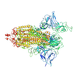 | | VAS5 Spike (1 RBD up) | | 分子名称: | 2-acetamido-2-deoxy-beta-D-glucopyranose, 2-acetamido-2-deoxy-beta-D-glucopyranose-(1-4)-2-acetamido-2-deoxy-beta-D-glucopyranose, Spike glycoprotein | | 著者 | Zhen, C, Wang, X. | | 登録日 | 2021-10-25 | | 公開日 | 2022-06-22 | | 実験手法 | ELECTRON MICROSCOPY (4.2 Å) | | 主引用文献 | VAS5 Spike(1 RBD up)
To Be Published
|
|
7XGL
 | | Quinolinate Phosphoribosyl Transferase (QAPRTase) from Streptomyces pyridomyceticus NRRL B-2517 in Apo form | | 分子名称: | CHLORIDE ION, GLYCEROL, Quinolinate Phosphoribosyl Transferase, ... | | 著者 | Zhou, Z, Yang, X, Huang, T, Wang, X, Liang, R, Zheng, J, Dai, S, Lin, S, Deng, Z. | | 登録日 | 2022-04-05 | | 公開日 | 2023-03-22 | | 最終更新日 | 2023-11-29 | | 実験手法 | X-RAY DIFFRACTION (2.11 Å) | | 主引用文献 | Bifunctional NadC Homologue PyrZ Catalyzes Nicotinic Acid Formation in Pyridomycin Biosynthesis.
Acs Chem.Biol., 18, 2023
|
|
7XGN
 | | Quinolinate Phosphoribosyl Transferase (QAPRTase) from Streptomyces pyridomyceticus NRRL B-2517 in complex with Nicotinic Acid (NA) | | 分子名称: | CHLORIDE ION, NICOTINIC ACID, Quinolinate Phosphoribosyl Transferase, ... | | 著者 | Zhou, Z, Yang, X, Huang, T, Wang, X, Liang, R, Zheng, J, Dai, S, Lin, S, Deng, Z. | | 登録日 | 2022-04-05 | | 公開日 | 2023-03-22 | | 最終更新日 | 2023-11-29 | | 実験手法 | X-RAY DIFFRACTION (2.6 Å) | | 主引用文献 | Bifunctional NadC Homologue PyrZ Catalyzes Nicotinic Acid Formation in Pyridomycin Biosynthesis.
Acs Chem.Biol., 18, 2023
|
|


