6UI4
 
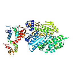 | | Crystal structure of phenamacril-bound F. graminearum myosin I | | 分子名称: | Calmodulin, MAGNESIUM ION, PHOSPHOTHIOPHOSPHORIC ACID-ADENYLATE ESTER, ... | | 著者 | Zhou, Y, Zhou, X.E, Gong, Y, Zhu, Y, Xu, H.E, Zhou, M, Melcher, K, Zhang, F. | | 登録日 | 2019-09-30 | | 公開日 | 2020-03-25 | | 最終更新日 | 2024-10-23 | | 実験手法 | X-RAY DIFFRACTION (2.65 Å) | | 主引用文献 | Structural basis of Fusarium myosin I inhibition by phenamacril.
Plos Pathog., 16, 2020
|
|
1R3J
 
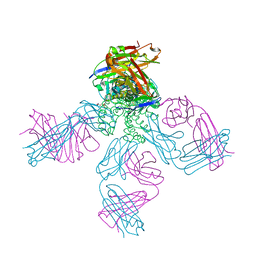 | | potassium channel KcsA-Fab complex in high concentration of Tl+ | | 分子名称: | Antibody Fab fragment heavy chain, Antibody Fab fragment light chain, DIACYL GLYCEROL, ... | | 著者 | Zhou, Y, MacKinnon, R. | | 登録日 | 2003-10-02 | | 公開日 | 2003-11-25 | | 最終更新日 | 2024-10-09 | | 実験手法 | X-RAY DIFFRACTION (1.9 Å) | | 主引用文献 | The occupancy of ions in the K+ selectivity filter: Charge balance and coupling of ion binding to a protein conformational change underlie high conduction rates
J.Mol.Biol., 333, 2003
|
|
6LT9
 
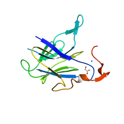 | |
7M42
 
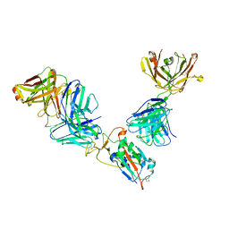 | | Complex of SARS-CoV-2 receptor binding domain with the Fab fragments of neutralizing antibodies REGN10985 and REGN10989 | | 分子名称: | REGN10985 antibody Fab fragment heavy chain, REGN10985 antibody Fab fragment light chain, REGN10989 antibody Fab fragment heavy chain, ... | | 著者 | Zhou, Y, Romero Hernandez, A, Saotome, K, Franklin, M.C. | | 登録日 | 2021-03-19 | | 公開日 | 2021-07-28 | | 最終更新日 | 2021-08-25 | | 実験手法 | ELECTRON MICROSCOPY (3.3 Å) | | 主引用文献 | The monoclonal antibody combination REGEN-COV protects against SARS-CoV-2 mutational escape in preclinical and human studies.
Cell, 184, 2021
|
|
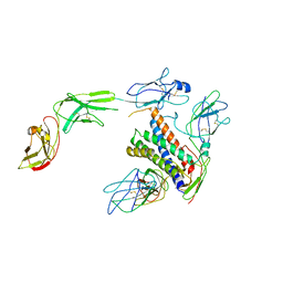
8D85
 
 | |
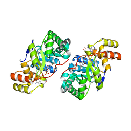
3D3F
 
 | |
6LQK
 
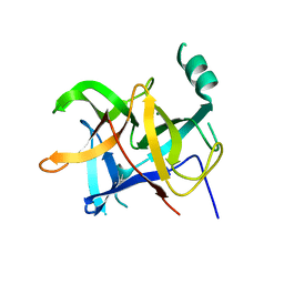 | | Crystal structure of honeybee RyR NTD | | 分子名称: | MAGNESIUM ION, ryanodine receptor | | 著者 | Zhou, Y, Lin, L, Yuchi, Z. | | 登録日 | 2020-01-13 | | 公開日 | 2021-01-20 | | 最終更新日 | 2024-10-23 | | 実験手法 | X-RAY DIFFRACTION (2.499 Å) | | 主引用文献 | Crystal structure of the N-terminal domain of ryanodine receptor from the honeybee, Apis mellifera.
Insect Biochem.Mol.Biol., 125, 2020
|
|
3GXB
 
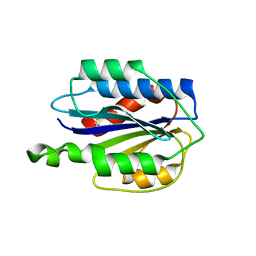 | | Crystal structure of VWF A2 domain | | 分子名称: | 2-acetamido-2-deoxy-beta-D-glucopyranose, 2-acetamido-2-deoxy-beta-D-glucopyranose-(1-4)-2-acetamido-2-deoxy-beta-D-glucopyranose, SULFATE ION, ... | | 著者 | Zhou, Y.F, Springer, T.A. | | 登録日 | 2009-04-02 | | 公開日 | 2009-05-05 | | 最終更新日 | 2023-09-06 | | 実験手法 | X-RAY DIFFRACTION (1.9 Å) | | 主引用文献 | Structural specializations of A2, a force-sensing domain in the ultralarge vascular protein von Willebrand factor.
Proc.Natl.Acad.Sci.USA, 106, 2009
|
|
6VC9
 
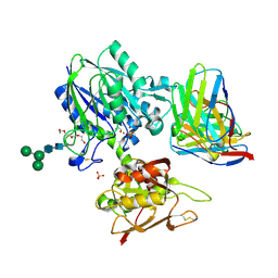 | | TB19 complex | | 分子名称: | 1,2-ETHANEDIOL, 5'-nucleotidase, ecto (CD73), ... | | 著者 | Zhou, Y.F, Lord, D.M. | | 登録日 | 2019-12-20 | | 公開日 | 2020-11-11 | | 最終更新日 | 2024-10-23 | | 実験手法 | X-RAY DIFFRACTION (2.25 Å) | | 主引用文献 | A highly potent CD73 biparatopic antibody blocks organization of the enzyme active site through dual mechanisms.
J.Biol.Chem., 295, 2020
|
|
6VCA
 
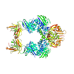 | | TB38 complex | | 分子名称: | 2-acetamido-2-deoxy-beta-D-glucopyranose, 2-acetamido-2-deoxy-beta-D-glucopyranose-(1-4)-2-acetamido-2-deoxy-beta-D-glucopyranose, 5'-nucleotidase, ... | | 著者 | Zhou, Y.F, Lord, D.M. | | 登録日 | 2019-12-20 | | 公開日 | 2020-11-11 | | 最終更新日 | 2024-10-30 | | 実験手法 | X-RAY DIFFRACTION (3.73 Å) | | 主引用文献 | A highly potent CD73 biparatopic antibody blocks organization of the enzyme active site through dual mechanisms.
J.Biol.Chem., 295, 2020
|
|
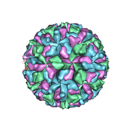
3M8L
 
 | |
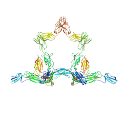
8D82
 
 | |
6N8K
 
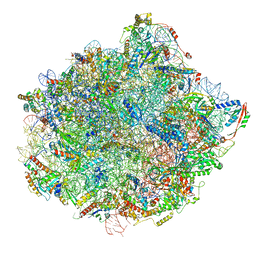 | | Cryo-EM structure of early cytoplasmic-immediate (ECI) pre-60S ribosomal subunit | | 分子名称: | 5.8S rRNA, 5S rRNA, 60S ribosomal export protein NMD3, ... | | 著者 | Zhou, Y, Musalgaonkar, S, Johnson, A.W, Taylor, D.W. | | 登録日 | 2018-11-29 | | 公開日 | 2019-03-13 | | 最終更新日 | 2020-01-08 | | 実験手法 | ELECTRON MICROSCOPY (3.6 Å) | | 主引用文献 | Tightly-orchestrated rearrangements govern catalytic center assembly of the ribosome.
Nat Commun, 10, 2019
|
|
6N8O
 
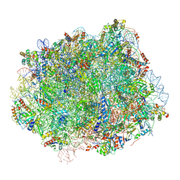 | | Cryo-EM structure of Rpl10-inserted (RI) pre-60S ribosomal subunit | | 分子名称: | 5.8S rRNA, 5S rRNA, 60S ribosomal export protein NMD3, ... | | 著者 | Zhou, Y, Musalgaonkar, S, Johnson, A.W, Taylor, D.W. | | 登録日 | 2018-11-29 | | 公開日 | 2019-03-13 | | 最終更新日 | 2020-01-08 | | 実験手法 | ELECTRON MICROSCOPY (3.5 Å) | | 主引用文献 | Tightly-orchestrated rearrangements govern catalytic center assembly of the ribosome.
Nat Commun, 10, 2019
|
|
6N8J
 
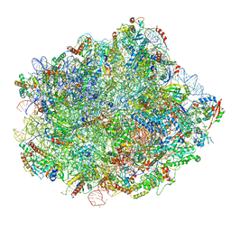 | | Cryo-EM structure of late nuclear (LN) pre-60S ribosomal subunit | | 分子名称: | 5.8S rRNA, 5S rRNA, 60S ribosomal protein L11-A, ... | | 著者 | Zhou, Y, Musalgaonkar, S, Johnson, A.W, Taylor, D.W. | | 登録日 | 2018-11-29 | | 公開日 | 2019-03-13 | | 最終更新日 | 2020-01-08 | | 実験手法 | ELECTRON MICROSCOPY (3.5 Å) | | 主引用文献 | Tightly-orchestrated rearrangements govern catalytic center assembly of the ribosome.
Nat Commun, 10, 2019
|
|
6N8L
 
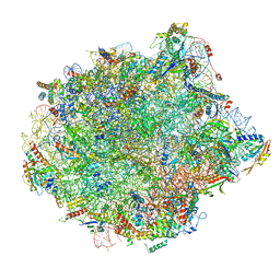 | | Cryo-EM structure of early cytoplasmic-late (ECL) pre-60S ribosomal subunit | | 分子名称: | 5.8S rRNA, 5S rRNA, 60S ribosomal export protein NMD3, ... | | 著者 | Zhou, Y, Musalgaonkar, S, Johnson, A.W, Taylor, D.W. | | 登録日 | 2018-11-29 | | 公開日 | 2019-03-13 | | 最終更新日 | 2020-01-08 | | 実験手法 | ELECTRON MICROSCOPY (3.6 Å) | | 主引用文献 | Tightly-orchestrated rearrangements govern catalytic center assembly of the ribosome.
Nat Commun, 10, 2019
|
|
6N8M
 
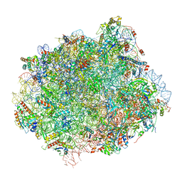 | | Cryo-EM structure of pre-Lsg1 (PL) pre-60S ribosomal subunit | | 分子名称: | 5.8S RNA, 5S rRNA, 60S ribosomal export protein NMD3, ... | | 著者 | Zhou, Y, Musalgaonkar, S, Johnson, A.W, Taylor, D.W. | | 登録日 | 2018-11-29 | | 公開日 | 2019-03-13 | | 最終更新日 | 2020-01-08 | | 実験手法 | ELECTRON MICROSCOPY (3.5 Å) | | 主引用文献 | Tightly-orchestrated rearrangements govern catalytic center assembly of the ribosome.
Nat Commun, 10, 2019
|
|
6N8N
 
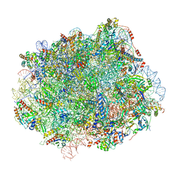 | | Cryo-EM structure of Lsg1-engaged (LE) pre-60S ribosomal subunit | | 分子名称: | 5.8S rRNA, 5S rRNA, 60S ribosomal export protein NMD3, ... | | 著者 | Zhou, Y, Musalgaonkar, S, Johnson, A.W, Taylor, D.W. | | 登録日 | 2018-11-29 | | 公開日 | 2019-03-13 | | 最終更新日 | 2024-10-30 | | 実験手法 | ELECTRON MICROSCOPY (3.8 Å) | | 主引用文献 | Tightly-orchestrated rearrangements govern catalytic center assembly of the ribosome.
Nat Commun, 10, 2019
|
|
1R3L
 
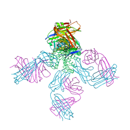 | | potassium channel KcsA-Fab complex in Cs+ | | 分子名称: | Antibody Fab fragment heavy chain, Antibody Fab fragment light chain, CESIUM ION, ... | | 著者 | Zhou, Y, MacKinnon, R. | | 登録日 | 2003-10-02 | | 公開日 | 2003-11-25 | | 最終更新日 | 2024-10-30 | | 実験手法 | X-RAY DIFFRACTION (2.41 Å) | | 主引用文献 | The occupancy of ions in the K+ selectivity filter: Charge balance and coupling of ion binding to a protein conformational change underlie high conduction rates
J.Mol.Biol., 333, 2003
|
|
1R3K
 
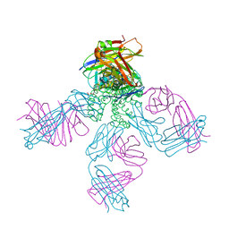 | | potassium channel KcsA-Fab complex in low concentration of Tl+ | | 分子名称: | Antibody Fab fragment heavy chain, Antibody Fab fragment light chain, DIACYL GLYCEROL, ... | | 著者 | Zhou, Y, MacKinnon, R. | | 登録日 | 2003-10-02 | | 公開日 | 2003-11-25 | | 最終更新日 | 2024-10-30 | | 実験手法 | X-RAY DIFFRACTION (2.8 Å) | | 主引用文献 | The occupancy of ions in the K+ selectivity filter: Charge balance and coupling of ion binding to a protein conformational change underlie high conduction rates
J.Mol.Biol., 333, 2003
|
|
1R3I
 
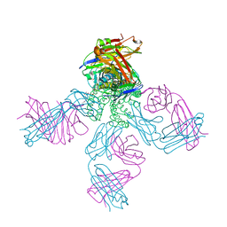 | | potassium channel KcsA-Fab complex in Rb+ | | 分子名称: | Antibody Fab fragment heavy chain, Antibody Fab fragment light chain, DIACYL GLYCEROL, ... | | 著者 | Zhou, Y, MacKinnon, R. | | 登録日 | 2003-10-02 | | 公開日 | 2003-11-25 | | 最終更新日 | 2024-10-30 | | 実験手法 | X-RAY DIFFRACTION (2.4 Å) | | 主引用文献 | The occupancy of ions in the K+ selectivity filter: Charge balance and coupling of ion binding to a protein conformational change underlie high conduction rates
J.Mol.Biol., 333, 2003
|
|
3F7J
 
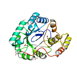 | | B.subtilis YvgN | | 分子名称: | NITRATE ION, POTASSIUM ION, YvgN protein | | 著者 | Zhou, Y.F, Lei, J, Liang, Y.H, Su, X.-D. | | 登録日 | 2008-11-09 | | 公開日 | 2008-11-25 | | 最終更新日 | 2023-12-27 | | 実験手法 | X-RAY DIFFRACTION (1.7 Å) | | 主引用文献 | Structural and biochemical analyses of YvgN and YtbE from Bacillus subtilis
Protein Sci., 18, 2009
|
|
8CYR
 
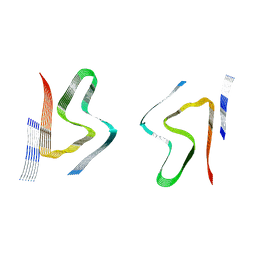 | | Alpha-synuclein fibril from spontaneous control | | 分子名称: | Alpha-synuclein | | 著者 | Zhou, Y, Sokratian, A, Xu, E, Viverette, E, Dillard, L, Yuan, Y, Li, J.Y, Matarangas, A, Bouvette, J, Borgnia, M, Bartesaghi, A, West, A. | | 登録日 | 2022-05-24 | | 公開日 | 2023-05-31 | | 最終更新日 | 2024-06-12 | | 実験手法 | ELECTRON MICROSCOPY (4.2 Å) | | 主引用文献 | Distinct cryo-EM structures and functions of alpha-synuclein fibrils amplified from cerebrospinal fluid
To Be Published
|
|
2K73
 
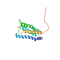 | | Solution NMR structure of integral membrane protein DsbB | | 分子名称: | Disulfide bond formation protein B | | 著者 | Zhou, Y, Cierpicki, T, Flores Jimenez, R.H, Lukasik, S.M, Ellena, J.F, Cafiso, D.S, Kadokura, H, Beckwith, J, Bushweller, J.H. | | 登録日 | 2008-08-01 | | 公開日 | 2008-10-07 | | 最終更新日 | 2021-10-20 | | 実験手法 | SOLUTION NMR | | 主引用文献 | NMR solution structure of the integral membrane enzyme DsbB: functional insights into DsbB-catalyzed disulfide bond formation.
Mol.Cell, 31, 2008
|
|
2K74
 
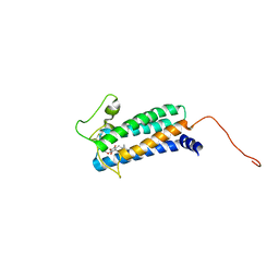 | | Solution NMR structure of DsbB-ubiquinone complex | | 分子名称: | Disulfide bond formation protein B, UBIQUINONE-2 | | 著者 | Zhou, Y, Cierpicki, T, Flores Jimenez, R.H, Lukasik, S.M, Ellena, J.F, Cafiso, D.S, Kadokura, H, Beckwith, J, Bushweller, J.H. | | 登録日 | 2008-08-01 | | 公開日 | 2008-10-07 | | 最終更新日 | 2021-10-20 | | 実験手法 | SOLUTION NMR | | 主引用文献 | NMR solution structure of the integral membrane enzyme DsbB: functional insights into DsbB-catalyzed disulfide bond formation.
Mol.Cell, 31, 2008
|
|
