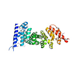
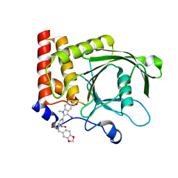
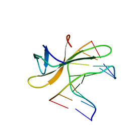
7YTP
 
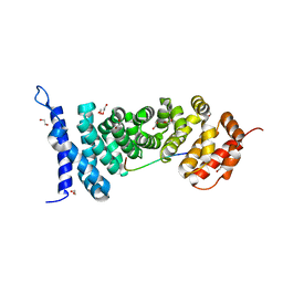
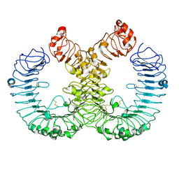 | | TLR7 in complex with an inhibitor | | 分子名称: | (2R,6R)-4-(8-cyanoquinolin-5-yl)-N-[(3S,4R)-4-fluoranylpyrrolidin-3-yl]-6-methyl-morpholine-2-carboxamide, 2-acetamido-2-deoxy-beta-D-glucopyranose, 2-acetamido-2-deoxy-beta-D-glucopyranose-(1-4)-2-acetamido-2-deoxy-beta-D-glucopyranose, ... | | 著者 | Zhang, Z, Ohto, U, Shimizu, T. | | 登録日 | 2022-08-15 | | 公開日 | 2023-11-15 | | 実験手法 | ELECTRON MICROSCOPY (2.77 Å) | | 主引用文献 | TLR7 in complex with an inhibitor
To Be Published
|
|
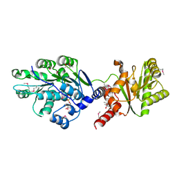
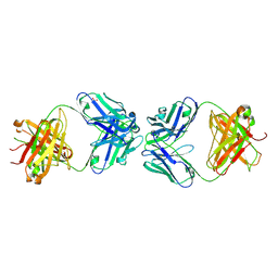
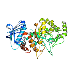
5C6D
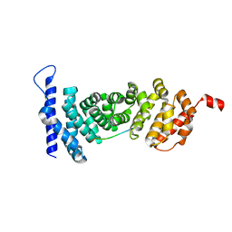
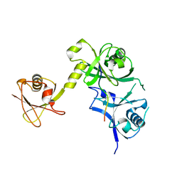 | | Crystal structure of USP7 in complex with UHRF1 | | 分子名称: | E3 ubiquitin-protein ligase UHRF1, Ubiquitin carboxyl-terminal hydrolase 7 | | 著者 | Zhang, Z.-M, Song, J. | | 登録日 | 2015-06-22 | | 公開日 | 2015-09-09 | | 最終更新日 | 2023-09-27 | | 実験手法 | X-RAY DIFFRACTION (2.292 Å) | | 主引用文献 | An Allosteric Interaction Links USP7 to Deubiquitination and Chromatin Targeting of UHRF1.
Cell Rep, 12, 2015
|
|
3V9F
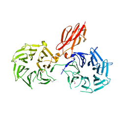 | |
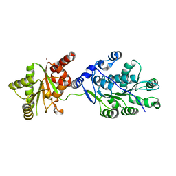
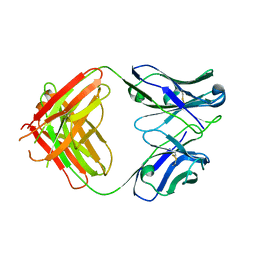
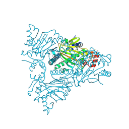
3NMX
 | | Crystal structure of APC complexed with Asef | | 分子名称: | APC variant protein, Rho guanine nucleotide exchange factor 4 | | 著者 | Zhang, Z, Chen, L, Gao, L, Lin, K, Wu, G. | | 登録日 | 2010-06-22 | | 公開日 | 2011-07-06 | | 最終更新日 | 2023-12-27 | | 実験手法 | X-RAY DIFFRACTION (2.3 Å) | | 主引用文献 | Structural basis for the recognition of Asef by adenomatous polyposis coli.
Cell Res., 22, 2012
|
|
3NMZ
 | | Crystal structure of APC complexed with Asef | | 分子名称: | APC variant protein, Rho guanine nucleotide exchange factor 4 | | 著者 | Zhang, Z, Chen, L, Gao, L, Lin, K, Wu, G. | | 登録日 | 2010-06-23 | | 公開日 | 2011-07-06 | | 最終更新日 | 2023-11-01 | | 実験手法 | X-RAY DIFFRACTION (3.01 Å) | | 主引用文献 | Structural basis for the recognition of Asef by adenomatous polyposis coli.
Cell Res., 22, 2012
|
|
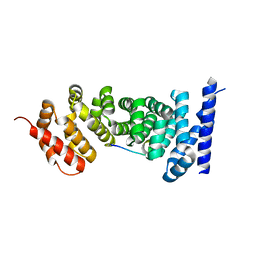
3NMW
 | | Crystal structure of armadillo repeats domain of APC | | 分子名称: | APC variant protein, SULFATE ION | | 著者 | Zhang, Z, Chen, L, Gao, L, Lin, K, Wu, G. | | 登録日 | 2010-06-22 | | 公開日 | 2011-07-06 | | 最終更新日 | 2023-12-27 | | 実験手法 | X-RAY DIFFRACTION (1.6 Å) | | 主引用文献 | Structural basis for the recognition of Asef by adenomatous polyposis coli.
Cell Res., 22, 2012
|
|
4LJY
 | | Crystal structure of RNA splicing effector Prp5 in complex with ADP | | 分子名称: | (4R)-2-METHYLPENTANE-2,4-DIOL, ADENOSINE-5'-DIPHOSPHATE, MAGNESIUM ION, ... | | 著者 | Zhang, Z.-M, Li, J, Yang, F, Xu, Y, Zhou, J. | | 登録日 | 2013-07-05 | | 公開日 | 2013-12-11 | | 最終更新日 | 2019-12-25 | | 実験手法 | X-RAY DIFFRACTION (1.95 Å) | | 主引用文献 | Crystal structure of Prp5p reveals interdomain interactions that impact spliceosome assembly.
Cell Rep, 5, 2013
|
|
4LK2
 | | Crystal structure of RNA splicing effector Prp5 | | 分子名称: | NICKEL (II) ION, Pre-mRNA-processing ATP-dependent RNA helicase PRP5 | | 著者 | Zhang, Z.-M, Li, J, Yang, F, Xu, Y, Zhou, J. | | 登録日 | 2013-07-05 | | 公開日 | 2013-12-11 | | 最終更新日 | 2024-03-20 | | 実験手法 | X-RAY DIFFRACTION (2.12 Å) | | 主引用文献 | Crystal structure of Prp5p reveals interdomain interactions that impact spliceosome assembly.
Cell Rep, 5, 2013
|
|
4N3Z
 | |
4YJL
 | | Crystal structure of APC-ARM in complexed with Amer1-A2 | | 分子名称: | 1,2-ETHANEDIOL, APC membrane recruitment protein 1, Adenomatous polyposis coli protein | | 著者 | Zhang, Z, Xiao, Y, Wu, G. | | 登録日 | 2015-03-03 | | 公開日 | 2016-03-09 | | 最終更新日 | 2023-11-08 | | 実験手法 | X-RAY DIFFRACTION (2.1 Å) | | 主引用文献 | Structures of the APC-ARM domain in complexes with discrete Amer1/WTX fragments reveal that it uses a consensus mode to recognize its binding partners
Cell Discov, 1, 2015
|
|
4N3X
 | |
4N3Y
 | |
4YK6
 | | Crystal structure of APC-ARM in complexed with Amer1-A4 | | 分子名称: | APC membrane recruitment protein 1, Adenomatous polyposis coli protein | | 著者 | Zhang, Z, Xiao, Y, Wu, G. | | 登録日 | 2015-03-04 | | 公開日 | 2016-03-09 | | 最終更新日 | 2023-11-08 | | 実験手法 | X-RAY DIFFRACTION (1.7 Å) | | 主引用文献 | Structures of the APC-ARM domain in complexes with discrete Amer1/WTX fragments reveal that it uses a consensus mode to recognize its binding partners
Cell Discov, 1, 2015
|
|
4YJE
 | | Crystal structure of APC-ARM in complexed with Amer1-A1 | | 分子名称: | APC membrane recruitment protein 1, Adenomatous polyposis coli protein | | 著者 | Zhang, Z, Xiao, Y, Wu, G. | | 登録日 | 2015-03-03 | | 公開日 | 2016-03-09 | | 最終更新日 | 2023-11-08 | | 実験手法 | X-RAY DIFFRACTION (1.9 Å) | | 主引用文献 | Structures of the APC-ARM domain in complexes with discrete Amer1/WTX fragments reveal that it uses a consensus mode to recognize its binding partners
Cell Discov, 1, 2015
|
|
4OV4
 | | Isopropylmalate synthase binding with ketoisovalerate | | 分子名称: | 2-isopropylmalate synthase, 3-METHYL-2-OXOBUTANOIC ACID, ZINC ION | | 著者 | Zhang, Z, Wu, J, Wang, C, Zhang, P. | | 登録日 | 2014-02-20 | | 公開日 | 2014-08-20 | | 最終更新日 | 2024-03-20 | | 実験手法 | X-RAY DIFFRACTION (2 Å) | | 主引用文献 | Subdomain II of alpha-isopropylmalate synthase is essential for activity: inferring a mechanism of feedback inhibition.
J.Biol.Chem., 289, 2014
|
|
4OV9
 | | Structure of isopropylmalate synthase binding with alpha-isopropylmalate | | 分子名称: | (2S)-2-hydroxy-2-(propan-2-yl)butanedioic acid, ZINC ION, isopropylmalate synthase | | 著者 | Zhang, Z, Wu, J, Wang, C, Zhang, P. | | 登録日 | 2014-02-20 | | 公開日 | 2014-08-20 | | 最終更新日 | 2024-03-20 | | 実験手法 | X-RAY DIFFRACTION (2.2 Å) | | 主引用文献 | Subdomain II of alpha-isopropylmalate synthase is essential for activity: inferring a mechanism of feedback inhibition.
J.Biol.Chem., 289, 2014
|
|
1MD2
 | | CHOLERA TOXIN B-PENTAMER WITH DECAVALENT LIGAND BMSC-0013 | | 分子名称: | 3-ETHYLAMINO-4-METHYLAMINO-CYCLOBUTANE-1,2-DIONE, CHOLERA TOXIN B SUBUNIT, CYANIDE ION, ... | | 著者 | Zhang, Z, Merritt, E.A, Ahn, M, Roach, C, Hol, W.G.J, Fan, E. | | 登録日 | 2002-08-06 | | 公開日 | 2002-12-11 | | 最終更新日 | 2017-10-11 | | 実験手法 | X-RAY DIFFRACTION (1.45 Å) | | 主引用文献 | Solution and crystallographic studies of branched multivalent ligands that inhibit the receptor-binding of cholera toxin.
J.Am.Chem.Soc., 124, 2002
|
|
4PVG
 | |
5JO4
 | | Antibody Fab Fragment Complex | | 分子名称: | D80 Fab Heavy Chain, D80 Fab Light Chain, G6 Fab Heavy Chain, ... | | 著者 | Zhang, Z, Prachanronarong, K.P, Marasco, W.A, Schiffer, C.A.S. | | 登録日 | 2016-05-01 | | 公開日 | 2017-11-08 | | 最終更新日 | 2019-11-27 | | 実験手法 | X-RAY DIFFRACTION (2.53 Å) | | 主引用文献 | Structural Basis of an Influenza Hemagglutinin Stem-Directed Antibody Retaining the G6 Idiotype
To Be Published
|
|
5JQD
 | | Antibody Fab Fragment | | 分子名称: | D80 Fab Fragment Heavy Chain, D80 Fab Fragment Light Chain | | 著者 | Zhang, Z, Prachanronarong, K, Gellatly, K, Marasco, W.A, Schiffer, C.A. | | 登録日 | 2016-05-04 | | 公開日 | 2017-11-08 | | 最終更新日 | 2019-11-27 | | 実験手法 | X-RAY DIFFRACTION (2.591 Å) | | 主引用文献 | Structural Basis of an Influenza Hemagglutinin Stem-Directed Antibody Retaining the G6 Idiotype
To Be Published
|
|
5K07
 | | Crystal structure of CREN7-DSDNA (GTAATTGC) complex | | 分子名称: | Chromatin protein Cren7, DNA (5'-D(*GP*TP*AP*AP*TP*TP*GP*C)-3') | | 著者 | Zhang, Z.F, Gong, Y. | | 登録日 | 2016-05-17 | | 公開日 | 2017-05-24 | | 最終更新日 | 2023-11-08 | | 実験手法 | X-RAY DIFFRACTION (2 Å) | | 主引用文献 | Sequence-Dependent T:G Base Pair Opening in DNA Double Helix Bound by Cren7, a Chromatin Protein Conserved among Crenarchaea
PLoS ONE, 11, 2016
|
|
5K17
 | | Crystal structure of CREN7-DSDNA (GTGATCGC) complex | | 分子名称: | Chromatin protein Cren7, DNA (5'-D(*GP*TP*GP*AP*TP*CP*GP*C)-3') | | 著者 | Zhang, Z.F, Gong, Y. | | 登録日 | 2016-05-17 | | 公開日 | 2017-05-24 | | 最終更新日 | 2023-11-08 | | 実験手法 | X-RAY DIFFRACTION (2.1 Å) | | 主引用文献 | Sequence-Dependent T:G Base Pair Opening in DNA Double Helix Bound by Cren7, a Chromatin Protein Conserved among Crenarchaea
PLoS ONE, 11, 2016
|
|
8BDP
 | |
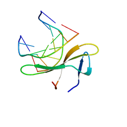
2JX0
 | | The paxillin-binding domain (PBD) of G Protein Coupled Receptor (GPCR)-kinase (GRK) interacting protein 1 (GIT1) | | 分子名称: | ARF GTPase-activating protein GIT1 | | 著者 | Zhang, Z, Guibao, C.D, Simmerman, J.A, Zheng, J. | | 登録日 | 2007-11-01 | | 公開日 | 2008-04-29 | | 最終更新日 | 2024-05-29 | | 実験手法 | SOLUTION NMR | | 主引用文献 | GIT1 paxillin-binding domain is a four-helix bundle, and it binds to both paxillin LD2 and LD4 motifs.
J.Biol.Chem., 283, 2008
|
|
1W9Y
 | | The structure of ACC oxidase | | 分子名称: | 1-AMINOCYCLOPROPANE-1-CARBOXYLATE OXIDASE 1, SULFATE ION | | 著者 | Zhang, Z, Ren, J.-S, Clifton, I.J, Schofield, C.J. | | 登録日 | 2004-10-20 | | 公開日 | 2005-10-26 | | 最終更新日 | 2011-07-13 | | 実験手法 | X-RAY DIFFRACTION (2.1 Å) | | 主引用文献 | Crystal Structure and Mechanistic Implications of 1-Aminocyclopropane-1-Carboxylic Acid Oxidase (the Ethyling Forming Enzyme)
Chem.Biol., 11, 2004
|
|


