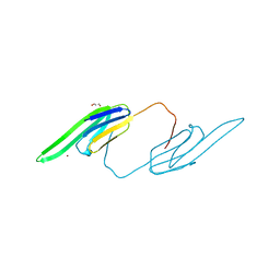
4DGI
 
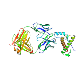 | | Structure of POM1 FAB fragment complexed with human PrPc Fragment 120-230 | | 分子名称: | Major prion protein, POM1 Fab Heavy chain, POM1 Fab Light chain, ... | | 著者 | Baral, P.K, Wieland, B, Swayampakula, M, James, M.N. | | 登録日 | 2012-01-26 | | 公開日 | 2012-10-31 | | 最終更新日 | 2013-01-23 | | 実験手法 | X-RAY DIFFRACTION (2.4 Å) | | 主引用文献 | Structural studies on the folded domain of the human prion protein bound to the Fab fragment of the antibody POM1.
Acta Crystallogr.,Sect.D, 68, 2012
|
|
5WB4
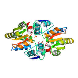 | | Crystal structure of the TarA wall teichoic acid glycosyltransferase | | 分子名称: | N-acetylglucosaminyldiphosphoundecaprenol N-acetyl-beta-D-mannosaminyltransferase, SULFATE ION | | 著者 | Kattke, M.D, Cascio, D, Sawaya, M.R, Clubb, R.T. | | 登録日 | 2017-06-27 | | 公開日 | 2019-01-16 | | 最終更新日 | 2019-07-31 | | 実験手法 | X-RAY DIFFRACTION (2 Å) | | 主引用文献 | Structure and mechanism of TagA, a novel membrane-associated glycosyltransferase that produces wall teichoic acids in pathogenic bacteria.
Plos Pathog., 15, 2019
|
|
1PRQ
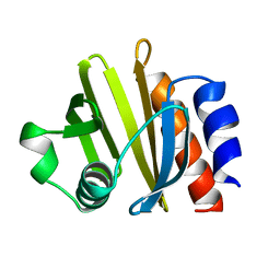 | | ACANTHAMOEBA CASTELLANII PROFILIN IA | | 分子名称: | PROFILIN IA | | 著者 | Fedorov, A.A, Pollard, T.D, Way, M, Lattman, E.E, Almo, S.C. | | 登録日 | 1997-08-18 | | 公開日 | 1997-12-24 | | 最終更新日 | 2024-05-22 | | 実験手法 | X-RAY DIFFRACTION (2.5 Å) | | 主引用文献 | Crystal packing induces a conformational change in profilin-I from Acanthamoeba castellanii.
J.Struct.Biol., 123, 1998
|
|
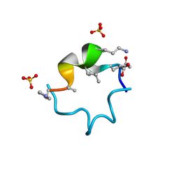
5WOR
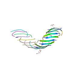 | |
4E0O
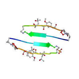 | | SVQIVYK segment from human Tau (305-311) displayed on 54-membered macrocycle scaffold (form III) | | 分子名称: | (4S)-2-METHYL-2,4-PENTANEDIOL, Cyclic pseudo-peptide SVQIVYK(ORN)EF(HAO)(4BF)K(ORN), PHOSPHATE ION | | 著者 | Zhao, M, Liu, C, Sawaya, M.R, Eisenberg, D. | | 登録日 | 2012-03-04 | | 公開日 | 2012-12-19 | | 最終更新日 | 2023-11-15 | | 実験手法 | X-RAY DIFFRACTION (1.82 Å) | | 主引用文献 | Out-of-register beta-sheets suggest a pathway to toxic amyloid aggregates.
Proc.Natl.Acad.Sci.USA, 109, 2012
|
|
4MA7
 | | Crystal structure of mouse prion protein complexed with Promazine | | 分子名称: | Major prion protein, POM1 heavy chain, POM1 light chain, ... | | 著者 | Baral, P.K, Swayampakula, M, James, M.N.G. | | 登録日 | 2013-08-15 | | 公開日 | 2014-01-22 | | 最終更新日 | 2023-09-20 | | 実験手法 | X-RAY DIFFRACTION (1.97 Å) | | 主引用文献 | Structural basis of prion inhibition by phenothiazine compounds.
Structure, 22, 2014
|
|
4MA8
 | | Crystal structure of mouse prion protein complexed with Chlorpromazine | | 分子名称: | 3-(2-chloro-10H-phenothiazin-10-yl)-N,N-dimethylpropan-1-amine, Major prion protein, POM1 heavy chain, ... | | 著者 | Baral, P.K, Swayampakula, M, James, M.N.G. | | 登録日 | 2013-08-15 | | 公開日 | 2014-01-22 | | 最終更新日 | 2023-09-20 | | 実験手法 | X-RAY DIFFRACTION (2.2 Å) | | 主引用文献 | Structural basis of prion inhibition by phenothiazine compounds.
Structure, 22, 2014
|
|
4E0K
 | | Crystal Structure of the amyloid-fibril forming peptide KDWSFY derived from human Beta 2 Microglobulin (58-63) | | 分子名称: | 2-{2-[2-(2-{2-[2-(2-ETHOXY-ETHOXY)-ETHOXY]-ETHOXY}-ETHOXY)-ETHOXY]-ETHOXY}-ETHANOL, Amyloidogenic peptide segment KDWSFY, SODIUM ION | | 著者 | Zhao, M, Liu, C, Sawaya, M.R, Eisenberg, D. | | 登録日 | 2012-03-04 | | 公開日 | 2012-12-19 | | 最終更新日 | 2024-02-28 | | 実験手法 | X-RAY DIFFRACTION (0.97 Å) | | 主引用文献 | Out-of-register beta-sheets suggest a pathway to toxic amyloid aggregates.
Proc.Natl.Acad.Sci.USA, 109, 2012
|
|
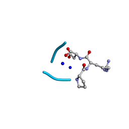
5WMJ
 | |
3L1E
 | | Bovine AlphaA crystallin Zinc Bound | | 分子名称: | Alpha-crystallin A chain, GLYCEROL, ZINC ION | | 著者 | Laganowsky, A, Sawaya, M.R, Cascio, D, Eisenberg, D. | | 登録日 | 2009-12-11 | | 公開日 | 2010-05-12 | | 最終更新日 | 2024-02-21 | | 実験手法 | X-RAY DIFFRACTION (1.15 Å) | | 主引用文献 | Crystal structures of truncated alphaA and alphaB crystallins reveal structural mechanisms of polydispersity important for eye lens function.
Protein Sci., 19, 2010
|
|
5WKD
 | | Crystal structure of the segment, GNNQGSN, from the low complexity domain of TDP-43, residues 300-306 | | 分子名称: | TAR DNA-binding protein 43 | | 著者 | Guenther, E.L, Trinh, H, Sawaya, M.R, Cascio, D, Eisenberg, D.S. | | 登録日 | 2017-07-25 | | 公開日 | 2018-04-18 | | 最終更新日 | 2024-04-03 | | 実験手法 | X-RAY DIFFRACTION (1.8 Å) | | 主引用文献 | Atomic structures of TDP-43 LCD segments and insights into reversible or pathogenic aggregation.
Nat. Struct. Mol. Biol., 25, 2018
|
|
5WHN
 | | Crystal structure of the segment, NFGAFS, from the low complexity domain of TDP-43, residues 312-317 | | 分子名称: | Segment of TAR DNA-binding protein 43 | | 著者 | Guenther, E.L, Sawaya, M.R, Eisenberg, D.S. | | 登録日 | 2017-07-17 | | 公開日 | 2018-04-25 | | 最終更新日 | 2023-10-04 | | 実験手法 | X-RAY DIFFRACTION (1.1 Å) | | 主引用文献 | Atomic structures of TDP-43 LCD segments and insights into reversible or pathogenic aggregation.
Nat. Struct. Mol. Biol., 25, 2018
|
|
5WHP
 | | Crystal structure of the segment, NFGTFS, from the A315T familial variant of the low complexity domain of TDP-43, residues 312-317 | | 分子名称: | Segment of TAR DNA-binding protein 43 | | 著者 | Guenther, E.L, Sawaya, M.R, Eisenberg, D.S. | | 登録日 | 2017-07-17 | | 公開日 | 2018-05-23 | | 最終更新日 | 2024-03-13 | | 実験手法 | X-RAY DIFFRACTION (1 Å) | | 主引用文献 | Atomic structures of TDP-43 LCD segments and insights into reversible or pathogenic aggregation.
Nat. Struct. Mol. Biol., 25, 2018
|
|
5WIQ
 | | Crystal structure of the segment, GFNGGFG, from the low complexity domain of TDP-43, residues 396-402 | | 分子名称: | TAR DNA-binding protein 43 | | 著者 | Guenther, E.L, Sawaya, M.R, Eisenberg, D.S. | | 登録日 | 2017-07-19 | | 公開日 | 2018-04-18 | | 最終更新日 | 2023-10-04 | | 実験手法 | X-RAY DIFFRACTION (1.25 Å) | | 主引用文献 | Atomic structures of TDP-43 LCD segments and insights into reversible or pathogenic aggregation.
Nat. Struct. Mol. Biol., 25, 2018
|
|
3L1F
 | | Bovine AlphaA crystallin | | 分子名称: | (4S)-2-METHYL-2,4-PENTANEDIOL, Alpha-crystallin A chain | | 著者 | Laganowsky, A, Sawaya, M.R, Cascio, D, Eisenberg, D. | | 登録日 | 2009-12-11 | | 公開日 | 2010-05-12 | | 最終更新日 | 2024-02-21 | | 実験手法 | X-RAY DIFFRACTION (1.53 Å) | | 主引用文献 | Crystal structures of truncated alphaA and alphaB crystallins reveal structural mechanisms of polydispersity important for eye lens function.
Protein Sci., 19, 2010
|
|
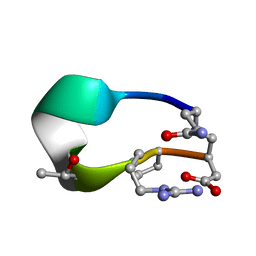
3KX9
 | |
3L1G
 | | Human AlphaB crystallin | | 分子名称: | Alpha-crystallin B chain, SULFATE ION | | 著者 | Laganowsky, A, Sawaya, M.R, Cascio, D, Eisenberg, D. | | 登録日 | 2009-12-11 | | 公開日 | 2010-05-12 | | 最終更新日 | 2024-02-21 | | 実験手法 | X-RAY DIFFRACTION (3.32 Å) | | 主引用文献 | Crystal structures of truncated alphaA and alphaB crystallins reveal structural mechanisms of polydispersity important for eye lens function.
Protein Sci., 19, 2010
|
|
4M5S
 | | Human alphaB crystallin core domain in complex with C-terminal peptide | | 分子名称: | Alpha-crystallin B chain, SUCCINIC ACID | | 著者 | Laganowsky, A, Cascio, D, Sawaya, M.R, Eisenberg, D. | | 登録日 | 2013-08-08 | | 公開日 | 2014-04-09 | | 最終更新日 | 2024-02-28 | | 実験手法 | X-RAY DIFFRACTION (1.37 Å) | | 主引用文献 | The structured core domain of alpha B-crystallin can prevent amyloid fibrillation and associated toxicity.
Proc.Natl.Acad.Sci.USA, 111, 2014
|
|
2QW7
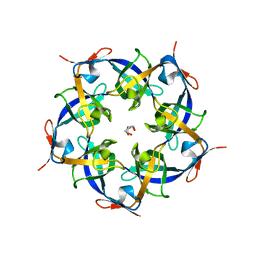 | | Carboxysome Subunit, CcmL | | 分子名称: | Carbon dioxide concentrating mechanism protein ccmL, GLYCEROL | | 著者 | Tanaka, S, Sawaya, M.R, Kerfeld, C.A, Yeates, T.O. | | 登録日 | 2007-08-09 | | 公開日 | 2008-03-04 | | 最終更新日 | 2024-02-21 | | 実験手法 | X-RAY DIFFRACTION (2.4 Å) | | 主引用文献 | Atomic-level models of the bacterial carboxysome shell.
Science, 319, 2008
|
|
6UG6
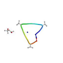 | | C3 symmetric peptide design number 1, Sporty, crystal form 2 | | 分子名称: | (4R)-2-METHYLPENTANE-2,4-DIOL, (4S)-2-METHYL-2,4-PENTANEDIOL, C3-1, ... | | 著者 | Mulligan, V.K, Kang, C.S, Antselovich, I, Sawaya, M.R, Yeates, T.O, Baker, D. | | 登録日 | 2019-09-25 | | 公開日 | 2020-12-02 | | 実験手法 | X-RAY DIFFRACTION (1.1 Å) | | 主引用文献 | Computational design of mixed chirality peptide macrocycles with internal symmetry.
Protein Sci., 29, 2020
|
|
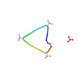
6UGC
 | | C3 symmetric peptide design number 3 | | 分子名称: | C3-3 cyclic peptide design, CADMIUM ION, SODIUM ION | | 著者 | Mulligan, V.K, Kang, C.S, Antselovich, I, Sawaya, M.R, Yeates, T.O, Baker, D. | | 登録日 | 2019-09-26 | | 公開日 | 2020-12-02 | | 実験手法 | X-RAY DIFFRACTION (0.9 Å) | | 主引用文献 | Computational design of mixed chirality peptide macrocycles with internal symmetry.
Protein Sci., 29, 2020
|
|
6UG3
 | | C3 symmetric peptide design number 1, Sporty, crystal form 1 | | 分子名称: | C3-1, Sporty, crystal form 1, ... | | 著者 | Mulligan, V.K, Kang, C.S, Antselovich, I, Sawaya, M.R, Yeates, T.O, Baker, D. | | 登録日 | 2019-09-25 | | 公開日 | 2020-12-02 | | 実験手法 | X-RAY DIFFRACTION (1.1 Å) | | 主引用文献 | Computational design of mixed chirality peptide macrocycles with internal symmetry.
Protein Sci., 29, 2020
|
|
6UF9
 | | S4 symmetric peptide design number 1, Tim apo form | | 分子名称: | S4-1, Tim apo-form, SULFATE ION | | 著者 | Mulligan, V.K, Kang, C.S, Antselovich, I, Sawaya, M.R, Yeates, T.O, Baker, D. | | 登録日 | 2019-09-24 | | 公開日 | 2020-12-02 | | 実験手法 | X-RAY DIFFRACTION (1.1 Å) | | 主引用文献 | Computational design of mixed chirality peptide macrocycles with internal symmetry.
Protein Sci., 29, 2020
|
|
6UGB
 | | C3 symmetric peptide design number 2, Baby Basil | | 分子名称: | C3 symmetric peptide design number 2, Baby Basil, CHLORIDE ION, ... | | 著者 | Mulligan, V.K, Kang, C.S, Antselovich, I, Sawaya, M.R, Yeates, T.O, Baker, D. | | 登録日 | 2019-09-26 | | 公開日 | 2020-12-02 | | 実験手法 | X-RAY DIFFRACTION (0.95 Å) | | 主引用文献 | Computational design of mixed chirality peptide macrocycles with internal symmetry.
Protein Sci., 29, 2020
|
|
6UDW
 | | S2 symmetric peptide design number 3 crystal form 2, Lurch | | 分子名称: | S2-3, Lurch crystal form 2 | | 著者 | Mulligan, V.K, Kang, C.S, Antselovich, I, Sawaya, M.R, Yeates, T.O, Baker, D. | | 登録日 | 2019-09-19 | | 公開日 | 2020-09-23 | | 最終更新日 | 2024-10-09 | | 実験手法 | X-RAY DIFFRACTION (1.1 Å) | | 主引用文献 | Computational design of mixed chirality peptide macrocycles with internal symmetry.
Protein Sci., 29, 2020
|
|


