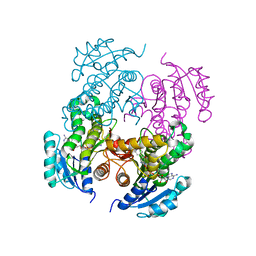
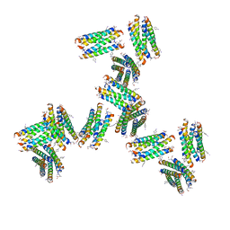
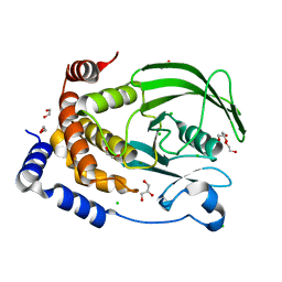
4K0B
 
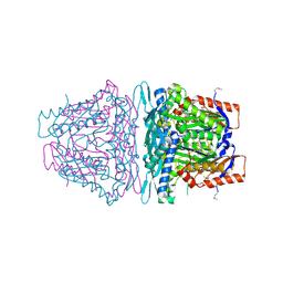 | | Crystal structure of S-Adenosylmethionine synthetase from Sulfolobus solfataricus complexed with SAM and PPi | | 分子名称: | DIPHOSPHATE, MAGNESIUM ION, PHOSPHATE ION, ... | | 著者 | Wang, F, Hurley, K.A, Helmich, K.E, Singh, S, Bingman, C.A, Thorson, J.S, Phillips Jr, G.N, Enzyme Discovery for Natural Product Biosynthesis (NatPro) | | 登録日 | 2013-04-03 | | 公開日 | 2013-05-01 | | 最終更新日 | 2014-10-08 | | 実験手法 | X-RAY DIFFRACTION (2.39 Å) | | 主引用文献 | Understanding molecular recognition of promiscuity of thermophilic methionine adenosyltransferase sMAT from Sulfolobus solfataricus.
Febs J., 281, 2014
|
|
8FOF
 | |
3FNH
 | | Crystal structure of InhA bound to triclosan derivative | | 分子名称: | 2-(2,4-DICHLOROPHENOXY)-5-(2-PHENYLETHYL)PHENOL, Enoyl-[acyl-carrier-protein] reductase [NADH], NICOTINAMIDE-ADENINE-DINUCLEOTIDE | | 著者 | Wang, F. | | 登録日 | 2008-12-24 | | 公開日 | 2009-01-20 | | 最終更新日 | 2023-09-06 | | 実験手法 | X-RAY DIFFRACTION (2.8 Å) | | 主引用文献 | Triclosan Derivatives: Towards Potent Inhibitors of Drug-Sensitive and Drug-Resistant Mycobacterium tuberculosis.
Chemmedchem, 4, 2009
|
|
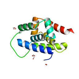
7T6E
 | |
5EU8
 | | Structure of FIPV main protease in complex with dual inhibitors | | 分子名称: | 1,2-ETHANEDIOL, N-[(5-METHYLISOXAZOL-3-YL)CARBONYL]ALANYL-L-VALYL-N~1~-((1R,2Z)-4-(BENZYLOXY)-4-OXO-1-{[(3R)-2-OXOPYRROLIDIN-3-YL]METHYL}BUT-2-ENYL)-L-LEUCINAMIDE, ZINC ION, ... | | 著者 | Wang, F, Chen, C, Liu, X, Yang, K, Xu, X, Yang, H. | | 登録日 | 2015-11-18 | | 公開日 | 2015-12-30 | | 最終更新日 | 2023-11-15 | | 実験手法 | X-RAY DIFFRACTION (2.447 Å) | | 主引用文献 | Crystal Structure of Feline Infectious Peritonitis Virus Main Protease in Complex with Synergetic Dual Inhibitors
J.Virol., 90, 2015
|
|
2NTJ
 | | Mycobacterium tuberculosis InhA bound with PTH-NAD adduct | | 分子名称: | Enoyl-[acyl-carrier-protein] reductase [NADH, {(2R,3S,4R,5R)-5-[(4S)-3-(AMINOCARBONYL)-4-(2-PROPYLISONICOTINOYL)PYRIDIN-1(4H)-YL]-3,4-DIHYDROXYTETRAHYDROFURAN-2-YL}M ETHYL [(2R,3S,4R,5R)-5-(6-AMINO-9H-PURIN-9-YL)-3,4-DIHYDROXYTETRAHYDROFURAN-2-YL]METHYL DIHYDROGEN DIPHOSPHATE | | 著者 | Wang, F, Sacchettini, J.C. | | 登録日 | 2006-11-07 | | 公開日 | 2007-01-30 | | 最終更新日 | 2023-08-30 | | 実験手法 | X-RAY DIFFRACTION (2.5 Å) | | 主引用文献 | Mechanism of thioamide drug action against tuberculosis and leprosy.
J.Exp.Med., 204, 2007
|
|
3FNG
 | | Crystal structure of InhA bound to triclosan derivative | | 分子名称: | 5-(cyclohexylmethyl)-2-(2,4-dichlorophenoxy)phenol, Enoyl-[acyl-carrier-protein] reductase [NADH], NICOTINAMIDE-ADENINE-DINUCLEOTIDE | | 著者 | Wang, F. | | 登録日 | 2008-12-24 | | 公開日 | 2009-01-20 | | 最終更新日 | 2023-09-06 | | 実験手法 | X-RAY DIFFRACTION (1.97 Å) | | 主引用文献 | Triclosan Derivatives: Towards Potent Inhibitors of Drug-Sensitive and Drug-Resistant Mycobacterium tuberculosis.
Chemmedchem, 4, 2009
|
|
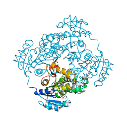
4L7I
 | | Crystal structure of S-Adenosylmethionine synthase from Sulfolobus solfataricus complexed with SAM and PPi | | 分子名称: | DIPHOSPHATE, MAGNESIUM ION, PHOSPHATE ION, ... | | 著者 | Wang, F, Hurley, K.A, Helmich, K.E, Singh, S, Bingman, C.A, Thorson, J.S, Phillips Jr, G.N, Enzyme Discovery for Natural Product Biosynthesis (NatPro) | | 登録日 | 2013-06-13 | | 公開日 | 2013-07-03 | | 最終更新日 | 2023-12-06 | | 実験手法 | X-RAY DIFFRACTION (2.189 Å) | | 主引用文献 | Understanding molecular recognition of promiscuity of thermophilic methionine adenosyltransferase sMAT from Sulfolobus solfataricus.
Febs J., 281, 2014
|
|
5GWY
 | | Structure of Main Protease from Human Coronavirus NL63: Insights for Wide Spectrum Anti-Coronavirus Drug Design | | 分子名称: | N-[(5-METHYLISOXAZOL-3-YL)CARBONYL]ALANYL-L-VALYL-N~1~-((1R,2Z)-4-(BENZYLOXY)-4-OXO-1-{[(3R)-2-OXOPYRROLIDIN-3-YL]METHYL}BUT-2-ENYL)-L-LEUCINAMIDE, main protease | | 著者 | Wang, F, Chen, C, Tan, W, Yang, K, Yang, H. | | 登録日 | 2016-09-14 | | 公開日 | 2017-09-20 | | 最終更新日 | 2023-11-15 | | 実験手法 | X-RAY DIFFRACTION (2.852 Å) | | 主引用文献 | Structure of Main Protease from Human Coronavirus NL63: Insights for Wide Spectrum Anti-Coronavirus Drug Design.
Sci Rep, 6, 2016
|
|
7UIT
 | |
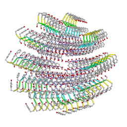
4M83
 | | Ensemble refinement of protein crystal structure (2IYF) of macrolide glycosyltransferases OleD complexed with UDP and Erythromycin A | | 分子名称: | ERYTHROMYCIN A, MAGNESIUM ION, Oleandomycin glycosyltransferase, ... | | 著者 | Wang, F, Helmich, K.E, Xu, W, Singh, S, Olmos Jr, J.L, Martinez iii, E, Bingman, C.A, Thorson, J.S, Phillips Jr, G.N, Enzyme Discovery for Natural Product Biosynthesis (NatPro) | | 登録日 | 2013-08-12 | | 公開日 | 2013-09-11 | | 最終更新日 | 2024-02-28 | | 実験手法 | X-RAY DIFFRACTION (1.698 Å) | | 主引用文献 | Crystal structure of macrolide glycosyltransferases OleD
To be Published
|
|
4M7P
 | | Ensemble refinement of protein crystal structure of macrolide glycosyltransferases OleD | | 分子名称: | Oleandomycin glycosyltransferase, SODIUM ION | | 著者 | Wang, F, Helmich, K.E, Xu, W, Singh, S, Olmos Jr, J.L, Martinez iii, E, Bingman, C.A, Thorson, J.S, Phillips Jr, G.N, Enzyme Discovery for Natural Product Biosynthesis (NatPro) | | 登録日 | 2013-08-12 | | 公開日 | 2013-09-11 | | 実験手法 | X-RAY DIFFRACTION (1.77 Å) | | 主引用文献 | Crystal structure of macrolide glycosyltransferases OleD
To be Published
|
|
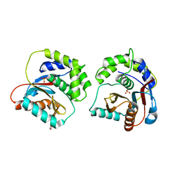
2HVC
 | | The Crystal Structure of Ligand-binding Domain (LBD) of human Androgen Receptor in Complex with a selective modulator LGD2226 | | 分子名称: | 6-[BIS(2,2,2-TRIFLUOROETHYL)AMINO]-4-(TRIFLUOROMETHYL)QUINOLIN-2(1H)-ONE, Androgen receptor | | 著者 | Wang, F, Liu, X.-Q, Li, H, Liang, K.-N, Miner, J.N, Hong, M, Kallel, E.A, van Oeveren, A, Zhi, L, Jiang, T. | | 登録日 | 2006-07-28 | | 公開日 | 2007-07-31 | | 最終更新日 | 2023-10-25 | | 実験手法 | X-RAY DIFFRACTION (2.1 Å) | | 主引用文献 | Structure of the ligand-binding domain (LBD) of human androgen receptor in complex with a selective modulator LGD2226
ACTA CRYSTALLOGR.,SECT.F, 62, 2006
|
|
8WTF
 | | The Crystal Structure of IRAK4 from Biortus | | 分子名称: | 1,2-ETHANEDIOL, 1-{[(2S,3S,4S)-3-ethyl-4-fluoro-5-oxopyrrolidin-2-yl]methoxy}-7-methoxyisoquinoline-6-carboxamide, Interleukin-1 receptor-associated kinase 4, ... | | 著者 | Wang, F, Cheng, W, Lv, Z, Meng, Q, Xu, Y. | | 登録日 | 2023-10-18 | | 公開日 | 2023-11-15 | | 実験手法 | X-RAY DIFFRACTION (2 Å) | | 主引用文献 | The Crystal Structure of IRAK4 from Biortus
To Be Published
|
|
8X2A
 | | The Crystal Structure of BMX from Biortus. | | 分子名称: | 1,2-ETHANEDIOL, 4-[(3S)-3-{[(2E)-but-2-enoyl]amino}piperidin-1-yl]-5-fluoro-2,3-dimethyl-1H-indole-7-carboxamide, CHLORIDE ION, ... | | 著者 | Wang, F, Cheng, W, Yuan, Z, Lin, D, Pan, W. | | 登録日 | 2023-11-09 | | 公開日 | 2023-12-27 | | 実験手法 | X-RAY DIFFRACTION (1.3 Å) | | 主引用文献 | The Crystal Structure of BMX from Biortus.
To Be Published
|
|
7UEG
 | |
7UUQ
 | |
8XLD
 | | Structure of the GFP:GFP-nanobody complex from Biortus. | | 分子名称: | 1,2-ETHANEDIOL, Nanobody(Staygold-S2G10)-Nanobody(Staygold-S4F1), ZINC ION, ... | | 著者 | Wang, F, Cheng, W, Yuan, Z, Lin, D, Bao, C. | | 登録日 | 2023-12-25 | | 公開日 | 2024-03-06 | | 実験手法 | X-RAY DIFFRACTION (2.1 Å) | | 主引用文献 | Structure of the GFP:GFP-nanobody complex from Biortus.
To Be Published
|
|
8XOV
 | | The Crystal Structure of N-terminal kinase domain of human RSK-1 from Biortus. | | 分子名称: | 1,2-ETHANEDIOL, PHOSPHOAMINOPHOSPHONIC ACID-ADENYLATE ESTER, Ribosomal protein S6 kinase alpha-1 | | 著者 | Wang, F, Cheng, W, Lv, Z, Meng, Q, Zhang, B. | | 登録日 | 2024-01-02 | | 公開日 | 2024-03-06 | | 実験手法 | X-RAY DIFFRACTION (2.55 Å) | | 主引用文献 | The Crystal Structure of N-terminal kinase domain of human RSK-1 from Biortus.
To Be Published
|
|
8XQU
 | | The Crystal Structure of ClpC1-NTD from Biortus. | | 分子名称: | 1,2-ETHANEDIOL, ATP-dependent Clp protease ATP-binding subunit ClpC1, DI(HYDROXYETHYL)ETHER, ... | | 著者 | Wang, F, Cheng, W, Lv, Z, Ju, C, Ni, C. | | 登録日 | 2024-01-05 | | 公開日 | 2024-03-06 | | 実験手法 | X-RAY DIFFRACTION (1.85 Å) | | 主引用文献 | The Crystal Structure of ClpC1-NTD from Biortus.
To Be Published
|
|
8XPG
 | | The Crystal Structure of polo box domain of Plk4 from Biortus. | | 分子名称: | SULFATE ION, Serine/threonine-protein kinase PLK4 | | 著者 | Wang, F, Cheng, W, Lv, Z, Meng, Q, Zhang, B. | | 登録日 | 2024-01-03 | | 公開日 | 2024-03-06 | | 実験手法 | X-RAY DIFFRACTION (2.7 Å) | | 主引用文献 | The Crystal Structure of polo box domain of Plk4 from Biortus.
To Be Published
|
|
8XPX
 | | The Crystal Structure of PARP12 from Biortus. | | 分子名称: | 1,2-ETHANEDIOL, ACETATE ION, DI(HYDROXYETHYL)ETHER, ... | | 著者 | Wang, F, Cheng, W, Lv, Z, Qi, J, Shen, Z. | | 登録日 | 2024-01-04 | | 公開日 | 2024-03-06 | | 実験手法 | X-RAY DIFFRACTION (1.75 Å) | | 主引用文献 | The Crystal Structure of PARP12 from Biortus.
To Be Published
|
|
8XI7
 | | The Crystal Structure of UCHL1 from Biortus. | | 分子名称: | 1,2-ETHANEDIOL, SULFATE ION, Ubiquitin carboxyl-terminal hydrolase isozyme L1 | | 著者 | Wang, F, Cheng, W, Yuan, Z, Qi, J, Shen, Z. | | 登録日 | 2023-12-19 | | 公開日 | 2024-03-06 | | 実験手法 | X-RAY DIFFRACTION (1.95 Å) | | 主引用文献 | The Crystal Structure of UCHL1 from Biortus.
To Be Published
|
|
8XQK
 | | The Crystal Structure of Apaf from Biortus. | | 分子名称: | Apoptotic protease-activating factor 1, PHOSPHATE ION | | 著者 | Wang, F, Cheng, W, Lv, Z, Ju, C, Ni, C. | | 登録日 | 2024-01-05 | | 公開日 | 2024-03-06 | | 実験手法 | X-RAY DIFFRACTION (2.85 Å) | | 主引用文献 | The Crystal Structure of Apaf from Biortus.
To Be Published
|
|
8XOY
 | | The Crystal Structure of PTP1B from Biortus. | | 分子名称: | 1,2-ETHANEDIOL, CHLORIDE ION, GLYCEROL, ... | | 著者 | Wang, F, Cheng, W, Lv, Z, Meng, Q, Li, J. | | 登録日 | 2024-01-02 | | 公開日 | 2024-01-24 | | 実験手法 | X-RAY DIFFRACTION (1.55 Å) | | 主引用文献 | The Crystal Structure of PTP1B from Biortus.
To Be Published
|
|


