1KFM
 
 | |
3IT9
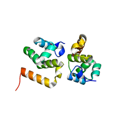
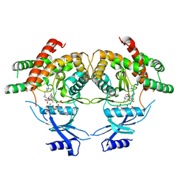
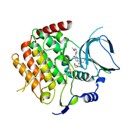
 | | Crystal structure of Penicillin-Binding Protein 6 (PBP6) from E. coli in apo state | | 分子名称: | D-alanyl-D-alanine carboxypeptidase dacC, SULFATE ION, beta-D-fructofuranose-(2-1)-alpha-D-glucopyranose | | 著者 | Chen, Y, Zhang, W, Shi, Q, Hesek, D, Lee, M, Mobashery, S, Shoichet, B.K. | | 登録日 | 2009-08-27 | | 公開日 | 2009-10-20 | | 最終更新日 | 2023-09-06 | | 実験手法 | X-RAY DIFFRACTION (2.1 Å) | | 主引用文献 | Crystal structures of penicillin-binding protein 6 from Escherichia coli.
J.Am.Chem.Soc., 131, 2009
|
|
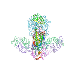
7E2I
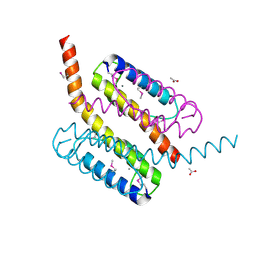
 | | Cryo-EM structure of hDisp1NNN-ShhN | | 分子名称: | 2-acetamido-2-deoxy-beta-D-glucopyranose, CHOLESTEROL HEMISUCCINATE, Protein dispatched homolog 1, ... | | 著者 | Li, W, Wang, L, Gong, X. | | 登録日 | 2021-02-05 | | 公開日 | 2021-12-08 | | 実験手法 | ELECTRON MICROSCOPY (4.07 Å) | | 主引用文献 | Structural insights into proteolytic activation of the human Dispatched1 transporter for Hedgehog morphogen release.
Nat Commun, 12, 2021
|
|
3M9W
 | |
5H5D
 | |
3IW3
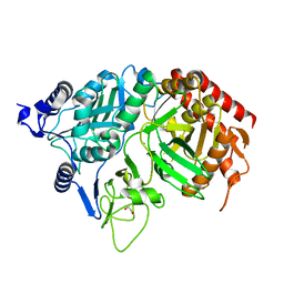
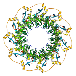
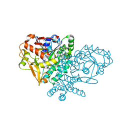
 | | Crystal structure of hyperthermophilic nitrilase | | 分子名称: | ACETIC ACID, MAGNESIUM ION, Nitrilase | | 著者 | Raczynska, J, Vorgias, C, Antranikian, G, Rypniewski, W. | | 登録日 | 2009-09-02 | | 公開日 | 2010-09-22 | | 最終更新日 | 2023-09-06 | | 実験手法 | X-RAY DIFFRACTION (1.8 Å) | | 主引用文献 | Crystallographic analysis of a thermoactive nitrilase.
J.Struct.Biol., 173, 2010
|
|
3MAB
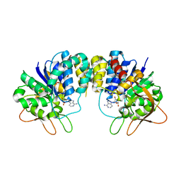
 | | CRYSTAL STRUCTURE OF AN UNCHARACTERIZED PROTEIN FROM LISTERIA MONOCYTOGENES, Triclinic FORM | | 分子名称: | uncharacterized protein | | 著者 | Madegowda, M, Chruszcz, M, Minor, W, Burley, S.K, Swaminathan, S, New York SGX Research Center for Structural Genomics (NYSGXRC) | | 登録日 | 2010-03-23 | | 公開日 | 2010-04-14 | | 最終更新日 | 2023-09-06 | | 実験手法 | X-RAY DIFFRACTION (1.42 Å) | | 主引用文献 | Crystal structure of an uncharacterized protein from listeria monocytogenes
To be Published
|
|
1KHG
 | | PEPCK | | 分子名称: | 1,2-ETHANEDIOL, MANGANESE (II) ION, Phosphoenolpyruvate carboxykinase, ... | | 著者 | Dunten, P, Belunis, C, Crowther, R, Hollfelder, K, Kammlott, U, Levin, W, Michel, H, Ramsey, G.B, Swain, A, Weber, D, Wertheimer, S.J. | | 登録日 | 2001-11-29 | | 公開日 | 2002-02-27 | | 最終更新日 | 2024-02-14 | | 実験手法 | X-RAY DIFFRACTION (2.34 Å) | | 主引用文献 | Crystal structure of human cytosolic phosphoenolpyruvate carboxykinase reveals a new GTP-binding site.
J.Mol.Biol., 316, 2002
|
|
2GF4
 | | Crystal structure of Vng1086c from Halobacterium salinarium (Halobacterium halobium). Northeast Structural Genomics Target HsR14 | | 分子名称: | ACETATE ION, CALCIUM ION, Protein Vng1086c | | 著者 | Benach, J, Zhou, W, Jayaraman, S, Forouhar, F.F, Janjua, H, Xiao, R, Ma, L.-C, Cunningham, K, Wang, D, Acton, T.B, Montelione, G.T, Tong, L, Hunt, J.F, Northeast Structural Genomics Consortium (NESG) | | 登録日 | 2006-03-21 | | 公開日 | 2006-04-18 | | 最終更新日 | 2017-10-18 | | 実験手法 | X-RAY DIFFRACTION (2.07 Å) | | 主引用文献 | Crystal structure of Vng1086c from Halobacterium salinarium (Halobacterium halobium). Northeast Structural Genomics Target HsR14
To be Published
|
|
1KIE
 | | Inosine-adenosine-guanosine preferring nucleoside hydrolase from Trypanosoma vivax: Asp10Ala mutant in complex with 3-deaza-adenosine | | 分子名称: | 3-DEAZA-ADENOSINE, CALCIUM ION, NICKEL (II) ION, ... | | 著者 | Versees, W, Decanniere, K, Van Holsbeke, E, Devroede, N, Steyaert, J. | | 登録日 | 2001-12-03 | | 公開日 | 2002-05-15 | | 最終更新日 | 2023-08-16 | | 実験手法 | X-RAY DIFFRACTION (2 Å) | | 主引用文献 | Enzyme-substrate interactions in the purine-specific nucleoside hydrolase from Trypanosoma vivax.
J.Biol.Chem., 277, 2002
|
|
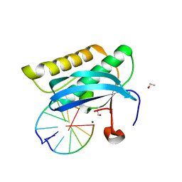
2G5M
 | | Spinophilin PDZ domain | | 分子名称: | Neurabin-2 | | 著者 | Kelker, M.S, Peti, W. | | 登録日 | 2006-02-23 | | 公開日 | 2007-01-09 | | 最終更新日 | 2024-05-29 | | 実験手法 | SOLUTION NMR | | 主引用文献 | Structural basis for spinophilin-neurabin receptor interaction.
Biochemistry, 46, 2007
|
|
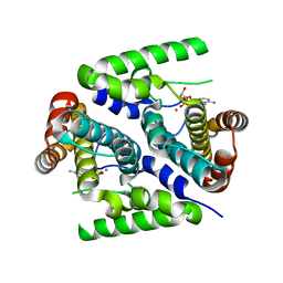
3TDN
 | | Computationally designed two-fold symmetric Tim-barrel protein, FLR | | 分子名称: | FLR SYMMETRIC ALPHA-BETA TIM BARREL | | 著者 | Harp, J.M, Fortenberry, C, Bowman, E, Profitt, W, Dorr, B, Mizoue, L, Meiler, J. | | 登録日 | 2011-08-11 | | 公開日 | 2011-11-16 | | 最終更新日 | 2024-04-03 | | 実験手法 | X-RAY DIFFRACTION (1.4 Å) | | 主引用文献 | Exploring symmetry as an avenue to the computational design of large protein domains.
J.Am.Chem.Soc., 133, 2011
|
|
1KIC
 | | Inosine-adenosine-guanosine preferring nucleoside hydrolase from Trypanosoma vivax: Asp10Ala mutant in complex with inosine | | 分子名称: | CALCIUM ION, INOSINE, NICKEL (II) ION, ... | | 著者 | Versees, W, Decanniere, K, Van Holsbeke, E, Devroede, N, Steyaert, J. | | 登録日 | 2001-12-03 | | 公開日 | 2002-05-15 | | 最終更新日 | 2023-08-16 | | 実験手法 | X-RAY DIFFRACTION (1.6 Å) | | 主引用文献 | Enzyme-substrate interactions in the purine-specific nucleoside hydrolase from Trypanosoma vivax.
J.Biol.Chem., 277, 2002
|
|
5GL8
 | |
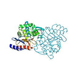
3MES
 | | Crystal structure of choline kinase from Cryptosporidium parvum Iowa II, cgd3_2030 | | 分子名称: | ADENOSINE-5'-DIPHOSPHATE, Choline kinase, DECAMETHONIUM ION, ... | | 著者 | Qiu, W, Wernimont, A, Hills, T, Lew, J, Artz, J.D, Xiao, T, Allali-Hassani, A, Vedadi, M, Kozieradzki, I, Cossar, D, Bountra, C, Weigelt, J, Arrowsmith, C.H, Edwards, A.M, Hui, R, Ma, D, Structural Genomics Consortium (SGC) | | 登録日 | 2010-03-31 | | 公開日 | 2010-04-07 | | 最終更新日 | 2023-09-06 | | 実験手法 | X-RAY DIFFRACTION (2.35 Å) | | 主引用文献 | Crystal structure of choline kinase from Cryptosporidium parvum Iowa II, cgd3_2030
TO BE PUBLISHED
|
|
2PAU
 | | Crystal structure of the 5'-deoxynucleotidase YfbR mutant E72A complexed with Co(2+) and dAMP | | 分子名称: | 2'-DEOXYADENOSINE-5'-MONOPHOSPHATE, 5'-deoxynucleotidase YfbR, COBALT (II) ION, ... | | 著者 | Zimmerman, M.D, Proudfoot, M, Yakunin, A, Minor, W. | | 登録日 | 2007-03-27 | | 公開日 | 2008-03-04 | | 最終更新日 | 2023-08-30 | | 実験手法 | X-RAY DIFFRACTION (2.1 Å) | | 主引用文献 | Structural insight into the mechanism of substrate specificity and catalytic activity of an HD-domain phosphohydrolase: the 5'-deoxyribonucleotidase YfbR from Escherichia coli.
J.Mol.Biol., 378, 2008
|
|
2G8V
 | |
1KN0
 | | Crystal Structure of the human Rad52 protein | | 分子名称: | Rad52 | | 著者 | Kagawa, W, Kurumizaka, H, Ishitani, R, Fukai, S, Nureki, O, Shibata, T, Yokoyama, S, RIKEN Structural Genomics/Proteomics Initiative (RSGI) | | 登録日 | 2001-12-18 | | 公開日 | 2002-09-04 | | 最終更新日 | 2024-03-13 | | 実験手法 | X-RAY DIFFRACTION (2.85 Å) | | 主引用文献 | Crystal structure of the homologous-pairing domain from the human Rad52 recombinase in the undecameric form.
Mol.Cell, 10, 2002
|
|
5GJS
 | | Crystal structure of H1 hemagglutinin from A/California/04/2009 in complex with a neutralizing antibody 3E1 | | 分子名称: | 2-acetamido-2-deoxy-beta-D-glucopyranose, 2-acetamido-2-deoxy-beta-D-glucopyranose-(1-4)-2-acetamido-2-deoxy-beta-D-glucopyranose, Hemagglutinin, ... | | 著者 | Wang, W, Zhang, T, Ding, J. | | 登録日 | 2016-07-01 | | 公開日 | 2016-12-07 | | 最終更新日 | 2023-11-08 | | 実験手法 | X-RAY DIFFRACTION (2.9 Å) | | 主引用文献 | Human antibody 3E1 targets the HA stem region of H1N1 and H5N6 influenza A viruses
Nat Commun, 7, 2016
|
|
3IQ1
 | | Crystal structure of DPS protein from Vibrio cholerae O1, a member of a broad superfamily of ferritin-like diiron-carboxylate proteins | | 分子名称: | CHLORIDE ION, DPS family protein | | 著者 | Nocek, B, Peterson, S, Gu, M, Otwinowski, Z, Anderson, W, Joachimiak, A, Center for Structural Genomics of Infectious Diseases (CSGID) | | 登録日 | 2009-08-18 | | 公開日 | 2009-09-08 | | 最終更新日 | 2011-07-13 | | 実験手法 | X-RAY DIFFRACTION (1.67 Å) | | 主引用文献 | Crystal structure of DPS protein from Vibrio cholerae O1, a member of a broad superfamily of ferritin-like diiron-carboxylate proteins
TO BE PUBLISHED
|
|
6F7B
 | | Crystal structure of the human Bub1 kinase domain in complex with BAY 1816032 | | 分子名称: | 2-[3,5-bis(fluoranyl)-4-[[3-[5-methoxy-4-[(3-methoxypyridin-4-yl)amino]pyrimidin-2-yl]indazol-1-yl]methyl]phenoxy]ethanol, MAGNESIUM ION, Mitotic checkpoint serine/threonine-protein kinase BUB1 | | 著者 | Holton, S.J, Siemeister, G, Mengel, A, Bone, W, Schroeder, J, Zitzmann-Kolbe, S, Briem, H, Fernandez-Montalvan, A, Prechtl, S, Moenning, U, von Ahsen, O, Johanssen, J, Cleve, A, Puetter, V, Hitchcock, M, von Nussbaum, F, Brands, M, Mumberg, D, Ziegelbauer, K. | | 登録日 | 2017-12-08 | | 公開日 | 2018-12-19 | | 最終更新日 | 2021-05-05 | | 実験手法 | X-RAY DIFFRACTION (2 Å) | | 主引用文献 | Inhibition of BUB1 Kinase by BAY 1816032 Sensitizes Tumor Cells toward Taxanes, ATR, and PARP InhibitorsIn VitroandIn Vivo.
Clin.Cancer Res., 25, 2019
|
|
1KJX
 | | IMP Complex of E. Coli Adenylosuccinate Synthetase | | 分子名称: | Adenylosuccinate Synthetase, INOSINIC ACID | | 著者 | Hou, Z, Wang, W, Fromm, H.J, Honzatko, R.B. | | 登録日 | 2001-12-05 | | 公開日 | 2002-03-20 | | 最終更新日 | 2024-02-14 | | 実験手法 | X-RAY DIFFRACTION (2.6 Å) | | 主引用文献 | IMP Alone Organizes the Active Site of Adenylosuccinate Synthetase from Escherichia coli.
J.Biol.Chem., 277, 2002
|
|
1KLJ
 | | Crystal structure of uninhibited factor VIIa | | 分子名称: | 1,2-ETHANEDIOL, CALCIUM ION, factor VIIa | | 著者 | Sichler, K, Banner, D, D'Arcy, A, Hopfner, K.P, Huber, R, Bode, W, Kresse, G.B, Kopetzki, E, Brandstetter, H. | | 登録日 | 2001-12-12 | | 公開日 | 2002-10-09 | | 最終更新日 | 2023-08-16 | | 実験手法 | X-RAY DIFFRACTION (2.44 Å) | | 主引用文献 | Crystal structures of uninhibited factor VIIa link its cofactor and substrate-assisted activation to specific interactions.
J.Mol.Biol., 322, 2002
|
|
5GS8
 | | Crystal structure of TLA-3 extended-spectrum beta-lactamase | | 分子名称: | Beta-lactamase, CHLORIDE ION, SODIUM ION, ... | | 著者 | Wachino, J, Jin, W, Arakawa, Y. | | 登録日 | 2016-08-14 | | 公開日 | 2017-07-12 | | 最終更新日 | 2023-11-08 | | 実験手法 | X-RAY DIFFRACTION (1.59 Å) | | 主引用文献 | Structural Insights into the TLA-3 Extended-Spectrum beta-Lactamase and Its Inhibition by Avibactam and OP0595.
Antimicrob. Agents Chemother., 61, 2017
|
|
2GD4
 | | Crystal Structure of the Antithrombin-S195A Factor Xa-Pentasaccharide Complex | | 分子名称: | 2-acetamido-2-deoxy-beta-D-glucopyranose, 2-acetamido-2-deoxy-beta-D-glucopyranose-(1-4)-2-acetamido-2-deoxy-beta-D-glucopyranose, 2-deoxy-6-O-sulfo-2-(sulfoamino)-alpha-D-glucopyranose-(1-4)-beta-D-glucopyranuronic acid-(1-4)-2-deoxy-3,6-di-O-sulfo-2-(sulfoamino)-alpha-D-glucopyranose-(1-4)-2-O-sulfo-alpha-L-idopyranuronic acid-(1-4)-methyl 2-deoxy-6-O-sulfo-2-(sulfoamino)-alpha-D-glucopyranoside, ... | | 著者 | Johnson, D.J, Li, W, Adams, T.E, Huntington, J.A. | | 登録日 | 2006-03-15 | | 公開日 | 2006-05-09 | | 最終更新日 | 2024-03-13 | | 実験手法 | X-RAY DIFFRACTION (3.3 Å) | | 主引用文献 | Antithrombin-S195A factor Xa-heparin structure reveals the allosteric mechanism of antithrombin activation.
Embo J., 25, 2006
|
|


