Loading
PDBj
MenuPDBj@FacebookPDBj@TwitterPDBj@YouTubewwPDB FoundationwwPDB
RCSB PDBPDBeBMRBAdv. SearchSearch help
PDB: 301 results

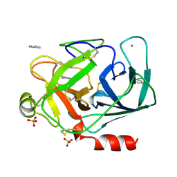
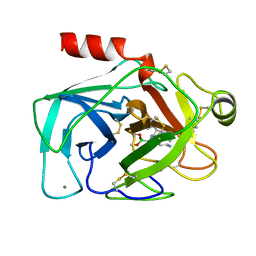
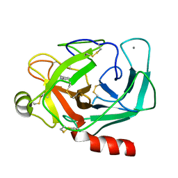
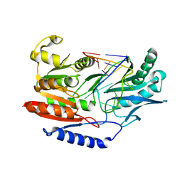
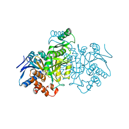
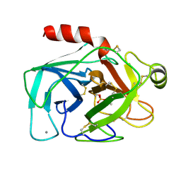
5PTP
DownloadVisualize
BU of 5ptp by Molmil
STRUCTURE OF HYDROLASE (SERINE PROTEINASE)
Descriptor: BETA TRYPSIN, CALCIUM ION
Authors:Stroud, R.M, Finer-Moore, J.
Deposit date:1997-03-31
Release date:1997-07-07
Last modified:2017-11-29
Method:X-RAY DIFFRACTION (1.34 Å)
Cite:Solvent structure in crystals of trypsin determined by X-ray and neutron diffraction.
Proteins, 12, 1992
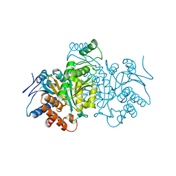
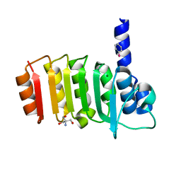
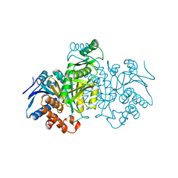
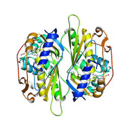
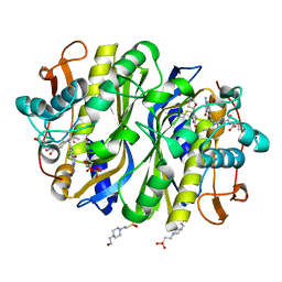
6NNR
DownloadVisualize
BU of 6nnr by Molmil
high-resolution structure of wild-type E. coli thymidylate synthase
Descriptor: 10-PROPARGYL-5,8-DIDEAZAFOLIC ACID, 2'-deoxy-5'-uridylic acid, 4-(2-HYDROXYETHYL)-1-PIPERAZINE ETHANESULFONIC ACID, ...
Authors:Stroud, R.M, Finer-Moore, J.
Deposit date:2019-01-15
Release date:2019-01-30
Last modified:2023-10-11
Method:X-RAY DIFFRACTION (1.05 Å)
Cite:Mg2+ binds to the surface of thymidylate synthase and affects hydride transfer at the interior active site.
J. Am. Chem. Soc., 135, 2013
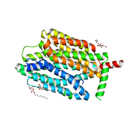
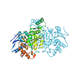
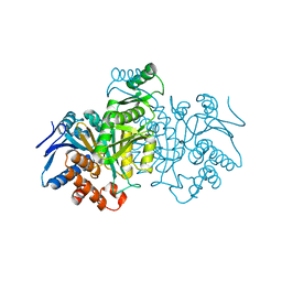
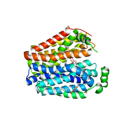
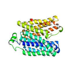
7SP5
DownloadVisualize
BU of 7sp5 by Molmil
Crystal Structure of a Eukaryotic Phosphate Transporter
Descriptor: PHOSPHATE ION, Phosphate transporter, nonyl beta-D-glucopyranoside
Authors:Stroud, R.M, Pedersen, B.P, Kumar, H, Waight, A.B, Risenmay, A.J, Roe-Zurz, Z, Chau, B.H, Schlessinger, A, Bonomi, M, Harries, W, Sali, A, Johri, A.K, Finer-Moore, J.
Deposit date:2021-11-02
Release date:2021-11-17
Last modified:2024-05-22
Method:X-RAY DIFFRACTION (2.9 Å)
Cite:Crystal structure of a eukaryotic phosphate transporter.
Nature, 496, 2013
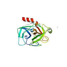
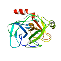
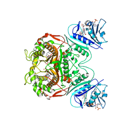
1DPO
DownloadVisualize
BU of 1dpo by Molmil
STRUCTURE OF RAT TRYPSIN
Descriptor: BENZAMIDINE, CALCIUM ION, SULFATE ION, ...
Authors:Stroud, R.M.
Deposit date:1997-03-31
Release date:1997-07-07
Last modified:2021-11-03
Method:X-RAY DIFFRACTION (1.59 Å)
Cite:1.59 A structure of trypsin at 120 K: comparison of low temperature and room temperature structures.
Proteins, 10, 1991
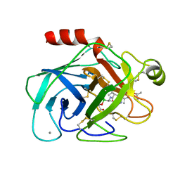
1BTX
DownloadVisualize
BU of 1btx by Molmil
Episelection: Novel Ki ~Nanomolar Inhibitors of Serine Proteases Selected by Binding or Chemistry on an Enzyme Surface
Descriptor: BETA-TRYPSIN, CALCIUM ION, N-(tert-butoxycarbonyl)-L-alanyl-N-[(1S)-5-amino-1-(diethoxyboranyl)pentyl]-L-valinamide
Authors:Stroud, R.M, Katz, B.A, Finer-Moore, J.
Deposit date:1995-05-17
Release date:1995-10-15
Last modified:2012-12-12
Method:X-RAY DIFFRACTION (1.7 Å)
Cite:Episelection: novel Ki approximately nanomolar inhibitors of serine proteases selected by binding or chemistry on an enzyme surface.
Biochemistry, 34, 1995
1BTW
DownloadVisualize
BU of 1btw by Molmil
Episelection: novel KI ~nanomolar inhibitors of serine proteases selected by binding or chemistry on an enzyme surface
Descriptor: BETA-TRYPSIN, CALCIUM ION, N-(tert-butoxycarbonyl)-L-alanyl-N-{(1S)-5-ammonio-1-[hydroxy(3-hydroxypropoxy)boranyl]pentyl}-L-valinamide
Authors:Stroud, R.M, Katz, B.A, Finer-Moore, J.
Deposit date:1995-05-17
Release date:1995-10-15
Last modified:2024-10-23
Method:X-RAY DIFFRACTION (1.7 Å)
Cite:Episelection: novel Ki approximately nanomolar inhibitors of serine proteases selected by binding or chemistry on an enzyme surface.
Biochemistry, 34, 1995
1BTZ
DownloadVisualize
BU of 1btz by Molmil
Episelection: novel KI ~nanomolar inhibitors of serine proteases selected by binding or chemistry on an enzyme surface
Descriptor: BETA-TRYPSIN, CALCIUM ION, N-(tert-butoxycarbonyl)-L-alanyl-N-{(1R)-5-ammonio-1-[hydroxy(methoxy)boranyl]pentyl}-L-valinamide
Authors:Stroud, R.M, Katz, B.A, Finer-Moore, J.
Deposit date:1995-05-17
Release date:1995-10-15
Last modified:2024-10-16
Method:X-RAY DIFFRACTION (2 Å)
Cite:Episelection: novel Ki approximately nanomolar inhibitors of serine proteases selected by binding or chemistry on an enzyme surface.
Biochemistry, 34, 1995
1BTY
DownloadVisualize
BU of 1bty by Molmil
Crystal structure of beta-trypsin in complex with benzamidine
Descriptor: BENZAMIDINE, BETA-TRYPSIN, CALCIUM ION
Authors:Stroud, R.M, Katz, B.A, Finer-Moore, J.
Deposit date:1995-05-17
Release date:1995-10-15
Last modified:2011-07-13
Method:X-RAY DIFFRACTION (1.5 Å)
Cite:Episelection: novel Ki approximately nanomolar inhibitors of serine proteases selected by binding or chemistry on an enzyme surface.
Biochemistry, 34, 1995
1CW1
DownloadVisualize
BU of 1cw1 by Molmil
CRYSTAL STRUCTURE OF ISOCITRATE DEHYDROGENASE MUTANT K230M BOUND TO ISOCITRATE AND MN2+
Descriptor: ISOCITRATE DEHYDROGENASE, ISOCITRIC ACID, MANGANESE (II) ION, ...
Authors:Stroud, R, Finer-Moore, J.
Deposit date:1999-08-25
Release date:1999-09-01
Last modified:2024-02-07
Method:X-RAY DIFFRACTION (2.1 Å)
Cite:Active site water molecules revealed in the 2.1 A resolution structure of a site-directed mutant of isocitrate dehydrogenase.
J.Mol.Biol., 295, 2000
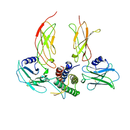
1CN4
DownloadVisualize
BU of 1cn4 by Molmil
ERYTHROPOIETIN COMPLEXED WITH EXTRACELLULAR DOMAINS OF ERYTHROPOIETIN RECEPTOR
Descriptor: PROTEIN (ERYTHROPOIETIN RECEPTOR), PROTEIN (ERYTHROPOIETIN)
Authors:Stroud, R.M, Reid, S.W.
Deposit date:1999-05-25
Release date:1999-08-11
Last modified:2024-10-09
Method:X-RAY DIFFRACTION (2.8 Å)
Cite:Efficiency of signalling through cytokine receptors depends critically on receptor orientation.
Nature, 395, 1998
2TRM
DownloadVisualize
BU of 2trm by Molmil
THE THREE-DIMENSIONAL STRUCTURE OF ASN102 MUTANT OF TRYPSIN. ROLE OF ASP102 IN SERINE PROTEASE CATALYSIS
Descriptor: BENZAMIDINE, CALCIUM ION, TRYPSIN
Authors:Stroud, R.M, Finer-Moore, J.
Deposit date:1988-04-25
Release date:1988-07-16
Last modified:2017-11-29
Method:X-RAY DIFFRACTION (2.8 Å)
Cite:The three-dimensional structure of Asn102 mutant of trypsin: role of Asp102 in serine protease catalysis.
Science, 237, 1987
3E4G
DownloadVisualize
BU of 3e4g by Molmil
Crystal structure of bovine coupling Factor B, G28E mutant
Descriptor: 2-AMINO-2-HYDROXYMETHYL-PROPANE-1,3-DIOL, ATP synthase subunit s, mitochondrial, ...
Authors:Stroud, R.M, Lee, J.K, Belogrudov, G.I.
Deposit date:2008-08-11
Release date:2008-08-26
Last modified:2023-08-30
Method:X-RAY DIFFRACTION (0.96 Å)
Cite:Crystal structure of bovine mitochondrial factor B at 0.96-A resolution.
Proc.Natl.Acad.Sci.Usa, 105, 2008
3E2J
DownloadVisualize
BU of 3e2j by Molmil
Crystal structure of bovine coupling factor B
Descriptor: ATP synthase subunit s, mitochondrial, MAGNESIUM ION
Authors:Stroud, R.M, Lee, J.K, Belogrudov, G.I.
Deposit date:2008-08-05
Release date:2008-08-19
Last modified:2023-08-30
Method:X-RAY DIFFRACTION (2.9 Å)
Cite:Crystal structure of bovine mitochondrial factor B at 0.96-A resolution.
Proc.Natl.Acad.Sci.Usa, 105, 2008
3E3Z
DownloadVisualize
BU of 3e3z by Molmil
Crystal structure of bovine coupling Factor B bound with phenylarsine oxide
Descriptor: 2-AMINO-2-HYDROXYMETHYL-PROPANE-1,3-DIOL, ATP synthase subunit s, mitochondrial, ...
Authors:Stroud, R.M, Lee, J.K, Belogrudov, G.I.
Deposit date:2008-08-08
Release date:2008-08-26
Last modified:2023-08-30
Method:X-RAY DIFFRACTION (1.7 Å)
Cite:Crystal structure of bovine mitochondrial factor B at 0.96-A resolution.
Proc.Natl.Acad.Sci.Usa, 105, 2008
9ICD
DownloadVisualize
BU of 9icd by Molmil
CATALYTIC MECHANISM OF NADP+-DEPENDENT ISOCITRATE DEHYDROGENASE: IMPLICATIONS FROM THE STRUCTURES OF MAGNESIUM-ISOCITRATE AND NADP+ COMPLEXES
Descriptor: ISOCITRATE DEHYDROGENASE, NADP NICOTINAMIDE-ADENINE-DINUCLEOTIDE PHOSPHATE
Authors:Hurley, J.H, Dean, A.M, Koshland Jr, D.E, Stroud, R.M.
Deposit date:1991-07-29
Release date:1991-10-15
Last modified:2024-02-14
Method:X-RAY DIFFRACTION (2.5 Å)
Cite:Catalytic mechanism of NADP(+)-dependent isocitrate dehydrogenase: implications from the structures of magnesium-isocitrate and NADP+ complexes.
Biochemistry, 30, 1991
1QZF
DownloadVisualize
BU of 1qzf by Molmil
Crystal structure of DHFR-TS from Cryptosporidium hominis
Descriptor: 10-PROPARGYL-5,8-DIDEAZAFOLIC ACID, 2'-DEOXYURIDINE 5'-MONOPHOSPHATE, FOLIC ACID, ...
Authors:O'Neil, R.H, Lilien, R.H, Donald, B.R, Stroud, R.M, Anderson, A.C.
Deposit date:2003-09-16
Release date:2003-11-11
Last modified:2023-08-23
Method:X-RAY DIFFRACTION (2.8 Å)
Cite:Phylogenetic classification of protozoa based on the structure of the linker domain in the bifunctional enzyme, dihydrofolate reductase-thymidylate synthase
J.Biol.Chem., 278, 2003
3BT7
DownloadVisualize
BU of 3bt7 by Molmil
Structure of E. coli 5-Methyluridine Methyltransferase TrmA in complex with 19 nucleotide T-arm analogue
Descriptor: RNA (5'-D(P*GP*CP*UP*GP*UP*GP*(5MU)P*UP*CP*GP*AP*UP*CP*CP*AP*CP*AP*GP*C)-3'), tRNA (uracil-5-)-methyltransferase
Authors:Alian, A, Stroud, R.M, Finer-Moore, J.
Deposit date:2007-12-27
Release date:2008-04-29
Last modified:2023-08-30
Method:X-RAY DIFFRACTION (2.43 Å)
Cite:Structure of a TrmA-RNA complex: A consensus RNA fold contributes to substrate selectivity and catalysis in m5U methyltransferases.
Proc.Natl.Acad.Sci.Usa, 105, 2008
8ICD
DownloadVisualize
BU of 8icd by Molmil
REGULATION OF AN ENZYME BY PHOSPHORYLATION AT THE ACTIVE SITE
Descriptor: ISOCITRATE DEHYDROGENASE, ISOCITRIC ACID, MAGNESIUM ION
Authors:Hurley, J.H, Dean, A.M, Sohl, J.L, Koshlandjunior, D.E, Stroud, R.M.
Deposit date:1990-05-30
Release date:1991-10-15
Last modified:2024-02-14
Method:X-RAY DIFFRACTION (2.5 Å)
Cite:Regulation of an enzyme by phosphorylation at the active site.
Science, 249, 1990
6ICD
DownloadVisualize
BU of 6icd by Molmil
REGULATION OF AN ENZYME BY PHOSPHORYLATION AT THE ACTIVE SITE
Descriptor: ISOCITRATE DEHYDROGENASE
Authors:Hurley, J.H, Dean, A.M, Sohl, J.L, Koshlandjunior, D.E, Stroud, R.M.
Deposit date:1990-05-30
Release date:1991-10-15
Last modified:2024-03-13
Method:X-RAY DIFFRACTION (2.8 Å)
Cite:Regulation of an enzyme by phosphorylation at the active site.
Science, 249, 1990
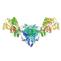
7UK4
DownloadVisualize
BU of 7uk4 by Molmil
KS-AT di-domain of mycobacterial Pks13 with endogenous KS ligand bound
Descriptor: Polyketide synthase PKS13, UNKNOWN LIGAND
Authors:Kim, S.K, Dickinson, M.S, Finer-Moore, J.S, Rosenberg, O.S, Stroud, R.M.
Deposit date:2022-03-31
Release date:2023-02-15
Last modified:2024-10-23
Method:ELECTRON MICROSCOPY (1.94 Å)
Cite:Structure and dynamics of the essential endogenous mycobacterial polyketide synthase Pks13.
Nat.Struct.Mol.Biol., 30, 2023
5ICD
DownloadVisualize
BU of 5icd by Molmil
REGULATION OF AN ENZYME BY PHOSPHORYLATION AT THE ACTIVE SITE
Descriptor: ISOCITRATE DEHYDROGENASE, ISOCITRIC ACID, MAGNESIUM ION
Authors:Hurley, J.H, Dean, A.M, Sohl, J.L, Koshlandjunior, D.E, Stroud, R.M.
Deposit date:1990-05-30
Release date:1991-10-15
Last modified:2024-03-06
Method:X-RAY DIFFRACTION (2.5 Å)
Cite:Regulation of an enzyme by phosphorylation at the active site.
Science, 249, 1990
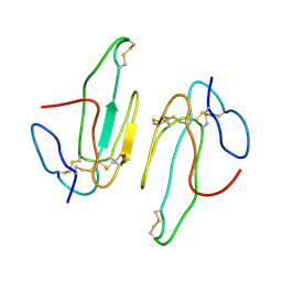
2AAZ
DownloadVisualize
BU of 2aaz by Molmil
Cryptococcus neoformans thymidylate synthase complexed with substrate and an antifolate
Descriptor: 10-PROPARGYL-5,8-DIDEAZAFOLIC ACID, 2'-DEOXYURIDINE 5'-MONOPHOSPHATE, Thymidylate synthase
Authors:Finer-Moore, J.S, Anderson, A.C, O'Neil, R.H, Costi, M.P, Ferrari, S, Krucinski, J, Stroud, R.M.
Deposit date:2005-07-14
Release date:2005-12-06
Last modified:2024-10-09
Method:X-RAY DIFFRACTION (2.08 Å)
Cite:The structure of Cryptococcus neoformans thymidylate synthase suggests strategies for using target dynamics for species-specific inhibition.
Acta Crystallogr.,Sect.D, 61, 2005
2ABX
DownloadVisualize
BU of 2abx by Molmil
THE CRYSTAL STRUCTURE OF ALPHA-BUNGAROTOXIN AT 2.5 ANGSTROMS RESOLUTION. RELATION TO SOLUTION STRUCTURE AND BINDING TO ACETYLCHOLINE RECEPTOR
Descriptor: ALPHA-BUNGAROTOXIN
Authors:Love, R, Stroud, R.
Deposit date:1986-02-19
Release date:1986-05-07
Last modified:2024-10-09
Method:X-RAY DIFFRACTION (2.5 Å)
Cite:The crystal structure of alpha-bungarotoxin at 2.5 A resolution: relation to solution structure and binding to acetylcholine receptor.
Protein Eng., 1, 1986
6E9N
DownloadVisualize
BU of 6e9n by Molmil
E. coli D-galactonate:proton symporter in the inward open form
Descriptor: D-galactonate transport, D-gluconic acid, nonyl beta-D-glucopyranoside
Authors:Leano, J.B, Edwards, R.H, Stroud, R.M.
Deposit date:2018-08-01
Release date:2019-05-22
Last modified:2023-10-11
Method:X-RAY DIFFRACTION (2.915 Å)
Cite:Structures suggest a mechanism for energy coupling by a family of organic anion transporters.
Plos Biol., 17, 2019
6E9O
DownloadVisualize
BU of 6e9o by Molmil
E. coli D-galactonate:proton symporter mutant E133Q in the outward substrate-bound form
Descriptor: D-galactonate transport, D-galactonic acid
Authors:Leano, J.B, Edwards, R.H, Stroud, R.M.
Deposit date:2018-08-01
Release date:2019-05-22
Last modified:2023-10-11
Method:X-RAY DIFFRACTION (3.501 Å)
Cite:Structures suggest a mechanism for energy coupling by a family of organic anion transporters.
Plos Biol., 17, 2019

226707

数据于2024-10-30公开中

PDB statisticsPDBj update infoContact PDBjnumon