Loading
PDBj
MenuPDBj@FacebookPDBj@TwitterPDBj@YouTubewwPDB FoundationwwPDB
RCSB PDBPDBeBMRBAdv. SearchSearch help
PDB: 167 results

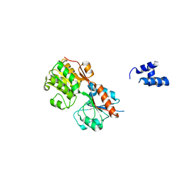
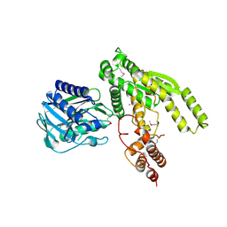
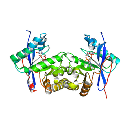
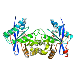
3D3A
DownloadVisualize
BU of 3d3a by Molmil
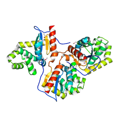
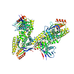
Crystal structure of a beta-galactosidase from Bacteroides thetaiotaomicron
Descriptor: Beta-galactosidase
Authors:Palani, K, Kumaran, D, Burley, S.K, Swaminathan, S, New York SGX Research Center for Structural Genomics (NYSGXRC)
Deposit date:2008-05-09
Release date:2008-05-27
Last modified:2021-02-03
Method:X-RAY DIFFRACTION (2.15 Å)
Cite:Crystal structure of a beta-galactosidase from Bacteroides thetaiotaomicron.
To be Published
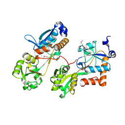
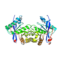
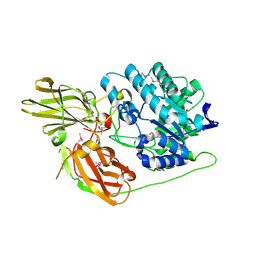
3FJY
DownloadVisualize
BU of 3fjy by Molmil
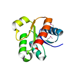
Crystal structure of a probable MutT1 protein from Bifidobacterium adolescentis
Descriptor: GLYCEROL, Probable MutT1 protein
Authors:Palani, K, Burley, S.K, Swaminathan, S, New York SGX Research Center for Structural Genomics (NYSGXRC)
Deposit date:2008-12-15
Release date:2009-01-13
Last modified:2024-10-30
Method:X-RAY DIFFRACTION (2.15 Å)
Cite:Crystal structure of a probable MutT1 protein from Bifidobacterium adolescentis
To be Published
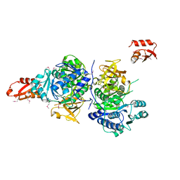
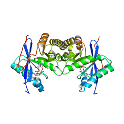
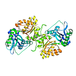
3F6A
DownloadVisualize
BU of 3f6a by Molmil
Crystal structure of a hydrolase, NUDIX family from Clostridium perfringens
Descriptor: Hydrolase, NUDIX family
Authors:Palani, K, Burley, S.K, Swaminathan, S, New York SGX Research Center for Structural Genomics (NYSGXRC)
Deposit date:2008-11-05
Release date:2008-11-25
Last modified:2023-12-27
Method:X-RAY DIFFRACTION (2.02 Å)
Cite:Crystal structure of a hydrolase, NUDIX family from Clostridium perfringens
To be Published
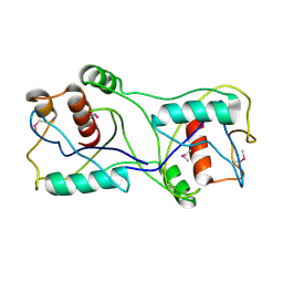
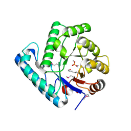
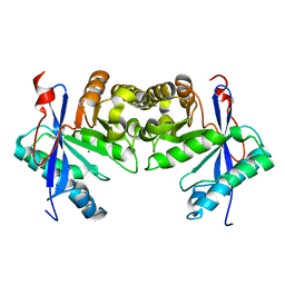
3H5T
DownloadVisualize
BU of 3h5t by Molmil
Crystal structure of a transcriptional regulator, Lacl family protein from Corynebacterium glutamicum
Descriptor: Transcriptional regulator, LacI family
Authors:Palani, K, Burley, S.K, Swaminathan, S, New York SGX Research Center for Structural Genomics (NYSGXRC)
Deposit date:2009-04-22
Release date:2009-05-05
Last modified:2024-02-21
Method:X-RAY DIFFRACTION (2.53 Å)
Cite:Crystal structure of a transcriptional regulator, Lacl family protein from Corynebacterium glutamicum
To be Published
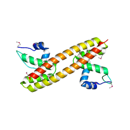
3GPV
DownloadVisualize
BU of 3gpv by Molmil
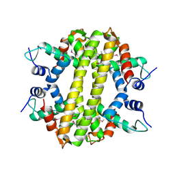
Crystal structure of a transcriptional regulator, MerR family from Bacillus thuringiensis
Descriptor: Transcriptional regulator, MerR family
Authors:Palani, K, Kumaran, D, Burley, S.K, Swaminathan, S, New York SGX Research Center for Structural Genomics (NYSGXRC)
Deposit date:2009-03-23
Release date:2009-04-14
Last modified:2021-02-10
Method:X-RAY DIFFRACTION (1.9 Å)
Cite:Crystal structure of a transcriptional regulator, MerR family from Bacillus thuringiensis
To be Published
3GYB
DownloadVisualize
BU of 3gyb by Molmil
Crystal structure of a LacI-family transcriptional regulatory protein from Corynebacterium glutamicum
Descriptor: MAGNESIUM ION, Transcriptional regulators (LACI-FAMILY TRANSCRIPTIONAL REGULATORY PROTEIN)
Authors:Palani, K, Burley, S.K, Swaminathan, S, New York SGX Research Center for Structural Genomics (NYSGXRC)
Deposit date:2009-04-03
Release date:2009-04-14
Last modified:2021-02-10
Method:X-RAY DIFFRACTION (1.6 Å)
Cite:Crystal structure of a LacI-family transcriptional regulatory protein from Corynebacterium glutamicum
To be Published
3GRC
DownloadVisualize
BU of 3grc by Molmil
Crystal structure of a sensor protein from Polaromonas sp. JS666
Descriptor: Sensor protein, Kinase
Authors:Palani, K, Kumaran, D, Burley, S.K, Swaminathan, S, New York SGX Research Center for Structural Genomics (NYSGXRC)
Deposit date:2009-03-25
Release date:2009-04-14
Last modified:2021-02-10
Method:X-RAY DIFFRACTION (2.21 Å)
Cite:Crystal structure of a sensor protein from Polaromonas sp. JS666
To be Published
3HH0
DownloadVisualize
BU of 3hh0 by Molmil
Crystal structure of a transcriptional regulator, MerR family from Bacillus cereus
Descriptor: Transcriptional regulator, MerR family
Authors:Palani, K, Zhang, Z, Burley, S.K, Swaminathan, S, New York SGX Research Center for Structural Genomics, New York SGX Research Center for Structural Genomics (NYSGXRC)
Deposit date:2009-05-14
Release date:2009-05-26
Last modified:2024-10-30
Method:X-RAY DIFFRACTION (2.67 Å)
Cite:Crystal structure of a transcriptional regulator, MerR family from Bacillus cereus
To be Published
3HD5
DownloadVisualize
BU of 3hd5 by Molmil
Crystal structure of a thiol:disulfide interchange protein dsbA from Bordetella parapertussis
Descriptor: Thiol:disulfide interchange protein dsbA
Authors:Palani, K, Burley, S.K, Swaminathan, S, New York SGX Research Center for Structural Genomics (NYSGXRC)
Deposit date:2009-05-06
Release date:2009-05-19
Last modified:2021-02-10
Method:X-RAY DIFFRACTION (2.35 Å)
Cite:Crystal structure of a thiol:disulfide interchange protein dsbA from Bordetella parapertussis
To be Published
3ID9
DownloadVisualize
BU of 3id9 by Molmil
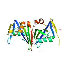
Crystal structure of a MutT/NUDIX family protein from Bacillus thuringiensis
Descriptor: CHLORIDE ION, MutT/NUDIX family protein, SULFATE ION
Authors:Palani, K, Burley, S.K, Swaminathan, S, New York SGX Research Center for Structural Genomics (NYSGXRC)
Deposit date:2009-07-20
Release date:2009-09-08
Last modified:2021-02-10
Method:X-RAY DIFFRACTION (2.55 Å)
Cite:Crystal structure of a MutT/NUDIX family protein from Bacillus thuringiensis
To be Published
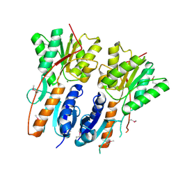
3ILV
DownloadVisualize
BU of 3ilv by Molmil
Crystal structure of a glutamine-dependent NAD(+) synthetase from Cytophaga hutchinsonii
Descriptor: Glutamine-dependent NAD(+) synthetase
Authors:Palani, K, Burley, S.K, Swaminathan, S, New York SGX Research Center for Structural Genomics (NYSGXRC)
Deposit date:2009-08-07
Release date:2009-08-18
Last modified:2021-02-10
Method:X-RAY DIFFRACTION (1.79 Å)
Cite:Crystal structure of a glutamine-dependent NAD(+) synthetase from Cytophaga hutchinsonii
To be Published
3HV1
DownloadVisualize
BU of 3hv1 by Molmil
Crystal structure of a polar amino acid ABC uptake transporter substrate binding protein from Streptococcus thermophilus
Descriptor: Polar amino acid ABC uptake transporter substrate binding protein
Authors:Palani, K, Burley, S.K, Swaminathan, S, New York SGX Research Center for Structural Genomics (NYSGXRC)
Deposit date:2009-06-15
Release date:2009-06-23
Last modified:2021-02-10
Method:X-RAY DIFFRACTION (1.9 Å)
Cite:Crystal structure of a polar amino acid ABC uptake transporter substrate binding protein from Streptococcus thermophilus
To be Published
3G7S
DownloadVisualize
BU of 3g7s by Molmil
Crystal structure of a long-chain-fatty-acid-CoA ligase (FadD1) from Archaeoglobus fulgidus
Descriptor: Long-chain-fatty-acid--CoA ligase (FadD-1)
Authors:Palani, K, Kumaran, D, Burley, S.K, Swaminathan, S, New York SGX Research Center for Structural Genomics (NYSGXRC)
Deposit date:2009-02-10
Release date:2009-03-03
Last modified:2024-10-16
Method:X-RAY DIFFRACTION (2.15 Å)
Cite:Crystal structure of a long-chain-fatty-acid-CoA ligase (FadD1) from Archaeoglobus fulgidus
To be Published
2J6V
DownloadVisualize
BU of 2j6v by Molmil
Crystal structure of the DNA repair enzyme UV Damage Endonuclease
Descriptor: MANGANESE (II) ION, PHOSPHATE ION, UV ENDONUCLEASE
Authors:Paspaleva, K, Thomassen, E.A.J, Pannu, N.S, Goossen, N, Abrahams, J.P.
Deposit date:2006-10-04
Release date:2007-10-16
Last modified:2024-10-23
Method:X-RAY DIFFRACTION (1.55 Å)
Cite:Crystal Structure of the DNA Repair Enzyme Ultraviolet Damage Endonuclease.
Structure, 15, 2007
3K85
DownloadVisualize
BU of 3k85 by Molmil
Crystal structure of a D-glycero-D-manno-heptose 1-phosphate kinase from Bacteriodes thetaiotaomicron
Descriptor: D-glycero-D-manno-heptose 1-phosphate kinase
Authors:Palani, K, Kumaran, D, Burley, S.K, Swaminathan, S, New York SGX Research Center for Structural Genomics (NYSGXRC)
Deposit date:2009-10-13
Release date:2009-12-08
Last modified:2021-02-10
Method:X-RAY DIFFRACTION (2.28 Å)
Cite:Crystal structure of a D-glycero-D-manno-heptose 1-phosphate kinase from Bacteriodes thetaiotaomicron
To be Published
3K17
DownloadVisualize
BU of 3k17 by Molmil
Crystal structure of a Lin0012 protein from Listeria innocua
Descriptor: Lin0012 protein, TRIETHYLENE GLYCOL
Authors:Palani, K, Burley, S.K, Swaminathan, S, New York SGX Research Center for Structural Genomics (NYSGXRC)
Deposit date:2009-09-25
Release date:2009-10-06
Last modified:2021-02-10
Method:X-RAY DIFFRACTION (2.1 Å)
Cite:Crystal structure of a Lin0012 protein from Listeria innocua
To be Published
6PWC
DownloadVisualize
BU of 6pwc by Molmil
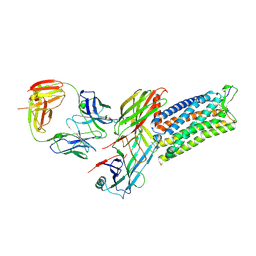
A complex structure of arrestin-2 bound to neurotensin receptor 1
Descriptor: Beta-arrestin-1, Fab30 heavy chain, Fab30 light chain, ...
Authors:Yin, W, Li, Z, Jin, M, Yin, Y.-L, de Waal, P.W, Pal, K, Gao, X, He, Y, Gao, J, Wang, X, Zhang, Y, Zhou, H, Melcher, K, Jiang, Y, Cong, Y, Zhou, X.E, Yu, X, Xu, H.E.
Deposit date:2019-07-22
Release date:2019-12-04
Last modified:2024-10-30
Method:ELECTRON MICROSCOPY (4.9 Å)
Cite:A complex structure of arrestin-2 bound to a G protein-coupled receptor.
Cell Res., 29, 2019
5W0P
DownloadVisualize
BU of 5w0p by Molmil
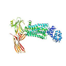
Crystal structure of rhodopsin bound to visual arrestin determined by X-ray free electron laser
Descriptor: 2-acetamido-2-deoxy-beta-D-glucopyranose-(1-4)-2-acetamido-2-deoxy-beta-D-glucopyranose, Endolysin,Rhodopsin,S-arrestin
Authors:Zhou, X.E, He, Y, de Waal, P.W, Gao, X, Kang, Y, Van Eps, N, Yin, Y, Pal, K, Goswami, D, White, T.A, Barty, A, Latorraca, N.R, Chapman, H.N, Hubbell, W.L, Dror, R.O, Stevens, R.C, Cherezov, V, Gurevich, V.V, Griffin, P.R, Ernst, O.P, Melcher, K, Xu, H.E.
Deposit date:2017-05-31
Release date:2017-08-09
Last modified:2024-10-30
Method:X-RAY DIFFRACTION (3.013 Å)
Cite:Identification of Phosphorylation Codes for Arrestin Recruitment by G Protein-Coupled Receptors.
Cell, 170, 2017
4ZWJ
DownloadVisualize
BU of 4zwj by Molmil
Crystal structure of rhodopsin bound to arrestin by femtosecond X-ray laser
Descriptor: Chimera protein of human Rhodopsin, mouse S-arrestin, and T4 Endolysin
Authors:Kang, Y, Zhou, X.E, Gao, X, He, Y, Liu, W, Ishchenko, A, Barty, A, White, T.A, Yefanov, O, Han, G.W, Xu, Q, de Waal, P.W, Ke, J, Tan, M.H.E, Zhang, C, Moeller, A, West, G.M, Pascal, B, Eps, N.V, Caro, L.N, Vishnivetskiy, S.A, Lee, R.J, Suino-Powell, K.M, Gu, X, Pal, K, Ma, J, Zhi, X, Boutet, S, Williams, G.J, Messerschmidt, M, Gati, C, Zatsepin, N.A, Wang, D, James, D, Basu, S, Roy-Chowdhury, S, Conrad, C, Coe, J, Liu, H, Lisova, S, Kupitz, C, Grotjohann, I, Fromme, R, Jiang, Y, Tan, M, Yang, H, Li, J, Wang, M, Zheng, Z, Li, D, Howe, N, Zhao, Y, Standfuss, J, Diederichs, K, Dong, Y, Potter, C.S, Carragher, B, Caffrey, M, Jiang, H, Chapman, H.N, Spence, J.C.H, Fromme, P, Weierstall, U, Ernst, O.P, Katritch, V, Gurevich, V.V, Griffin, P.R, Hubbell, W.L, Stevens, R.C, Cherezov, V, Melcher, K, Xu, H.E, GPCR Network (GPCR)
Deposit date:2015-05-19
Release date:2015-07-29
Last modified:2023-09-27
Method:X-RAY DIFFRACTION (3.302 Å)
Cite:Crystal structure of rhodopsin bound to arrestin by femtosecond X-ray laser.
Nature, 523, 2015
3TVD
DownloadVisualize
BU of 3tvd by Molmil
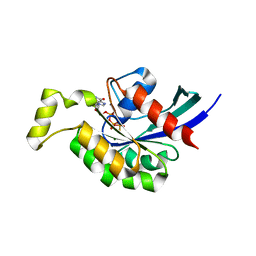
Crystal Structure of Mouse RhoA-GTP complex
Descriptor: 5'-GUANOSINE-DIPHOSPHATE-MONOTHIOPHOSPHATE, MAGNESIUM ION, Transforming protein RhoA
Authors:Swaminathan, K, Pal, K, Jobichen, C.
Deposit date:2011-09-20
Release date:2012-10-10
Last modified:2023-11-01
Method:X-RAY DIFFRACTION (2.989 Å)
Cite:Crystal structure of mouse RhoA:GTPgammaS complex in a centered lattice.
J.Struct.Funct.Genom., 13, 2012
6IH1
DownloadVisualize
BU of 6ih1 by Molmil
Crystal structure of a standalone versatile EAL protein from Vibrio cholerae O395 - c-di-GMP bound form
Descriptor: 9,9'-[(2R,3R,3aS,5S,7aR,9R,10R,10aS,12S,14aR)-3,5,10,12-tetrahydroxy-5,12-dioxidooctahydro-2H,7H-difuro[3,2-d:3',2'-j][1,3,7,9,2,8]tetraoxadiphosphacyclododecine-2,9-diyl]bis(2-amino-1,9-dihydro-6H-purin-6-one), CALCIUM ION, cyclic di nucleotide phoshodiesterase
Authors:Yadav, M, Pal, K, Sen, U.
Deposit date:2018-09-28
Release date:2019-10-02
Last modified:2024-03-27
Method:X-RAY DIFFRACTION (1.95 Å)
Cite:Structures of c-di-GMP/cGAMP degrading phosphodiesterase VcEAL: identification of a novel conformational switch and its implication.
Biochem.J., 476, 2019
6IJ2
DownloadVisualize
BU of 6ij2 by Molmil
Crystal structure of a standalone versatile EAL protein from Vibrio cholerae O395 - 5'-pGpG bound form
Descriptor: CALCIUM ION, EAL domain protein, RNA (5'-R(P*GP*G)-3')
Authors:Yadav, M, Pal, K, Sen, U.
Deposit date:2018-10-08
Release date:2019-10-09
Last modified:2023-11-22
Method:X-RAY DIFFRACTION (1.7 Å)
Cite:Structures of c-di-GMP/cGAMP degrading phosphodiesterase VcEAL: identification of a novel conformational switch and its implication.
Biochem.J., 476, 2019
6K6T
DownloadVisualize
BU of 6k6t by Molmil
Crystal structure of a standalone versatile EAL protein from Vibrio cholerae O395 - c-di-IMP bound form
Descriptor: 9-[(1R,6R,8R,9S,10R,15S,17R,18S)-3,9,12,18-tetrakis(oxidanyl)-3,12-bis(oxidanylidene)-17-(6-oxidanylidene-3H-purin-9-yl)-2,4,7,11,13,16-hexaoxa-3$l^{5},12$l^{5}-diphosphatricyclo[13.3.0.0^{6,10}]octadecan-8-yl]-3H-purin-6-one, CALCIUM ION, EAL domain protein
Authors:Yadav, M, Pal, K, Sen, U.
Deposit date:2019-06-04
Release date:2020-06-17
Last modified:2023-11-22
Method:X-RAY DIFFRACTION (2.2 Å)
Cite:Crystal structure of a standalone versatile EAL protein from Vibrio cholerae O395 - c-di-IMP bound form
To Be Published
6IFQ
DownloadVisualize
BU of 6ifq by Molmil
Crystal structure of a standalone versatile EAL protein from Vibrio cholerae O395 - Apo form
Descriptor: CALCIUM ION, cyclic di nucleotide phoshodiesterase
Authors:Yadav, M, Pal, K, Sen, U.
Deposit date:2018-09-20
Release date:2019-10-02
Last modified:2024-03-27
Method:X-RAY DIFFRACTION (1.95 Å)
Cite:Structures of c-di-GMP/cGAMP degrading phosphodiesterase VcEAL: identification of a novel conformational switch and its implication.
Biochem.J., 476, 2019
6IH7
DownloadVisualize
BU of 6ih7 by Molmil
Crystal structure of a standalone versatile EAL protein from Vibrio cholerae O395 - 3',3'-cGAMP bound form
Descriptor: 2-amino-9-[(2R,3R,3aS,5R,7aR,9R,10R,10aS,12R,14aR)-9-(6-amino-9H-purin-9-yl)-3,5,10,12-tetrahydroxy-5,12-dioxidooctahydro-2H,7H-difuro[3,2-d:3',2'-j][1,3,7,9,2,8]tetraoxadiphosphacyclododecin-2-yl]-1,9-dihydro-6H-purin-6-one, CALCIUM ION, cyclic di nucleotide phoshodiesterase
Authors:Yadav, M, Pal, K, Sen, U.
Deposit date:2018-09-28
Release date:2019-10-02
Last modified:2023-11-22
Method:X-RAY DIFFRACTION (2.25 Å)
Cite:Structures of c-di-GMP/cGAMP degrading phosphodiesterase VcEAL: identification of a novel conformational switch and its implication.
Biochem.J., 476, 2019

226707

PDB entries from 2024-10-30

PDB statisticsPDBj update infoContact PDBjnumon