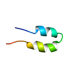
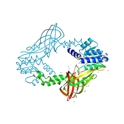
1DZ7
 
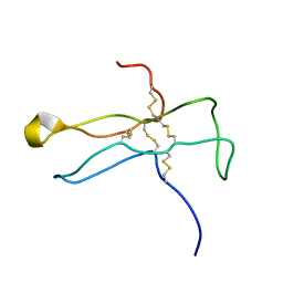 | | Solution structure of the a-subunit of human chorionic gonadotropin [modeled without carbohydrate residues] | | 分子名称: | CHORIONIC GONADOTROPIN | | 著者 | Erbel, P.J.A, Karimi-Nejad, Y, De Beer, T, Boelens, R, Kamerling, J.P, Vliegenthart, J.F.G. | | 登録日 | 2000-02-18 | | 公開日 | 2000-02-29 | | 最終更新日 | 2019-10-09 | | 実験手法 | SOLUTION NMR | | 主引用文献 | Solution structure of the alpha-subunit of human chorionic gonadotropin.
Eur.J.Biochem., 260, 1999
|
|
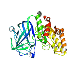
3L5H
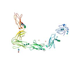 | | Crystal structure of the full ectodomain of human gp130: New insights into the molecular assembly of receptor complexes | | 分子名称: | 2-acetamido-2-deoxy-beta-D-glucopyranose-(1-4)-2-acetamido-2-deoxy-beta-D-glucopyranose, Interleukin-6 receptor subunit beta, SULFATE ION, ... | | 著者 | Xu, Y, Garrett, T.P.J, Zhang, J.G. | | 登録日 | 2009-12-21 | | 公開日 | 2010-05-19 | | 最終更新日 | 2020-07-29 | | 実験手法 | X-RAY DIFFRACTION (3.6 Å) | | 主引用文献 | Crystal structure of the entire ectodomain of gp130: insights into the molecular assembly of the tall cytokine receptor complexes.
J.Biol.Chem., 285, 2010
|
|
1E3T
 | | Solution Structure of the NADP(H) binding Component (dIII) of Proton-Translocating Transhydrogenase from Rhodospirillum rubrum | | 分子名称: | NADP NICOTINAMIDE-ADENINE-DINUCLEOTIDE PHOSPHATE, NICOTINAMIDE NUCLEOTIDE TRANSHYDROGENASE (SUBUNIT BETA) | | 著者 | Jeeves, M, Smith, K.J, Quirk, P.G, Cotton, N.P.J, Jackson, J.B. | | 登録日 | 2000-06-22 | | 公開日 | 2000-10-03 | | 最終更新日 | 2024-05-15 | | 実験手法 | SOLUTION NMR | | 主引用文献 | Solution Structure of the Nadp(H)-Binding Component (Diii) of Proton-Translocating Transhydrogenase from Rhodospirillum Rubrum
Biochim.Biophys.Acta, 1459, 2000
|
|
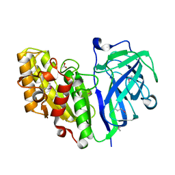
2F4W
 | | Human ubiquitin-conjugating enzyme E2 J2 | | 分子名称: | ubiquitin-conjugating enzyme E2, J2 | | 著者 | Walker, J.R, Avvakumov, G.V, Xue, S, Finerty Jr, P.J, Newman, E.M, Mackenzie, F, Weigelt, J, Sundstrom, M, Arrowsmith, C, Edwards, A, Bochkarev, A, Dhe-Paganon, S, Structural Genomics Consortium (SGC) | | 登録日 | 2005-11-24 | | 公開日 | 2005-12-27 | | 最終更新日 | 2023-08-23 | | 実験手法 | X-RAY DIFFRACTION (2 Å) | | 主引用文献 | A human ubiquitin conjugating enzyme (E2)-HECT E3 ligase structure-function screen.
Mol Cell Proteomics, 11, 2012
|
|
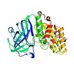
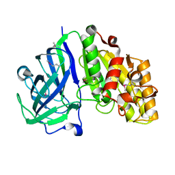
2OXE
 | | Structure of the Human Pancreatic Lipase-related Protein 2 | | 分子名称: | CALCIUM ION, CHLORIDE ION, Pancreatic lipase-related protein 2, ... | | 著者 | Walker, J.R, Davis, T, Seitova, A, Finerty Jr, P.J, Butler-Cole, C, Kozieradzki, I, Weigelt, J, Sundstrom, M, Arrowsmith, C.H, Edwards, A.M, Bochkarev, A, Dhe-Paganon, S, Structural Genomics Consortium (SGC) | | 登録日 | 2007-02-20 | | 公開日 | 2007-03-27 | | 最終更新日 | 2023-08-30 | | 実験手法 | X-RAY DIFFRACTION (2.8 Å) | | 主引用文献 | Structure of human pancreatic lipase-related protein 2 with the lid in an open conformation.
Biochemistry, 47, 2008
|
|
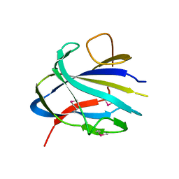
1E9J
 | | SOLUTION STRUCTURE OF THE A-SUBUNIT OF HUMAN CHORIONIC GONADOTROPIN [INCLUDING A SINGLE GLCNAC RESIDUE AT ASN52 AND ASN78] | | 分子名称: | 2-acetamido-2-deoxy-beta-D-glucopyranose, CHORIONIC GONADOTROPIN | | 著者 | Erbel, P.J.A, Karimi-Nejad, Y, van Kuik, J.A, Boelens, R, Kamerling, J.P, Vliegenthart, J.F.G. | | 登録日 | 2000-10-18 | | 公開日 | 2002-07-25 | | 最終更新日 | 2020-07-29 | | 実験手法 | SOLUTION NMR | | 主引用文献 | Effects of the N-linked glycans on the 3D structure of the free alpha-subunit of human chorionic gonadotropin.
Biochemistry, 39, 2000
|
|
2M5N
 | | Atomic-resolution structure of a cross-beta protofilament | | 分子名称: | Transthyretin | | 著者 | Fitzpatrick, A.W.P, Debelouchina, G.T, Bayro, M.J, Clare, D.K, Caporini, M.A, Bajaj, V.S, Jaroniec, C.P, Wang, L, Ladizhansky, V, Muller, S, MacPhee, C.E, Waudby, C.A, Mott, H.R, de Simone, A, Knowles, T.P.J, Saibil, H.R, Vendruscolo, M, Orlova, E.V, Griffin, R.G, Dobson, C.M. | | 登録日 | 2013-02-27 | | 公開日 | 2013-07-17 | | 最終更新日 | 2024-05-15 | | 実験手法 | SOLID-STATE NMR | | 主引用文献 | Atomic structure and hierarchical assembly of a cross-{beta} amyloid fibril.
Proc.Natl.Acad.Sci.USA, 110, 2013
|
|
7SPN
 | | Crystal structure of IS11, a thermophilic esterase | | 分子名称: | IS11 | | 著者 | Stogios, P.J, Evdokimova, E, Khusnutdinova, A, Yakunin, A.F, Savchenko, A. | | 登録日 | 2021-11-02 | | 公開日 | 2022-08-24 | | 最終更新日 | 2024-04-03 | | 実験手法 | X-RAY DIFFRACTION (2.92 Å) | | 主引用文献 | Crystal structure of IS11, a thermophilic esterase
To Be Published
|
|
2BN5
 | | P-Element Somatic Inhibitor Protein Complex with U1-70k proline-rich peptide | | 分子名称: | PSI, U1 SMALL NUCLEAR RIBONUCLEOPROTEIN 70 KDA | | 著者 | Ignjatovic, T, Yang, J.-C, Butler, P.J.G, Neuhaus, D, Nagai, K. | | 登録日 | 2005-03-21 | | 公開日 | 2005-07-06 | | 最終更新日 | 2024-05-15 | | 実験手法 | SOLUTION NMR | | 主引用文献 | Structural Basis of the Interaction between P-Element Somatic Inhibitor and U1-70K Essential for the Alternative Splicing of P-Element Transposase.
J.Mol.Biol., 351, 2005
|
|
2M5K
 | | Atomic-resolution structure of a doublet cross-beta amyloid fibril | | 分子名称: | Transthyretin | | 著者 | Fitzpatrick, A.W.P, Debelouchina, G.T, Bayro, M.J, Clare, D.K, Caporini, M.A, Bajaj, V.S, Jaroniec, C.P, Wang, L, Ladizhansky, V, Muller, S, MacPhee, C.E, Waudby, C.A, Mott, H.R, de Simone, A, Knowles, T.P.J, Saibil, H.R, Vendruscolo, M, Orlova, E.V, Griffin, R.G, Dobson, C.M. | | 登録日 | 2013-02-27 | | 公開日 | 2013-12-04 | | 最終更新日 | 2024-05-15 | | 実験手法 | ELECTRON MICROSCOPY (12.7 Å), SOLID-STATE NMR | | 主引用文献 | Atomic structure and hierarchical assembly of a cross-beta amyloid fibril.
Proc.Natl.Acad.Sci.USA, 110, 2013
|
|
2M5M
 | | Atomic-resolution structure of a triplet cross-beta amyloid fibril | | 分子名称: | Transthyretin | | 著者 | Fitzpatrick, A.W.P, Debelouchina, G.T, Bayro, M.J, Clare, D.K, Caporini, M.A, Bajaj, V.S, Jaroniec, C.P, Wang, L, Ladizhansky, V, Muller, S, MacPhee, C.E, Waudby, C.A, Mott, H.R, de Simone, A, Knowles, T.P.J, Saibil, H.R, Vendruscolo, M, Orlova, E.V, Griffin, R.G, Dobson, C.M. | | 登録日 | 2013-02-27 | | 公開日 | 2013-12-04 | | 最終更新日 | 2024-05-15 | | 実験手法 | ELECTRON MICROSCOPY (12.2 Å), SOLID-STATE NMR | | 主引用文献 | Atomic structure and hierarchical assembly of a cross-beta amyloid fibril.
Proc.Natl.Acad.Sci.USA, 110, 2013
|
|
2BN6
 | | P-Element Somatic Inhibitor Protein | | 分子名称: | PSI | | 著者 | Ignjatovic, T, Yang, J.C, Butler, P.J.G, Neuhaus, D, Nagai, K. | | 登録日 | 2005-03-21 | | 公開日 | 2005-07-06 | | 最終更新日 | 2024-05-15 | | 実験手法 | SOLUTION NMR | | 主引用文献 | Structural Basis of the Interaction between P-Element Somatic Inhibitor and U1-70K Essential for the Alternative Splicing of P-Element Transposase.
J.Mol.Biol., 351, 2005
|
|
7TOK
 | | Crystal structure of the CBM domain of carbohydrate esterase FjoAcXE | | 分子名称: | Acetylxylan esterase I | | 著者 | Stogios, P.J, Skarina, T, Di Leo, R, Jurak, E, Master, E. | | 登録日 | 2022-01-24 | | 公開日 | 2022-04-13 | | 最終更新日 | 2022-11-02 | | 実験手法 | X-RAY DIFFRACTION (2.45 Å) | | 主引用文献 | Elucidating Sequence and Structural Determinants of Carbohydrate Esterases for Complete Deacetylation of Substituted Xylans.
Molecules, 27, 2022
|
|
7TOG
 | | Crystal structure of carbohydrate esterase PbeAcXE, apoenzyme | | 分子名称: | SGNH hydrolase | | 著者 | Stogios, P.J, Skarina, T, Di Leo, R, Jurak, E, Master, E. | | 登録日 | 2022-01-24 | | 公開日 | 2022-04-13 | | 最終更新日 | 2022-11-02 | | 実験手法 | X-RAY DIFFRACTION (1.35 Å) | | 主引用文献 | Elucidating Sequence and Structural Determinants of Carbohydrate Esterases for Complete Deacetylation of Substituted Xylans.
Molecules, 27, 2022
|
|
7TOI
 | | Crystal structure of carbohydrate esterase PbeAcXE, in complex with acetate | | 分子名称: | ACETATE ION, SGNH hydrolase | | 著者 | Stogios, P.J, Skarina, T, Di Leo, R, Jurak, E, Master, E. | | 登録日 | 2022-01-24 | | 公開日 | 2022-04-13 | | 最終更新日 | 2022-11-02 | | 実験手法 | X-RAY DIFFRACTION (1.13 Å) | | 主引用文献 | Elucidating Sequence and Structural Determinants of Carbohydrate Esterases for Complete Deacetylation of Substituted Xylans.
Molecules, 27, 2022
|
|
7TOJ
 | | Crystal structure of carbohydrate esterase CspAcXE, apoenzyme | | 分子名称: | CHLORIDE ION, SGNH/GDSL hydrolase family protein | | 著者 | Stogios, P.J, Skarina, T, Di Leo, R, Jurak, E, Master, E. | | 登録日 | 2022-01-24 | | 公開日 | 2022-04-13 | | 最終更新日 | 2022-11-02 | | 実験手法 | X-RAY DIFFRACTION (1.3 Å) | | 主引用文献 | Elucidating Sequence and Structural Determinants of Carbohydrate Esterases for Complete Deacetylation of Substituted Xylans.
Molecules, 27, 2022
|
|
7TOH
 | | Crystal structure of carbohydrate esterase PbeAcXE, in complex with MeGlcpA-Xylp | | 分子名称: | 4-O-methyl-alpha-D-glucopyranuronic acid-(1-2)-beta-D-xylopyranose, SGNH hydrolase | | 著者 | Stogios, P.J, Skarina, T, Di Leo, R, Jurak, E, Master, E. | | 登録日 | 2022-01-24 | | 公開日 | 2022-04-13 | | 最終更新日 | 2022-11-02 | | 実験手法 | X-RAY DIFFRACTION (1.26 Å) | | 主引用文献 | Elucidating Sequence and Structural Determinants of Carbohydrate Esterases for Complete Deacetylation of Substituted Xylans.
Molecules, 27, 2022
|
|
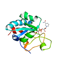
4QQK
 | | Human HMT1 hnRNP methyltransferase-like protein 6 (S. cerevisiae) with GMS | | 分子名称: | (5S)-5-{[(2R,3S,4R,5R)-5-(6-amino-9H-purin-9-yl)-3,4-dihydroxytetrahydrofuran-2-yl]methyl}-N~6~-carbamimidoyl-L-lysine, GLYCEROL, Protein arginine N-methyltransferase 6, ... | | 著者 | Dong, A, Zeng, H, He, H, Wernimont, A, Bountra, C, Arrowsmith, C.H, Edwards, A.M, Brown, P.J, Min, J, Luo, M, Wu, H, Structural Genomics Consortium (SGC) | | 登録日 | 2014-06-27 | | 公開日 | 2014-07-16 | | 最終更新日 | 2023-09-20 | | 実験手法 | X-RAY DIFFRACTION (1.88 Å) | | 主引用文献 | Structural basis of arginine asymmetrical dimethylation by PRMT6.
Biochem. J., 473, 2016
|
|
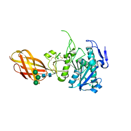
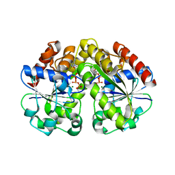
1S63
 | | Human protein farnesyltransferase complexed with L-778,123 and FPP | | 分子名称: | 4-[(5-{[4-(3-CHLOROPHENYL)-3-OXOPIPERAZIN-1-YL]METHYL}-1H-IMIDAZOL-1-YL)METHYL]BENZONITRILE, FARNESYL DIPHOSPHATE, Protein farnesyltransferase beta subunit, ... | | 著者 | Long, S.B, Casey, P.J, Beese, L.S. | | 登録日 | 2004-01-22 | | 公開日 | 2004-07-27 | | 最終更新日 | 2023-08-23 | | 実験手法 | X-RAY DIFFRACTION (1.9 Å) | | 主引用文献 | Crystallographic Analysis Reveals that Anticancer Clinical Candidate L-778,123 Inhibits Protein Farnesyltransferase and Geranylgeranyltransferase-I by Different Binding Modes.
Biochemistry, 43, 2004
|
|
2BEJ
 | |
2ODY
 | | Thrombin-bound boophilin displays a functional and accessible reactive-site loop | | 分子名称: | 2-acetamido-2-deoxy-beta-D-glucopyranose, Boophilin, PHOSPHATE ION, ... | | 著者 | Macedo-Ribeiro, S, Fuentes-Prior, P, Pereira, P.J.B. | | 登録日 | 2006-12-27 | | 公開日 | 2008-01-22 | | 最終更新日 | 2023-08-30 | | 実験手法 | X-RAY DIFFRACTION (2.35 Å) | | 主引用文献 | Isolation, cloning and structural characterisation of boophilin, a multifunctional kunitz-type proteinase inhibitor from the cattle tick.
Plos One, 3, 2008
|
|
2BEK
 | |
1GJT
 | | Solution structure of the Albumin binding domain of Streptococcal Protein G | | 分子名称: | IMMUNOGLOBULIN G BINDING PROTEIN G | | 著者 | Johansson, M.U, Frick, I.M, Nilsson, H, Kraulis, P.J, Hober, S, Jonasson, P, Nygren, A.P, Uhlen, M, Bjorck, L, Drakenberg, T, Forsen, S, Wikstrom, M. | | 登録日 | 2001-08-02 | | 公開日 | 2001-08-09 | | 最終更新日 | 2024-05-15 | | 実験手法 | SOLUTION NMR | | 主引用文献 | Structure, Specificity, and Mode of Interaction for Bacterial Albumin-Binding Modules
J.Biol.Chem., 277, 2002
|
|
1GJS
 | | Solution structure of the Albumin binding domain of Streptococcal Protein G | | 分子名称: | IMMUNOGLOBULIN G BINDING PROTEIN G | | 著者 | Johansson, M.U, Frick, I.M, Nilsson, H, Kraulis, P.J, Hober, S, Jonasson, P, Nygren, A.P, Uhlen, M, Bjorck, L, Drakenberg, T, Forsen, S, Wikstrom, M. | | 登録日 | 2001-08-02 | | 公開日 | 2001-08-09 | | 最終更新日 | 2024-05-15 | | 実験手法 | SOLUTION NMR | | 主引用文献 | Structure, Specificity, and Mode of Interaction for Bacterial Albumin-Binding Modules
J.Biol.Chem., 277, 2002
|
|
1GKA
 | | The molecular basis of the coloration mechanism in lobster shell. beta-crustacyanin at 3.2 A resolution | | 分子名称: | 2-AMINO-2-HYDROXYMETHYL-PROPANE-1,3-DIOL, 4-(2-HYDROXYETHYL)-1-PIPERAZINE ETHANESULFONIC ACID, ASTAXANTHIN, ... | | 著者 | Cianci, M, Rizkallah, P.J, Olczak, A, Raftery, J, Chayen, N.E, Zagalsky, P.F, Helliwell, J.R. | | 登録日 | 2001-08-10 | | 公開日 | 2002-08-08 | | 最終更新日 | 2023-12-13 | | 実験手法 | X-RAY DIFFRACTION (3.23 Å) | | 主引用文献 | The Molecular Basis of the Coloration Mechanism in Lobster Shell: Beta -Crustacyanin at 3.2-A Resolution
Proc.Natl.Acad.Sci.USA, 99, 2002
|
|


