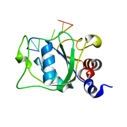
4AR5
 
 | | X-ray crystallographic structure of the oxidised form perdeuterated Pyrococcus furiosus rubredoxin in D2O at 295K (in quartz capillary) to 1.00 Angstrom resolution. | | 分子名称: | FE (III) ION, RUBREDOXIN | | 著者 | Cuypers, M.G, Mason, S.A, Blakeley, M.P, Mitchell, E.P, Haertlein, M, Forsyth, V.T. | | 登録日 | 2012-04-20 | | 公開日 | 2012-12-19 | | 最終更新日 | 2024-05-01 | | 実験手法 | X-RAY DIFFRACTION (1 Å) | | 主引用文献 | Near-Atomic Resolution Neutron Crystallography on Perdeuterated Pyrococcus Furiosus Rubredoxin: Implication of Hydronium Ions and Protonation Equilibria and Hydronium Ions in Redox Changes
Angew.Chem.Int.Ed.Engl., 52, 2013
|
|
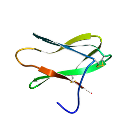
4A2L
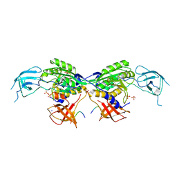
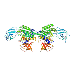
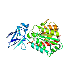
 | | Structure of the periplasmic domain of the heparin and heparan sulphate sensing hybrid two component system BT4663 in apo and ligand bound forms | | 分子名称: | 1,2-ETHANEDIOL, 2-(N-MORPHOLINO)-ETHANESULFONIC ACID, DI(HYDROXYETHYL)ETHER, ... | | 著者 | Lowe, E.C, Basle, A, Czjzek, M, Firbank, S.J, Bolam, D.N. | | 登録日 | 2011-09-27 | | 公開日 | 2012-05-02 | | 最終更新日 | 2024-05-08 | | 実験手法 | X-RAY DIFFRACTION (2.6 Å) | | 主引用文献 | A Scissor Blade-Like Closing Mechanism Implicated in Transmembrane Signaling in a Bacteroides Hybrid Two-Component System.
Proc.Natl.Acad.Sci.USA, 109, 2012
|
|
2N0B
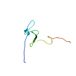
 | | NMR structure of Neuromedin C in aqueous solution | | 分子名称: | Neuromedin C (NMC) | | 著者 | Adrover, M, Sanchis, P, Vilanova, B, Pauwels, K, Martorell, G, Perez, J. | | 登録日 | 2015-03-05 | | 公開日 | 2015-10-14 | | 最終更新日 | 2024-05-15 | | 実験手法 | SOLUTION NMR | | 主引用文献 | Conformational ensembles of neuromedin C reveal a progressive coil-helix transition within a binding-induced folding mechanism.
RSC ADV, 5, 2015
|
|
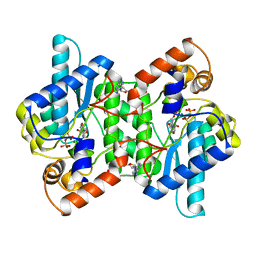
4AAP
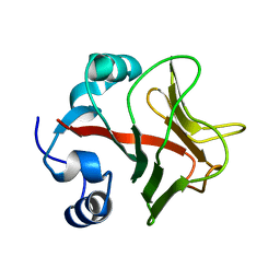
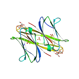
 | | Crystal structure of JMJD5 domain of human Lysine-specific demethylase 8 (KDM8) in complex with N-oxalylglycine (NOG) | | 分子名称: | LYSINE-SPECIFIC DEMETHYLASE 8, N-OXALYLGLYCINE, ZINC ION | | 著者 | Vollmar, M, Johansson, C, Krojer, T, Canning, P, Allerston, C, Gadhave, A, von Delft, F, Bountra, C, Arrowsmith, C.H, Weigelt, J, Edwards, A, Oppermann, U. | | 登録日 | 2011-12-05 | | 公開日 | 2012-02-29 | | 最終更新日 | 2024-05-08 | | 実験手法 | X-RAY DIFFRACTION (2.6 Å) | | 主引用文献 | Crystal Structure of Jmjd5 Domain of Human Lysine- Specific Demethylase 8 (Kdm8) in Complex with N- Oxalylglycine (Nog)
To be Published
|
|
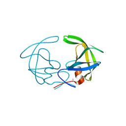
4ACJ
 | | Crystal structure of the TLDC domain of Oxidation resistance protein 2 from zebrafish | | 分子名称: | WU:FB25H12 PROTEIN, | | 著者 | Blaise, M, B Alsarraf, H.M.A, Wong, J.E.M.M, Midtgaard, S.R, Laroche, F, Schack, L, Spaink, H, Stougaard, J, Thirup, S. | | 登録日 | 2011-12-15 | | 公開日 | 2012-02-08 | | 最終更新日 | 2024-05-08 | | 実験手法 | X-RAY DIFFRACTION (0.97 Å) | | 主引用文献 | Crystal Structure of the Tldc Domain of Oxidation Resistance Protein 2 from Zebrafish.
Proteins, 80, 2012
|
|
8BVF
 | | MoeA2 from Corynebacterium glutamicum in complex with FtsZ-CTD | | 分子名称: | Cell division protein FtsZ, Molybdopterin molybdenumtransferase, SODIUM ION, ... | | 著者 | Martinez, M, Haouz, A, Wehenkel, A.M, Alzari, P.M. | | 登録日 | 2022-12-03 | | 公開日 | 2023-02-22 | | 最終更新日 | 2024-06-19 | | 実験手法 | X-RAY DIFFRACTION (2.68 Å) | | 主引用文献 | Eukaryotic-like gephyrin and cognate membrane receptor coordinate corynebacterial cell division and polar elongation.
Biorxiv, 2023
|
|
2MXW
 | |
8BVE
 | | MoeA2 from Corynebacterium glutamicum | | 分子名称: | CITRIC ACID, Molybdopterin molybdenumtransferase, SODIUM ION | | 著者 | Martinez, M, Haouz, A, Wehenkel, A.M, Alzari, P.M. | | 登録日 | 2022-12-03 | | 公開日 | 2023-02-22 | | 最終更新日 | 2024-06-19 | | 実験手法 | X-RAY DIFFRACTION (2.14 Å) | | 主引用文献 | Eukaryotic-like gephyrin and cognate membrane receptor coordinate corynebacterial cell division and polar elongation.
Biorxiv, 2023
|
|
4A37
 | | Metallo-carboxypeptidase from Pseudomonas Aeruginosa | | 分子名称: | METALLO-CARBOXYPEPTIDASE, ZINC ION | | 著者 | Otero, A, Rodriguez de la Vega, M, Tanco, S.M, Lorenzo, J, Aviles, F.X, Reverter, D. | | 登録日 | 2011-09-30 | | 公開日 | 2012-05-09 | | 最終更新日 | 2024-05-08 | | 実験手法 | X-RAY DIFFRACTION (1.6 Å) | | 主引用文献 | The Novel Structure of a Cytosolic M14 Metallocarboxypeptidase (Ccp) from Pseudomonas Aeruginosa: A Model for Mammalian Ccps.
Faseb J., 26, 2012
|
|
4AOC
 | | crystal structure of BC2L-A Lectin from Burkolderia cenocepacia in complex with methyl-heptoside | | 分子名称: | BC2L-A LECTIN, CALCIUM ION, SULFATE ION, ... | | 著者 | Marchetti, R, Malinovska, L, Lameignere, E, deCastro, C, Cioci, G, Kosma, P, Wimmerova, M, Molinaro, A, Imberty, A, Silipo, A. | | 登録日 | 2012-03-26 | | 公開日 | 2012-08-15 | | 最終更新日 | 2023-12-20 | | 実験手法 | X-RAY DIFFRACTION (2.7 Å) | | 主引用文献 | Burkholderia Cenocepacia Lectin a Binding to Heptoses from the Bacterial Lipopolysaccharide.
Glycobiology, 22, 2012
|
|
421D
 | |
8BQV
 | | Cryo-EM structure of alpha-synuclein singlet filament from Juvenile-onset synucleinopathy | | 分子名称: | Alpha-synuclein, Unknown protein | | 著者 | Yang, Y, Garringer, J.H, Shi, Y, Lovestam, S, Peak-Chew, S.Y, Zhang, X.J, Kotecha, A, Bacioglu, M, Koto, A, Takao, M, Spillantini, G.M, Ghetti, B, Vidal, R, Murzin, G.A, Scheres, H.W.S, Goedert, M. | | 登録日 | 2023-01-18 | | 公開日 | 2023-02-22 | | 最終更新日 | 2024-07-24 | | 実験手法 | ELECTRON MICROSCOPY (2 Å) | | 主引用文献 | New SNCA mutation and structures of alpha-synuclein filaments from juvenile-onset synucleinopathy.
Acta Neuropathol, 145, 2023
|
|
2MZ9
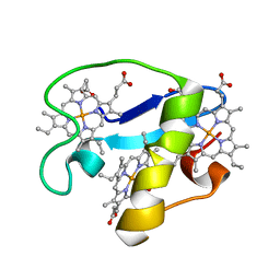 | | Solution structure of oxidized triheme cytochrome PpcA from Geobacter sulfurreducens | | 分子名称: | HEME C, PpcA | | 著者 | Morgado, L, Bruix, M, Pokkuluri, R, Salgueiro, C.A, Turner, D.L. | | 登録日 | 2015-02-08 | | 公開日 | 2016-02-10 | | 最終更新日 | 2024-05-01 | | 実験手法 | SOLUTION NMR | | 主引用文献 | Redox- and pH-linked conformational changes in triheme cytochrome PpcA from Geobacter sulfurreducens.
Biochem. J., 474, 2017
|
|
436D
 | |
2N26
 | |
2N6D
 | |
2MU8
 | | Residues belonging the n-terminal region derived of merozoite surface protein-2 of plasmodium falciparum | | 分子名称: | MSP-2 peptide | | 著者 | Cifuentes, G, Patarroyo, M.E, Urquiza, M, Ramirez, L.E, Reyes, C, Rodriguez, R. | | 登録日 | 2014-09-04 | | 公開日 | 2014-11-12 | | 最終更新日 | 2024-05-15 | | 実験手法 | SOLUTION NMR | | 主引用文献 | Distorting malaria peptide backbone structure to enable fitting into MHC class II molecules renders modified peptides immunogenic and protective.
J.Med.Chem., 46, 2003
|
|
7BC2
 | | BAZ2A bromodomain in complex with triazole compound MS04-TN04 | | 分子名称: | 2-(1-methyl-1H-1,2,3-triazol-5-yl)-5-((2-(pyridin-4-yl)pyrrolidin-1-yl)methyl)-1H-benzo[d]imidazole, Bromodomain adjacent to zinc finger domain protein 2A | | 著者 | Dalle Vedove, A, Cazzanelli, G, Sedykh, M, Caflisch, A, Lolli, G. | | 登録日 | 2020-12-18 | | 公開日 | 2021-10-20 | | 最終更新日 | 2024-01-31 | | 実験手法 | X-RAY DIFFRACTION (2 Å) | | 主引用文献 | Identification of a BAZ2A-Bromodomain Hit Compound by Fragment Growing.
Acs Med.Chem.Lett., 13, 2022
|
|
2MU6
 | |
4A2O
 | | STRUCTURE OF THE HUMAN EOSINOPHIL CATIONIC PROTEIN IN COMPLEX WITH SULFATE ANIONS | | 分子名称: | EOSINOPHIL CATIONIC PROTEIN, SULFATE ION | | 著者 | Boix, E, Pulido, D, Moussaoui, M, Nogues, V, Russi, S. | | 登録日 | 2011-09-28 | | 公開日 | 2012-06-27 | | 最終更新日 | 2023-12-20 | | 実験手法 | X-RAY DIFFRACTION (1.69 Å) | | 主引用文献 | The Sulfate-Binding Site Structure of the Human Eosinophil Cationic Protein as Revealed by a New Crystal Form.
J.Struct.Biol., 179, 2012
|
|
4APC
 | | Crystal Structure of Human NIMA-related Kinase 1 (NEK1) | | 分子名称: | CHLORIDE ION, SERINE/THREONINE-PROTEIN KINASE NEK1 | | 著者 | Elkins, J.M, Hanchuk, T.D.M, Lovato, D.V, Basei, F.L, Meirelles, G.V, Kobarg, J, Szklarz, M, Vollmar, M, Mahajan, P, Rellos, P, Zhang, Y, Krojer, T, Pike, A.C.W, Bountra, C, Arrowsmith, C, Edwards, A, Knapp, S. | | 登録日 | 2012-04-02 | | 公開日 | 2012-04-25 | | 最終更新日 | 2023-12-20 | | 実験手法 | X-RAY DIFFRACTION (2.1 Å) | | 主引用文献 | NEK1 kinase domain structure and its dynamic protein interactome after exposure to Cisplatin.
Sci Rep, 7, 2017
|
|
2MTV
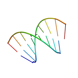
 | | Solution Structure of the YTH Domain of YT521-B in complex with N6-Methyladenosine containing RNA | | 分子名称: | RNA_(5'-R(*UP*GP*(6MZ)P*CP*AP*C)-3'), YTH domain-containing protein 1 | | 著者 | Theler, D, Dominguez, C, Blatter, M, Boudet, J, Allain, F.H.-T. | | 登録日 | 2014-09-01 | | 公開日 | 2014-11-26 | | 最終更新日 | 2024-05-01 | | 実験手法 | SOLUTION NMR | | 主引用文献 | Solution structure of the YTH domain in complex with N6-methyladenosine RNA: a reader of methylated RNA.
Nucleic Acids Res., 42, 2014
|
|
4AIT
 | |
1NSY
 | | CRYSTAL STRUCTURE OF NH3-DEPENDENT NAD+ SYNTHETASE FROM BACILLUS SUBTILIS | | 分子名称: | ADENOSINE MONOPHOSPHATE, ADENOSINE-5'-TRIPHOSPHATE, MAGNESIUM ION, ... | | 著者 | Rizzi, M, Nessi, C, Bolognesi, M, Galizzi, A, Coda, A. | | 登録日 | 1996-07-22 | | 公開日 | 1997-07-23 | | 最終更新日 | 2024-02-14 | | 実験手法 | X-RAY DIFFRACTION (2 Å) | | 主引用文献 | Crystal structure of NH3-dependent NAD+ synthetase from Bacillus subtilis.
EMBO J., 15, 1996
|
|
3HVP
 | |


