4NHI
 
 | |
2PL2
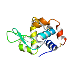
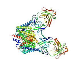
 | | Crystal structure of TTC0263: a thermophilic TPR protein in Thermus thermophilus HB27 | | 分子名称: | Hypothetical conserved protein TTC0263 | | 著者 | Lim, H, Kim, K, Han, D, Oh, J. | | 登録日 | 2007-04-18 | | 公開日 | 2008-03-04 | | 最終更新日 | 2024-03-13 | | 実験手法 | X-RAY DIFFRACTION (2.5 Å) | | 主引用文献 | Crystal structure of TTC0263, a thermophilic TPR protein from Thermus thermophilus HB27.
Mol.Cell, 24, 2007
|
|
3ZHK
 | |
3ZHD
 | |
3ZHL
 | |
7BXA
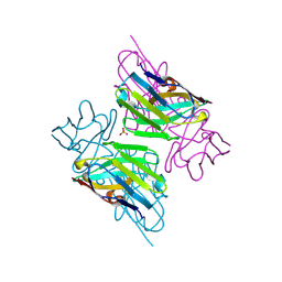
 | | Crystal structure of PD-1 in complex with tislelizumab Fab | | 分子名称: | Programmed cell death protein 1, heavy chain, light chain | | 著者 | Heo, Y.S, Lee, S.H, Lim, H, Lee, H.T, Kim, Y.J, Park, E.B. | | 登録日 | 2020-04-18 | | 公開日 | 2020-06-10 | | 最終更新日 | 2023-11-29 | | 実験手法 | X-RAY DIFFRACTION (3.32 Å) | | 主引用文献 | Crystal structure of PD-1 in complex with an antibody-drug tislelizumab used in tumor immune checkpoint therapy.
Biochem.Biophys.Res.Commun., 527, 2020
|
|
4WFQ
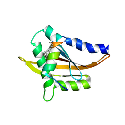
 | | Crystal structure of TFIIH subunit | | 分子名称: | GLYCEROL, SULFATE ION, Suppressor of stem-loop protein 1 | | 著者 | Cho, Y, Kim, J.S, Lim, H.S. | | 登録日 | 2014-09-17 | | 公開日 | 2015-02-18 | | 最終更新日 | 2024-03-20 | | 実験手法 | X-RAY DIFFRACTION (2.4 Å) | | 主引用文献 | Crystal structure of the Rad3/XPD regulatory domain of Ssl1/p44
J.Biol.Chem., 290, 2015
|
|
7CVT
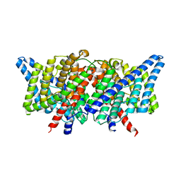
 | | Crystal structure of the C85A/L194A/H234C mutant CLC-ec1 with Fab fragment | | 分子名称: | CHLORIDE ION, H(+)/Cl(-) exchange transporter ClcA, antibody Fab fragment heavy chain, ... | | 著者 | Park, K, Mersch, K, Robertson, J, Lim, H.-H. | | 登録日 | 2020-08-27 | | 公開日 | 2021-09-01 | | 最終更新日 | 2023-11-29 | | 実験手法 | X-RAY DIFFRACTION (2.9 Å) | | 主引用文献 | Altering CLC stoichiometry by reducing non-polar side-chains at the dimerization interface.
J.Mol.Biol., 433, 2021
|
|
7CVS
 | | Crystal structure of the C85A/L194A mutant CLC-ec1 with Fab fragment | | 分子名称: | CHLORIDE ION, H(+)/Cl(-) exchange transporter ClcA, antibody Fab fragment heavy chain, ... | | 著者 | Park, K, Mersch, K, Robertson, J, Lim, H.-H. | | 登録日 | 2020-08-27 | | 公開日 | 2021-09-01 | | 最終更新日 | 2023-11-29 | | 実験手法 | X-RAY DIFFRACTION (3.01 Å) | | 主引用文献 | Altering CLC stoichiometry by reducing non-polar side-chains at the dimerization interface.
J.Mol.Biol., 433, 2021
|
|
5Y7W
 | | Crystal structure of the Nco-A1 PAS-B domain with YL-2 | | 分子名称: | Nuclear receptor coactivator 1, YL-2 peptide | | 著者 | Lee, Y.J, Yoon, H.S, Lee, J.H, Bae, J.H, Song, J.Y, Lim, H.S. | | 登録日 | 2017-08-18 | | 公開日 | 2017-11-15 | | 最終更新日 | 2023-11-22 | | 実験手法 | X-RAY DIFFRACTION (2.25 Å) | | 主引用文献 | Targeted Inhibition of the NCOA1/STAT6 Protein-Protein Interaction
J. Am. Chem. Soc., 139, 2017
|
|
2PYY
 | | Crystal Structure of the GluR0 ligand-binding core from Nostoc punctiforme in complex with (L)-glutamate | | 分子名称: | GLUTAMIC ACID, Ionotropic glutamate receptor bacterial homologue | | 著者 | Lee, J.H, Kang, G.B, Lim, H.-H, Ree, M, Park, C.-S, Eom, S.H. | | 登録日 | 2007-05-17 | | 公開日 | 2008-01-22 | | 最終更新日 | 2023-08-30 | | 実験手法 | X-RAY DIFFRACTION (2.1 Å) | | 主引用文献 | Crystal structure of the GluR0 ligand-binding core from Nostoc punctiforme in complex with L-glutamate: structural dissection of the ligand interaction and subunit interface.
J.Mol.Biol., 376, 2008
|
|
4KKL
 | | Structure of the E148A mutant of CLC-ec1 delta NC construct in 100mM fluoride | | 分子名称: | FLUORIDE ION, Fab, heavy chain, ... | | 著者 | Stockbridge, R.B, Lim, H.-H, Miller, C. | | 登録日 | 2013-05-06 | | 公開日 | 2013-08-21 | | 最終更新日 | 2013-12-04 | | 実験手法 | X-RAY DIFFRACTION (2.85 Å) | | 主引用文献 | Fluoride-dependent interruption of the transport cycle of a CLC Cl(-)/H(+) antiporter.
Nat.Chem.Biol., 9, 2013
|
|
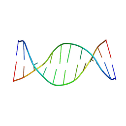
1ZYF
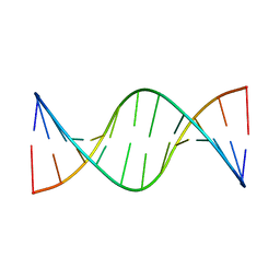 | | Structure of a Supercoiling Responsive DNA Site | | 分子名称: | 5'-D(*CP*AP*AP*CP*CP*AP*TP*GP*GP*TP*TP*G)-3' | | 著者 | Bae, S.H, Yun, S.H, Sun, D, Lim, H.M, Choi, B.S. | | 登録日 | 2005-06-10 | | 公開日 | 2006-05-23 | | 最終更新日 | 2024-05-29 | | 実験手法 | SOLUTION NMR | | 主引用文献 | Structural and dynamic basis of a supercoiling-responsive DNA element
Nucleic Acids Res., 34, 2006
|
|
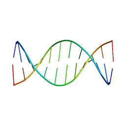
1ZYH
 | | Structure of a Supercoiling Responsive DNA site | | 分子名称: | 5'-D(*CP*AP*AP*CP*CP*AP*GP*GP*GP*TP*TP*G)-3', 5'-D(*CP*AP*AP*CP*CP*CP*TP*GP*GP*TP*TP*G)-3' | | 著者 | Bae, S.H, Yun, S.H, Sun, D, Lim, H.M, Choi, B.S. | | 登録日 | 2005-06-10 | | 公開日 | 2006-05-23 | | 最終更新日 | 2024-05-29 | | 実験手法 | SOLUTION NMR | | 主引用文献 | Structural and dynamic basis of a supercoiling-responsive DNA element
Nucleic Acids Res., 34, 2006
|
|
1ZYG
 | | Structure of a Supercoiling Responsive DNA Site | | 分子名称: | 5'-D(*CP*AP*AP*CP*CP*CP*GP*GP*GP*TP*TP*G)-3' | | 著者 | Bae, S.H, Yun, S.H, Sun, D, Lim, H.M, Choi, B.S. | | 登録日 | 2005-06-10 | | 公開日 | 2006-05-23 | | 最終更新日 | 2024-05-29 | | 実験手法 | SOLUTION NMR | | 主引用文献 | Structural and dynamic basis of a supercoiling-responsive DNA element
Nucleic Acids Res., 34, 2006
|
|
6K5F
 | |
6K5I
 | |
6K5A
 | |
6K5D
 | |
2BM2
 | | human beta-II tryptase in complex with 4-(3-Aminomethyl-phenyl)- piperidin-1-yl-(5-phenethyl- pyridin-3-yl)-methanone | | 分子名称: | 1-[3-(1-{[5-(2-PHENYLETHYL)PYRIDIN-3-YL]CARBONYL}PIPERIDIN-4-YL)PHENYL]METHANAMINE, HUMAN BETA2 TRYPTASE | | 著者 | Maignan, S, Guilloteau, J.-P, Dupuy, A, Levell, J, Astles, P, Eastwood, P, Cairns, J, Houille, O, Aldous, S, Merriman, G, Whiteley, B, Pribish, J, Czekaj, M, Liang, G, Davidson, J, Harrison, T, Morley, A, Watson, S, Fenton, G, Mccarthy, C, Romano, J, Mathew, R, Engers, D, Gardyan, M, Sides, K, Kwong, J, Tsay, J, Rebello, S, Shen, L, Wang, J, Luo, Y, Giardino, O, Lim, H.-K, Smith, K, Pauls, H. | | 登録日 | 2005-03-09 | | 公開日 | 2005-03-22 | | 最終更新日 | 2023-12-13 | | 実験手法 | X-RAY DIFFRACTION (2.2 Å) | | 主引用文献 | Structure Based Design of 4-(3-Aminomethylphenyl) Piperidinyl-1-Amides: Novel, Potent, Selective, and Orally Bioavailable Inhibitors of Bii Tryptase
Bioorg.Med.Chem., 13, 2005
|
|
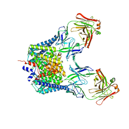
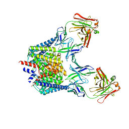
2EHO
 | | Crystal structure of human GINS complex | | 分子名称: | DNA replication complex GINS protein PSF1, DNA replication complex GINS protein PSF2, GINS complex subunit 3, ... | | 著者 | Choi, J.M, Lim, H.S, Kim, J.J, Song, O.K, Cho, Y. | | 登録日 | 2007-03-07 | | 公開日 | 2007-06-19 | | 最終更新日 | 2011-07-13 | | 実験手法 | X-RAY DIFFRACTION (3 Å) | | 主引用文献 | Crystal structure of the human GINS complex
Genes Dev., 21, 2007
|
|
2JQT
 | | Structure of the bacterial replication origin-associated protein Cnu | | 分子名称: | H-NS/stpA-binding protein 2 | | 著者 | Bae, S.H, Liu, D, Lim, H.M, Lee, Y, Choi, B.S. | | 登録日 | 2007-06-07 | | 公開日 | 2008-04-22 | | 最終更新日 | 2023-12-20 | | 実験手法 | SOLUTION NMR | | 主引用文献 | Structure of the nucleoid-associated protein Cnu reveals common binding sites for H-NS in Cnu and Hha.
Biochemistry, 47, 2008
|
|
2Q7F
 | |


