5E1T
 
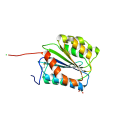
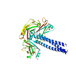 | | Crystal structure of TRAF1 TRAF domain | | 分子名称: | TNF receptor-associated factor 1 | | 著者 | Park, H.H, Kim, C.M. | | 登録日 | 2015-09-30 | | 公開日 | 2016-12-07 | | 最終更新日 | 2024-03-20 | | 実験手法 | X-RAY DIFFRACTION (2.8 Å) | | 主引用文献 | Crystal structure of TRAF1 TRAF domain and its implications in the TRAF1-mediated intracellular signaling pathway
Sci Rep, 6, 2016
|
|
3OCP
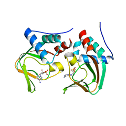 | | Crystal structure of cAMP bound cGMP-dependent protein kinase(92-227) | | 分子名称: | ADENOSINE-3',5'-CYCLIC-MONOPHOSPHATE, PRKG1 protein | | 著者 | Kim, J.J, Huang, G, Kwon, T.K, Zwart, P, Headd, J, Kim, C. | | 登録日 | 2010-08-10 | | 公開日 | 2011-05-11 | | 最終更新日 | 2023-09-06 | | 実験手法 | X-RAY DIFFRACTION (2.49 Å) | | 主引用文献 | Co-Crystal Structures of PKG Ibeta (92-227) with cGMP and cAMP Reveal the Molecular Details of Cyclic-Nucleotide Binding
Plos One, 6, 2011
|
|
4LMP
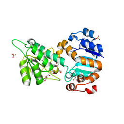 | | Mycobacterium tuberculosis L-alanine dehydrogenase x-ray structure in complex with N6-methyl adenosine | | 分子名称: | Alanine dehydrogenase, GLYCEROL, N-methyladenosine, ... | | 著者 | Kim, H.-B, Hung, L.-W, Goulding, C.W, Terwilliger, T.C, Kim, C.-Y, Structures of Mtb Proteins Conferring Susceptibility to Known Mtb Inhibitors (MTBI) | | 登録日 | 2013-07-10 | | 公開日 | 2013-11-06 | | 最終更新日 | 2023-09-20 | | 実験手法 | X-RAY DIFFRACTION (1.95 Å) | | 主引用文献 | Drug target analysis by dye-ligand affinity chromatography
To be Published
|
|
1XXU
 | | Crystal Structure of AhpE from Mycrobacterium tuberculosis, a 1-Cys peroxiredoxin | | 分子名称: | Hypothetical protein Rv2238c/MT2298 | | 著者 | Li, S, Peterson, N.A, Kim, M.Y, Kim, C.Y, Hung, L.W, Yu, M, Lekin, T, Segelke, B.W, Lott, J.S, Baker, E.N, TB Structural Genomics Consortium (TBSGC) | | 登録日 | 2004-11-08 | | 公開日 | 2005-02-22 | | 最終更新日 | 2023-10-25 | | 実験手法 | X-RAY DIFFRACTION (1.9 Å) | | 主引用文献 | Crystal Structure of AhpE from Mycobacterium tuberculosis, a 1-Cys Peroxiredoxin
J.Mol.Biol., 346, 2005
|
|
4O6G
 | | Rv3902c from M. tuberculosis | | 分子名称: | Uncharacterized protein | | 著者 | Reddy, B.G, Moates, D.B, Kim, H, Green, T.J, Kim, C, Terwilliger, T.J, Delucas, L.J, TB Structural Genomics Consortium (TBSGC) | | 登録日 | 2013-12-20 | | 公開日 | 2014-03-05 | | 最終更新日 | 2024-04-03 | | 実験手法 | X-RAY DIFFRACTION (1.55 Å) | | 主引用文献 | 1.55 angstrom resolution X-ray crystal structure of Rv3902c from Mycobacterium tuberculosis.
Acta Crystallogr F Struct Biol Commun, 70, 2014
|
|
3OD0
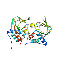 | | Crystal structure of cGMP bound cGMP-dependent protein kinase(92-227) | | 分子名称: | CYCLIC GUANOSINE MONOPHOSPHATE, PRKG1 protein | | 著者 | Kim, J.J, Huang, G, Kwon, T.K, Zwart, P, Headd, J, Kim, C. | | 登録日 | 2010-08-10 | | 公開日 | 2011-05-11 | | 最終更新日 | 2024-02-21 | | 実験手法 | X-RAY DIFFRACTION (2.9 Å) | | 主引用文献 | Co-Crystal Structures of PKG Ibeta (92-227) with cGMP and cAMP Reveal the Molecular Details of Cyclic-Nucleotide Binding
Plos One, 6, 2011
|
|
6NAZ
 | | Crystal structure of DIRAS 2/3 chimera in complex with GDP | | 分子名称: | 1,2-ETHANEDIOL, GLYCEROL, GTP-binding protein Di-Ras2,GTP-binding protein Di-Ras3,GTP-binding protein Di-Ras2, ... | | 著者 | Gilbert, Y.H, Reger, A, Sharma, R, Bast, J, Kim, C. | | 登録日 | 2018-12-06 | | 公開日 | 2019-12-18 | | 最終更新日 | 2023-10-11 | | 実験手法 | X-RAY DIFFRACTION (3.081 Å) | | 主引用文献 | Crystal structure of DIRAS 2/3 chimera in complex with GDP
To Be Published
|
|
6C0U
 | | Crystal structure of cAMP-dependent protein kinase Calpha subunit bound with N46 | | 分子名称: | DIMETHYL SULFOXIDE, N-[(3R,4R)-4-{[4-(2-fluoro-3-methoxy-6-propoxybenzene-1-carbonyl)benzene-1-carbonyl]amino}pyrrolidin-3-yl]-1H-indazole-5-carboxamide, PHOSPHATE ION, ... | | 著者 | Qin, L, Sankaran, B, Kim, C. | | 登録日 | 2018-01-02 | | 公開日 | 2018-05-30 | | 最終更新日 | 2023-10-04 | | 実験手法 | X-RAY DIFFRACTION (2.65 Å) | | 主引用文献 | Structural basis for selective inhibition of human PKG I alpha by the balanol-like compound N46.
J. Biol. Chem., 293, 2018
|
|
6C0T
 | | Crystal structure of cGMP-dependent protein kinase Ialpha (PKG Ialpha) catalytic domain bound with N46 | | 分子名称: | DIMETHYL SULFOXIDE, N-[(3R,4R)-4-{[4-(2-fluoro-3-methoxy-6-propoxybenzene-1-carbonyl)benzene-1-carbonyl]amino}pyrrolidin-3-yl]-1H-indazole-5-carboxamide, TRIETHYLENE GLYCOL, ... | | 著者 | Qin, L, Sankaran, B, Kim, C. | | 登録日 | 2018-01-02 | | 公開日 | 2018-05-30 | | 最終更新日 | 2023-10-04 | | 実験手法 | X-RAY DIFFRACTION (1.98 Å) | | 主引用文献 | Structural basis for selective inhibition of human PKG I alpha by the balanol-like compound N46.
J. Biol. Chem., 293, 2018
|
|
4RZM
 | | Crystal structure of the Lsd19-lasalocid A complex | | 分子名称: | CHLORIDE ION, Epoxide hydrolase LasB, FORMIC ACID, ... | | 著者 | Mathews, I.I, Hotta, K, Chen, X, Kim, C.-Y. | | 登録日 | 2014-12-22 | | 公開日 | 2015-01-21 | | 最終更新日 | 2023-09-20 | | 実験手法 | X-RAY DIFFRACTION (2.33 Å) | | 主引用文献 | Epoxide hydrolase-lasalocid a structure provides mechanistic insight into polyether natural product biosynthesis.
J.Am.Chem.Soc., 137, 2015
|
|
4OFF
 | |
4OFG
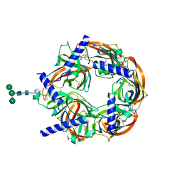
 | | Co-crystal structure of carboxy cGMP binding domain of Plasmodium falciparum PKG with cGMP | | 分子名称: | CGMP-dependent protein kinase, CYCLIC GUANOSINE MONOPHOSPHATE, ETHANOL, ... | | 著者 | Kim, J.J, Sanabria figueroa, E, Kim, C. | | 登録日 | 2014-01-14 | | 公開日 | 2015-01-21 | | 最終更新日 | 2024-02-28 | | 実験手法 | X-RAY DIFFRACTION (2 Å) | | 主引用文献 | Crystal Structures of the Carboxyl cGMP Binding Domain of the Plasmodium falciparum cGMP-dependent Protein Kinase Reveal a Novel Capping Triad Crucial for Merozoite Egress.
Plos Pathog., 11, 2015
|
|
6BG2
 | |
3IIA
 | | Crystal structure of apo (91-244) RIa subunit of cAMP-dependent protein kinase | | 分子名称: | GLYCEROL, cAMP-dependent protein kinase type I-alpha regulatory subunit | | 著者 | Sjoberg, T.J, Kim, C, Kornev, A.P, Taylor, S.S. | | 登録日 | 2009-07-31 | | 公開日 | 2010-08-11 | | 最終更新日 | 2023-09-06 | | 実験手法 | X-RAY DIFFRACTION (2.7 Å) | | 主引用文献 | Cyclic AMP analog blocks kinase activation by stabilizing inactive conformation: conformational selection highlights a new concept in allosteric inhibitor design.
Mol.Cell Proteomics, 10, 2011
|
|
4EZ1
 | | Crystal structure of acetylcholine binding protein (AChBP) from Aplysia Californica in complex with alpha-conotoxin BuIA | | 分子名称: | Alpha-conotoxin BuIA, MANGANESE (II) ION, Soluble acetylcholine receptor | | 著者 | Talley, T.T, Reger, A.S, Kim, C, Sankaran, B, Ho, K, Taylor, P, McIntosh, J.M. | | 登録日 | 2012-05-02 | | 公開日 | 2013-05-08 | | 最終更新日 | 2013-06-19 | | 実験手法 | X-RAY DIFFRACTION (2.49 Å) | | 主引用文献 | Pairwise interaction of alpha-conotoxin BuIA Pro6 with the beta subunit of nicotinic acetylcholine receptor
To be Published
|
|
4DBM
 | | Aplysia californica-AChBP in complex with triazole 18 | | 分子名称: | (3-exo)-8,8-dimethyl-3-(4-{[(1-methyl-2-oxo-1,2-dihydroquinolin-4-yl)oxy]methyl}-1H-1,2,3-triazol-1-yl)-8-azoniabicyclo[3.2.1]octane, 2-acetamido-2-deoxy-beta-D-glucopyranose, Soluble acetylcholine receptor, ... | | 著者 | Nemecz, A, Yamauchi, J.G, Kim, C. | | 登録日 | 2012-01-16 | | 公開日 | 2012-03-21 | | 最終更新日 | 2023-09-13 | | 実験手法 | X-RAY DIFFRACTION (2.3 Å) | | 主引用文献 | Generation of candidate ligands for nicotinic acetylcholine receptors via in situ click chemistry with a soluble acetylcholine binding protein template.
J.Am.Chem.Soc., 134, 2012
|
|
2HG4
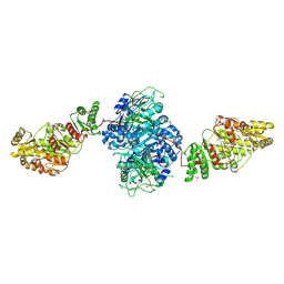 | | Structure of the ketosynthase-acyltransferase didomain of module 5 from DEBS. | | 分子名称: | 6-Deoxyerythronolide B Synthase, ACETATE ION, CHLORIDE ION, ... | | 著者 | Tang, Y, Kim, C.Y, Mathews, I.I, Cane, D.E, Khosla, C. | | 登録日 | 2006-06-26 | | 公開日 | 2006-07-11 | | 最終更新日 | 2011-07-13 | | 実験手法 | X-RAY DIFFRACTION (2.73 Å) | | 主引用文献 | The 2.7-A crystal structure of a 194-kDa homodimeric fragment of the 6-deoxyerythronolide B synthase.
Proc.Natl.Acad.Sci.Usa, 103, 2006
|
|
5BV8
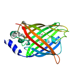
 | | G1324S mutation in von Willebrand Factor A1 domain | | 分子名称: | 1,2-ETHANEDIOL, CHLORIDE ION, von Willebrand factor | | 著者 | Campbell, J.C, Kim, C, Tischer, A, Auton, M. | | 登録日 | 2015-06-04 | | 公開日 | 2015-12-23 | | 最終更新日 | 2023-09-27 | | 実験手法 | X-RAY DIFFRACTION (1.59 Å) | | 主引用文献 | Mutational Constraints on Local Unfolding Inhibit the Rheological Adaptation of von Willebrand Factor.
J.Biol.Chem., 291, 2016
|
|
3DQ1
 | |
3DQA
 | |
3DQ6
 | |
3DQL
 | |
3DQ3
 | |
3DQD
 | |
3DQN
 | |


