8HVR
 
 | |
1XBS
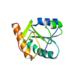 | | Crystal structure of human dim2: a dim1-like protein | | 分子名称: | Dim1-like protein | | 著者 | Simeoni, F, Arvai, A, Hopfner, K.-P, Bello, P, Gondeau, C, Heitz, F, Tainer, J, Divita, G. | | 登録日 | 2004-08-31 | | 公開日 | 2005-08-30 | | 最終更新日 | 2024-02-14 | | 実験手法 | X-RAY DIFFRACTION (2.5 Å) | | 主引用文献 | Biochemical Characterization and Crystal Structure of a Dim1 Family Associated Protein: Dim2
Biochemistry, 44, 2005
|
|
4PZD
 | |
1XD4
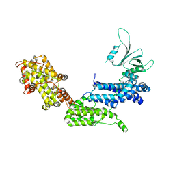 | | Crystal structure of the DH-PH-cat module of Son of Sevenless (SOS) | | 分子名称: | Son of sevenless protein homolog 1 | | 著者 | Sondermann, H, Soisson, S.M, Boykevisch, S, Yang, S.S, Bar-Sagi, D, Kuriyan, J. | | 登録日 | 2004-09-03 | | 公開日 | 2004-11-02 | | 最終更新日 | 2023-08-23 | | 実験手法 | X-RAY DIFFRACTION (3.64 Å) | | 主引用文献 | Structural analysis of autoinhibition in the ras activator son of sevenless.
Cell(Cambridge,Mass.), 119, 2004
|
|
4Q0C
 | | 3.1 A resolution crystal structure of the B. pertussis BvgS periplasmic domain | | 分子名称: | Virulence sensor protein BvgS | | 著者 | Dupre, E, Herrou, J, Lensink, M.F, Wintjens, R, Lebedev, A, Crosson, S, Villeret, V, Locht, C, Antoine, R, Jacob-Dubuisson, F. | | 登録日 | 2014-04-01 | | 公開日 | 2015-02-11 | | 最終更新日 | 2023-09-20 | | 実験手法 | X-RAY DIFFRACTION (3.1 Å) | | 主引用文献 | Virulence Regulation with Venus Flytrap Domains: Structure and Function of the Periplasmic Moiety of the Sensor-Kinase BvgS.
Plos Pathog., 11, 2015
|
|
5F3A
 | |
1XK2
 | | NADPH- and Ascorbate-Supported Heme Oxygenase Reactions are Distinct. Regiospecificity of Heme Cleavage by the R183E Mutant | | 分子名称: | Heme oxygenase 1, PROTOPORPHYRIN IX CONTAINING FE | | 著者 | Wang, J, Lad, L, Poulos, T.L, Ortiz de montellano, P.R. | | 登録日 | 2004-09-26 | | 公開日 | 2004-11-09 | | 最終更新日 | 2024-02-14 | | 実験手法 | X-RAY DIFFRACTION (2.2 Å) | | 主引用文献 | Regiospecificity determinants of human heme oxygenase: differential NADPH- and ascorbate-dependent heme cleavage by the R183E mutant.
J.Biol.Chem., 280, 2005
|
|
8HWA
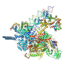
 | | D5 ATP-ADP-Apo-ssDNA IS1 | | 分子名称: | ADENOSINE-5'-DIPHOSPHATE, ADENOSINE-5'-TRIPHOSPHATE, DNA (5'-D(P*TP*TP*TP*TP*TP*T)-3'), ... | | 著者 | Li, Y.N, Zhu, J, Guo, Y.Y, Yan, R.H. | | 登録日 | 2022-12-29 | | 公開日 | 2024-01-10 | | 最終更新日 | 2024-01-31 | | 実験手法 | ELECTRON MICROSCOPY (3.7 Å) | | 主引用文献 | Structural insight into the assembly and working mechanism of helicase-primase D5 from Mpox virus.
Nat.Struct.Mol.Biol., 31, 2024
|
|
1XL5
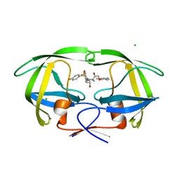 | | HIV-1 Protease in complex with amidhyroxysulfone | | 分子名称: | CHLORIDE ION, N-{(1S)-1-(3-BROMOBENZYL)-4-[(4-BROMOPHENYL)SULFONYL]-6-METHYL-2-OXOHEPTYL}-2-(2,6-DIMETHYLPHENOXY)ACETAMIDE, PROTEASE RETROPEPSIN | | 著者 | Boettcher, J, Specker, E, Heine, A, Klebe, G. | | 登録日 | 2004-09-30 | | 公開日 | 2005-06-07 | | 最終更新日 | 2023-08-23 | | 実験手法 | X-RAY DIFFRACTION (1.73 Å) | | 主引用文献 | An Old Target Revisited: Two New Privileged Skeletons and an Unexpected Binding Mode For HIV-Protease Inhibitors
Angew.Chem.Int.Ed.Engl., 44, 2005
|
|
8ILJ
 | |
5F0W
 | |
1XHD
 | | X-ray crystal structure of putative acetyltransferase, product of BC4754 gene [Bacillus cereus] | | 分子名称: | SULFATE ION, putative acetyltransferase/acyltransferase | | 著者 | Osipiuk, J, Zhou, M, Moy, S, Collart, F, Joachimiak, A, Midwest Center for Structural Genomics (MCSG) | | 登録日 | 2004-09-17 | | 公開日 | 2004-11-02 | | 最終更新日 | 2011-07-13 | | 実験手法 | X-RAY DIFFRACTION (1.9 Å) | | 主引用文献 | X-ray crystal structure of putative acetyltransferase, product of BC4754 gene from Bacillus cereus.
To be Published
|
|
8HWG
 | | D5 ATPrS-ADP-ssDNA form | | 分子名称: | ADENOSINE-5'-DIPHOSPHATE, DNA (5'-D(P*TP*TP*TP*TP*TP*T)-3'), MAGNESIUM ION, ... | | 著者 | Li, Y.N, Zhu, J, Guo, Y.Y, Yan, R.H. | | 登録日 | 2022-12-29 | | 公開日 | 2024-01-10 | | 最終更新日 | 2024-01-31 | | 実験手法 | ELECTRON MICROSCOPY (3 Å) | | 主引用文献 | Structural insight into the assembly and working mechanism of helicase-primase D5 from Mpox virus.
Nat.Struct.Mol.Biol., 31, 2024
|
|
5EV9
 | |
8HWE
 | | Cryo-EM Structure of D5 ATP-ADP form | | 分子名称: | ADENOSINE-5'-DIPHOSPHATE, ADENOSINE-5'-TRIPHOSPHATE, MAGNESIUM ION, ... | | 著者 | Li, Y.N, Zhu, J, Guo, Y.Y, Yan, R.H. | | 登録日 | 2022-12-29 | | 公開日 | 2024-01-10 | | 最終更新日 | 2024-01-31 | | 実験手法 | ELECTRON MICROSCOPY (3.3 Å) | | 主引用文献 | Structural insight into the assembly and working mechanism of helicase-primase D5 from Mpox virus.
Nat.Struct.Mol.Biol., 31, 2024
|
|
8I52
 | |
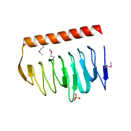
5EVE
 | | Crystal structure of Amb a 8 in complex with poly-Pro10 | | 分子名称: | Poly-Proline peptide, Profilin | | 著者 | Offermann, L.R, Schlachter, C.R, Garrett, J, Chruszcz, M. | | 登録日 | 2015-11-19 | | 公開日 | 2016-06-08 | | 最終更新日 | 2023-09-27 | | 実験手法 | X-RAY DIFFRACTION (2.55 Å) | | 主引用文献 | Structural, Functional, and Immunological Characterization of Profilin Panallergens Amb a 8, Art v 4, and Bet v 2.
J.Biol.Chem., 291, 2016
|
|
1XN2
 | | New substrate binding pockets for beta-secretase. | | 分子名称: | Beta-secretase 1, OM03-4 | | 著者 | Turner III, R.T, Hong, L, Koelsch, G, Ghosh, A.K, Tang, J. | | 登録日 | 2004-10-04 | | 公開日 | 2005-03-22 | | 最終更新日 | 2023-11-15 | | 実験手法 | X-RAY DIFFRACTION (1.9 Å) | | 主引用文献 | Structural locations and functional roles of new subsites S5, S6, and S7 in memapsin 2 (beta-secretase).
Biochemistry, 44, 2005
|
|
1AXV
 | | SOLUTION NMR STRUCTURE OF THE [BP]DA ADDUCT OPPOSITE DT IN A DNA DUPLEX, 6 STRUCTURES | | 分子名称: | 1,2,3-TRIHYDROXY-1,2,3,4-TETRAHYDROBENZO[A]PYRENE, DNA DUPLEX D(CTCTC-[BP]A-CTTCC)D(GGAAGTGAGAG) | | 著者 | Mao, B, Gu, Z, Gorin, A.A, Chen, J, Hingerty, B.E, Amid, S, Broyde, S, Geacintov, N.E, Patel, D.J. | | 登録日 | 1997-10-21 | | 公開日 | 1998-07-01 | | 最終更新日 | 2024-05-22 | | 実験手法 | SOLUTION NMR | | 主引用文献 | Solution structure of the (+)-cis-anti-benzo[a]pyrene-dA ([BP]dA) adduct opposite dT in a DNA duplex.
Biochemistry, 38, 1999
|
|
5EX3
 | | Crystal structure of human SMYD3 in complex with a VEGFR1 peptide | | 分子名称: | ACETIC ACID, Histone-lysine N-methyltransferase SMYD3, S-ADENOSYL-L-HOMOCYSTEINE, ... | | 著者 | Qiao, Q, Fu, W, Liu, N, Wang, M, Min, J, Zhu, B, Xu, R.M, Yang, N. | | 登録日 | 2015-11-23 | | 公開日 | 2016-03-09 | | 最終更新日 | 2017-10-18 | | 実験手法 | X-RAY DIFFRACTION (2.408 Å) | | 主引用文献 | Structural Basis for Substrate Preference of SMYD3, a SET Domain-containing Protein Lysine Methyltransferase
J.Biol.Chem., 291, 2016
|
|
5EWA
 | | Crystal structure of the metallo-beta-lactamase IMP-1 in complex with the bisthiazolidine inhibitor L-VC26 | | 分子名称: | (3~{R},5~{R},7~{a}~{S})-2,2-dimethyl-5-(sulfanylmethyl)-3,5,7,7~{a}-tetrahydro-[1,3]thiazolo[4,3-b][1,3]thiazole-3-carboxylic acid, 1,2-ETHANEDIOL, Beta-lactamase IMP-1, ... | | 著者 | Kosmopoulou, M, Hinchliffe, P, Spencer, J. | | 登録日 | 2015-11-20 | | 公開日 | 2016-06-01 | | 最終更新日 | 2024-01-10 | | 実験手法 | X-RAY DIFFRACTION (2.3 Å) | | 主引用文献 | Cross-class metallo-beta-lactamase inhibition by bisthiazolidines reveals multiple binding modes.
Proc.Natl.Acad.Sci.USA, 113, 2016
|
|
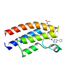
5EWW
 | | Crystal structure of the human BRPF1 bromodomain in complex with SEED21 | | 分子名称: | NITRATE ION, Peregrin, ~{N}-[(2~{R})-butan-2-yl]-1-methyl-[1,2,4]triazolo[4,3-a]quinoxalin-4-amine | | 著者 | Zhu, J, Wiedmer, L, Caflisch, A. | | 登録日 | 2015-11-21 | | 公開日 | 2016-11-02 | | 最終更新日 | 2024-01-10 | | 実験手法 | X-RAY DIFFRACTION (1.73 Å) | | 主引用文献 | Crystal structure of the human BRPF1 bromodomain in complex with SEED21
To Be Published
|
|
1AWE
 | |
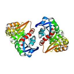
4Q6O
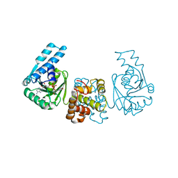
 | | Structural analysis of the mDAP-bound form of Helicobacter pylori Csd4, a D,L-carboxypeptidase | | 分子名称: | 2,6-DIAMINOPIMELIC ACID, CALCIUM ION, Conserved hypothetical secreted protein, ... | | 著者 | Kim, H.S, Kim, J, Im, H.N, An, D.R, Lee, M, Hesek, D, Mobashery, S, Kim, J.Y, Cho, K, Yoon, H.J, Han, B.W, Lee, B.I, Suh, S.W. | | 登録日 | 2014-04-23 | | 公開日 | 2014-11-05 | | 最終更新日 | 2023-11-15 | | 実験手法 | X-RAY DIFFRACTION (1.41 Å) | | 主引用文献 | Structural basis for the recognition of muramyltripeptide by Helicobacter pylori Csd4, a D,L-carboxypeptidase controlling the helical cell shape
Acta Crystallogr.,Sect.D, 70, 2014
|
|
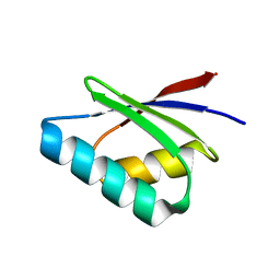
1XOY
 | | Solution structure of At3g04780.1, an Arabidopsis ortholog of the C-terminal domain of human thioredoxin-like protein | | 分子名称: | hypothetical protein At3g04780.1 | | 著者 | Song, J, Robert, C.T, Lee, M.S, Markley, J.L, Center for Eukaryotic Structural Genomics (CESG) | | 登録日 | 2004-10-07 | | 公開日 | 2004-10-12 | | 最終更新日 | 2024-05-22 | | 実験手法 | SOLUTION NMR | | 主引用文献 | Solution structure of At3g04780.1-des15, an Arabidopsis thaliana ortholog of the C-terminal domain of human thioredoxin-like protein.
Protein Sci., 14, 2005
|
|


