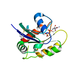
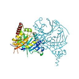
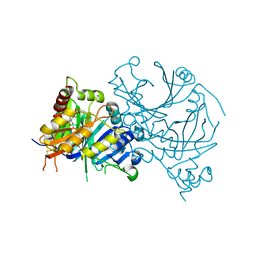
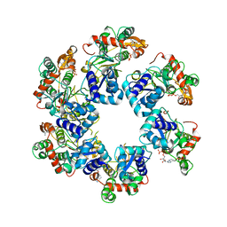
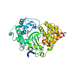
4PYM
 
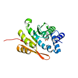 | | humanized rat apo-COMT bound to sulphate | | 分子名称: | Catechol O-methyltransferase, POTASSIUM ION, SULFATE ION | | 著者 | Ehler, A, Benz, J, Schlatter, D, Rudolph, M.G. | | 登録日 | 2014-03-27 | | 公開日 | 2014-06-11 | | 最終更新日 | 2024-02-28 | | 実験手法 | X-RAY DIFFRACTION (1.19 Å) | | 主引用文献 | Mapping the conformational space accessible to catechol-O-methyltransferase.
Acta Crystallogr.,Sect.D, 70, 2014
|
|
8I2S
 | | Crystal structure of AtHPPD-Y18979 complex | | 分子名称: | 1,5-dimethyl-3-(naphthalen-2-ylmethyl)-6-(2-oxidanyl-6-oxidanylidene-cyclohexen-1-yl)carbonyl-quinazoline-2,4-dione, 4-hydroxyphenylpyruvate dioxygenase, COBALT (II) ION | | 著者 | Dong, J, Lin, H.-Y, Yang, G.-F. | | 登録日 | 2023-01-15 | | 公開日 | 2023-12-20 | | 実験手法 | X-RAY DIFFRACTION (1.592 Å) | | 主引用文献 | Crystal structure of AtHPPD-Y18979 complex
To Be Published
|
|
5F4Z
 | | The crystal structure of an epoxide hydrolase from Streptomyces carzinostaticus subsp. neocarzinostaticus | | 分子名称: | (1~{R},2~{R})-2,3-dihydro-1~{H}-indene-1,2-diol, 2-AMINO-2-HYDROXYMETHYL-PROPANE-1,3-DIOL, ACETATE ION, ... | | 著者 | Tan, K, Li, H, Jedrzejczak, R, BABNIGG, G, BINGMAN, C.A, YENNAMALLI, R, LOHMAN, J, Chang, C.Y, Shen, B, Phillips Jr, G.N, Joachimiak, A, Midwest Center for Structural Genomics (MCSG), Enzyme Discovery for Natural Product Biosynthesis (NatPro) | | 登録日 | 2015-12-03 | | 公開日 | 2016-02-17 | | 最終更新日 | 2020-09-23 | | 実験手法 | X-RAY DIFFRACTION (1.82 Å) | | 主引用文献 | The crystal structure of an epoxide hydrolase from Streptomyces carzinostaticus subsp. neocarzinostaticus
To Be Published
|
|
1X9T
 | | The crystal structure of human adenovirus 2 penton base in complex with an ad2 N-terminal fibre peptide | | 分子名称: | N-DODECYL-N,N-DIMETHYL-3-AMMONIO-1-PROPANESULFONATE, N-terminal peptide of Fiber protein, Penton protein | | 著者 | Zubieta, C, Schoehn, G, Chroboczek, J, Cusack, S. | | 登録日 | 2004-08-24 | | 公開日 | 2005-01-18 | | 最終更新日 | 2024-04-03 | | 実験手法 | X-RAY DIFFRACTION (3.5 Å) | | 主引用文献 | The structure of the human adenovirus 2 penton
Mol.Cell, 17, 2005
|
|
8I8X
 | | Cryo-EM Structure of OmpC3-MlaA-MlaC Complex in MSP2N2 Nanodiscs | | 分子名称: | (2~{R},4~{R},5~{R},6~{R})-6-[(1~{R})-1,2-bis(oxidanyl)ethyl]-2-[(2~{R},4~{R},5~{R},6~{R})-6-[(1~{R})-1,2-bis(oxidanyl)ethyl]-2-carboxy-2-[[(2~{R},3~{S},4~{R},5~{R},6~{R})-5-[[(3~{R})-3-dodecanoyloxytetradecanoyl]amino]-6-[[(2~{R},3~{S},4~{R},5~{R},6~{R})-3-oxidanyl-5-[[(3~{R})-3-oxidanyltetradecanoyl]amino]-4-[(3~{R})-3-oxidanyltetradecanoyl]oxy-6-phosphonooxy-oxan-2-yl]methoxy]-3-phosphonooxy-4-[(3~{R})-3-tetradecanoyloxytetradecanoyl]oxy-oxan-2-yl]methoxy]-5-oxidanyl-oxan-4-yl]oxy-4,5-bis(oxidanyl)oxane-2-carboxylic acid, Intermembrane phospholipid transport system binding protein MlaC, Intermembrane phospholipid transport system lipoprotein MlaA, ... | | 著者 | Yeow, J, Luo, M, Chng, S.S. | | 登録日 | 2023-02-05 | | 公開日 | 2023-12-20 | | 最終更新日 | 2023-12-27 | | 実験手法 | ELECTRON MICROSCOPY (3.25 Å) | | 主引用文献 | Molecular mechanism of phospholipid transport at the bacterial outer membrane interface.
Nat Commun, 14, 2023
|
|
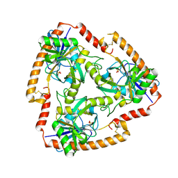
4PZ9
 | | The native structure of mycobacterial glucosyl-3-phosphoglycerate phosphatase Rv2419c | | 分子名称: | Glucosyl-3-phosphoglycerate phosphatase | | 著者 | Zhou, W.H, Zheng, Q.Q, Jiang, D.Q, Zhang, W, Zhang, Q.Q, Jin, J, Li, X, Yang, H.T, Shaw, N, Rao, Z. | | 登録日 | 2014-03-28 | | 公開日 | 2014-06-11 | | 最終更新日 | 2023-11-08 | | 実験手法 | X-RAY DIFFRACTION (1.94 Å) | | 主引用文献 | Mechanism of dephosphorylation of glucosyl-3-phosphoglycerate by a histidine phosphatase
J.Biol.Chem., 289, 2014
|
|
1X95
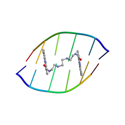 | | Solution structure of the DNA-hexamer ATGCAT complexed with DNA Bis-intercalating Anticancer Drug XR5944 (MLN944) | | 分子名称: | 1-METHYL-9-[12-(9-METHYLPHENAZIN-10-IUM-1-YL)-12-OXO-2,11-DIAZA-5,8-DIAZONIADODEC-1-ANOYL]PHENAZIN-10-IUM, 5'-D(*AP*TP*GP*CP*AP*T)-3' | | 著者 | Dai, J, Punchihewa, C, Mistry, P, Ooi, A.T, Yang, D. | | 登録日 | 2004-08-19 | | 公開日 | 2004-09-28 | | 最終更新日 | 2024-05-01 | | 実験手法 | SOLUTION NMR | | 主引用文献 | Novel DNA bis-intercalation by MLN944, a potent clinical bisphenazine anticancer drug.
J.Biol.Chem., 279, 2004
|
|
5F5R
 | | TRAP1N-ADPNP | | 分子名称: | Heat shock protein 75 kDa, mitochondrial, MAGNESIUM ION, ... | | 著者 | Tsai, F.T.F, Lee, S, Sung, N, Lee, J, Chang, C, Joachimiak, A. | | 登録日 | 2015-12-04 | | 公開日 | 2016-03-02 | | 最終更新日 | 2023-09-27 | | 実験手法 | X-RAY DIFFRACTION (1.85 Å) | | 主引用文献 | Mitochondrial Hsp90 is a ligand-activated molecular chaperone coupling ATP binding to dimer closure through a coiled-coil intermediate.
Proc.Natl.Acad.Sci.USA, 113, 2016
|
|
5EVA
 | |
1XAG
 | | CRYSTAL STRUCTURE OF STAPHLYOCOCCUS AUREUS 3-DEHYDROQUINATE SYNTHASE (DHQS) IN COMPLEX WITH ZN2+, NAD+ AND CARBAPHOSPHONATE | | 分子名称: | 3-dehydroquinate synthase, CHLORIDE ION, NICOTINAMIDE-ADENINE-DINUCLEOTIDE, ... | | 著者 | Nichols, C.E, Ren, J, Leslie, K, Dhaliwal, B, Lockyer, M, Charles, I, Hawkins, A.R, Stammers, D.K. | | 登録日 | 2004-08-25 | | 公開日 | 2005-03-01 | | 最終更新日 | 2023-08-23 | | 実験手法 | X-RAY DIFFRACTION (2.45 Å) | | 主引用文献 | Comparison of ligand induced conformational changes and domain closure mechanisms, between prokaryotic and eukaryotic dehydroquinate synthases.
J.Mol.Biol., 343, 2004
|
|
4Q01
 | | Second-site screening of K-Ras in the presence of covalently attached first-site ligands | | 分子名称: | GUANOSINE-5'-DIPHOSPHATE, K-Ras, MAGNESIUM ION, ... | | 著者 | Sun, Q, Phan, J, Friberg, A, Camper, D.V, Olejniczak, E.T, Fesik, S.W. | | 登録日 | 2014-03-31 | | 公開日 | 2014-09-10 | | 実験手法 | X-RAY DIFFRACTION (1.291 Å) | | 主引用文献 | A method for the second-site screening of K-Ras in the presence of a covalently attached first-site ligand.
J.Biomol.Nmr, 60, 2014
|
|
4PVQ
 | |
5EVK
 | | Crystal structure of the metallo-beta-lactamase L1 in complex with the bisthiazolidine inhibitor L-CS319 | | 分子名称: | (3R,5R,7aS)-5-(sulfanylmethyl)tetrahydro[1,3]thiazolo[4,3-b][1,3]thiazole-3-carboxylic acid, Metallo-beta-lactamase L1, SULFATE ION, ... | | 著者 | Hinchliffe, P, Spencer, J. | | 登録日 | 2015-11-19 | | 公開日 | 2016-06-01 | | 最終更新日 | 2024-01-10 | | 実験手法 | X-RAY DIFFRACTION (1.627 Å) | | 主引用文献 | Cross-class metallo-beta-lactamase inhibition by bisthiazolidines reveals multiple binding modes.
Proc.Natl.Acad.Sci.USA, 113, 2016
|
|
8HX2
 | | Crystal structure of AtHPPD-Y18405 complex | | 分子名称: | 3-[2-(3-chlorophenyl)ethyl]-1,5-dimethyl-6-(2-oxidanyl-6-oxidanylidene-cyclohexa-1,3-dien-1-yl)carbonyl-quinazoline-2,4-dione, 4-hydroxyphenylpyruvate dioxygenase, COBALT (II) ION | | 著者 | Dong, J, Lin, H.-Y, Yang, G.-F. | | 登録日 | 2023-01-03 | | 公開日 | 2023-12-20 | | 実験手法 | X-RAY DIFFRACTION (1.996 Å) | | 主引用文献 | Crystal structure of AtHPPD-Y18405 complex
To Be Published
|
|
8HZ9
 | | Crystal structure of AtHPPD-Y181136 complex | | 分子名称: | 4-hydroxyphenylpyruvate dioxygenase, 5-methyl-6-[(2-methyl-3-oxidanylidene-1H-pyrazol-4-yl)carbonyl]-3-propan-2-yl-1,2,3-benzotriazin-4-one, COBALT (II) ION | | 著者 | Dong, J, Lin, H.-Y, Yang, G.-F. | | 登録日 | 2023-01-08 | | 公開日 | 2023-12-20 | | 実験手法 | X-RAY DIFFRACTION (2.011 Å) | | 主引用文献 | Crystal structure of AtHPPD-Y181136 complex
To Be Published
|
|
5EWM
 | |
8HZ6
 | | Crystal structure of AtHPPD-QRY2089 complex | | 分子名称: | 1,5-dimethyl-6-(2-oxidanyl-6-oxidanylidene-cyclohexen-1-yl)carbonyl-3-prop-2-ynyl-quinazoline-2,4-dione, 4-hydroxyphenylpyruvate dioxygenase, COBALT (II) ION | | 著者 | Dong, J, Lin, H.-Y, Yang, G.-F. | | 登録日 | 2023-01-08 | | 公開日 | 2023-12-20 | | 実験手法 | X-RAY DIFFRACTION (1.605 Å) | | 主引用文献 | Crystal structure of AtHPPD-QRY2089 complex
To Be Published
|
|
1XCJ
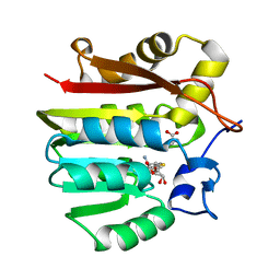 | | Guanidinoacetate methyltransferase containing S-adenosylhomocysteine and guanidinoacetate | | 分子名称: | GUANIDINO ACETATE, Guanidinoacetate N-methyltransferase, S-ADENOSYL-L-HOMOCYSTEINE | | 著者 | Komoto, J, Yamada, T, Takata, Y, Takusagawa, F. | | 登録日 | 2004-09-02 | | 公開日 | 2004-12-07 | | 最終更新日 | 2024-02-14 | | 実験手法 | X-RAY DIFFRACTION (2 Å) | | 主引用文献 | Catalytic mechanism of guanidinoacetate methyltransferase: crystal structures of guanidinoacetate methyltransferase ternary complexes.
Biochemistry, 43, 2004
|
|
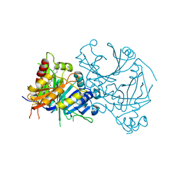
8HWD
 | | Cryo-EM Structure of D5 ADP form | | 分子名称: | ADENOSINE-5'-DIPHOSPHATE, Primase D5 | | 著者 | Li, Y.N, Zhu, J, Guo, Y.Y, Yan, R.H. | | 登録日 | 2022-12-29 | | 公開日 | 2024-01-10 | | 最終更新日 | 2024-01-31 | | 実験手法 | ELECTRON MICROSCOPY (3.3 Å) | | 主引用文献 | Structural insight into the assembly and working mechanism of helicase-primase D5 from Mpox virus.
Nat.Struct.Mol.Biol., 31, 2024
|
|
5F78
 | | Crystal structure of Mutant N87T of adenosine/Methylthioadenosine phosphorylase from Schistosoma mansoni in APO form | | 分子名称: | Methylthioadenosine phosphorylase, SULFATE ION | | 著者 | Torini, J.R.S, Brandao-Neto, J, DeMarco, R, Pereira, H.M. | | 登録日 | 2015-12-07 | | 公開日 | 2016-12-21 | | 最終更新日 | 2023-09-27 | | 実験手法 | X-RAY DIFFRACTION (1.8518 Å) | | 主引用文献 | Crystal Structure of Schistosoma mansoni Adenosine Phosphorylase/5'-Methylthioadenosine Phosphorylase and Its Importance on Adenosine Salvage Pathway.
PLoS Negl Trop Dis, 10, 2016
|
|
1X84
 | | IPP isomerase (wt) reacted with (S)-bromohydrine of IPP | | 分子名称: | (S)-4-BROMO-3-HYDROXY-3-METHYLBUTYL DIPHOSPHATE, Isopentenyl-diphosphate delta-isomerase, MAGNESIUM ION, ... | | 著者 | Wouters, J, Oldfield, E. | | 登録日 | 2004-08-17 | | 公開日 | 2005-01-25 | | 最終更新日 | 2011-07-13 | | 実験手法 | X-RAY DIFFRACTION (1.78 Å) | | 主引用文献 | A Crystallographic Investigation of Phosphoantigen Binding to Isopentenyl Pyrophosphate/Dimethylallyl Pyrophosphate Isomerase
J.Am.Chem.Soc., 127, 2005
|
|
4Q6P
 | | Structural analysis of the Zn-form I of Helicobacter pylori Csd4, a D,L-carboxypeptidase | | 分子名称: | 2,6-DIAMINOPIMELIC ACID, CALCIUM ION, Conserved hypothetical secreted protein, ... | | 著者 | Kim, H.S, Kim, J, Im, H.N, An, D.R, Lee, M, Hesek, D, Mobashery, S, Kim, J.Y, Cho, K, Yoon, H.J, Han, B.W, Lee, B.I, Suh, S.W. | | 登録日 | 2014-04-23 | | 公開日 | 2014-11-05 | | 最終更新日 | 2023-11-15 | | 実験手法 | X-RAY DIFFRACTION (2.62 Å) | | 主引用文献 | Structural basis for the recognition of muramyltripeptide by Helicobacter pylori Csd4, a D,L-carboxypeptidase controlling the helical cell shape
Acta Crystallogr.,Sect.D, 70, 2014
|
|
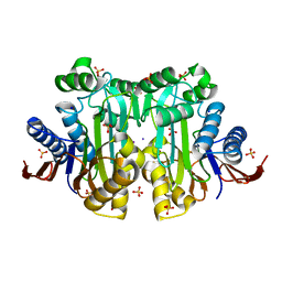
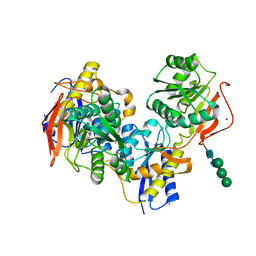
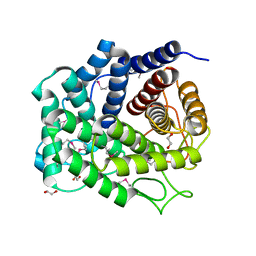
4PYK
 | | human COMT, double domain swap | | 分子名称: | CHLORIDE ION, Catechol O-methyltransferase, MAGNESIUM ION, ... | | 著者 | Ehler, A, Benz, J, Schlatter, D, Rudolph, M.G. | | 登録日 | 2014-03-27 | | 公開日 | 2014-06-11 | | 最終更新日 | 2024-04-03 | | 実験手法 | X-RAY DIFFRACTION (2.22 Å) | | 主引用文献 | Mapping the conformational space accessible to catechol-O-methyltransferase.
Acta Crystallogr.,Sect.D, 70, 2014
|
|
5EX0
 | | Crystal structure of human SMYD3 in complex with a MAP3K2 peptide | | 分子名称: | ACETIC ACID, Histone-lysine N-methyltransferase SMYD3, MAP3K2 peptide, ... | | 著者 | Fu, W, Liu, N, Qiao, Q, Wang, M, Min, J, Zhu, B, Xu, R.M, Yang, N. | | 登録日 | 2015-11-23 | | 公開日 | 2016-03-09 | | 最終更新日 | 2023-11-08 | | 実験手法 | X-RAY DIFFRACTION (2.7 Å) | | 主引用文献 | Structural Basis for Substrate Preference of SMYD3, a SET Domain-containing Protein Lysine Methyltransferase
J.Biol.Chem., 291, 2016
|
|
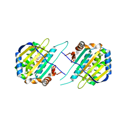
4Q88
 | | Glycosyl hydrolase family 88 from Bacteroides vulgatus | | 分子名称: | 1,2-ETHANEDIOL, SULFATE ION, Uncharacterized protein | | 著者 | Osipiuk, J, Li, H, Endres, M, Joachimiak, A, Midwest Center for Structural Genomics (MCSG) | | 登録日 | 2014-04-25 | | 公開日 | 2014-05-21 | | 最終更新日 | 2017-11-22 | | 実験手法 | X-RAY DIFFRACTION (1.73 Å) | | 主引用文献 | Glycosyl hydrolase Family 88 from Bacteroides vulgatus
To be Published
|
|


