Loading
PDBj
MenuPDBj@FacebookPDBj@TwitterPDBj@YouTubewwPDB FoundationwwPDB
RCSB PDBPDBeBMRBAdv. SearchSearch help
PDB: 134 results

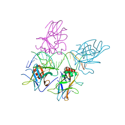
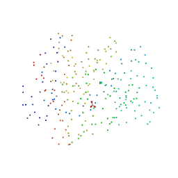
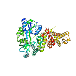
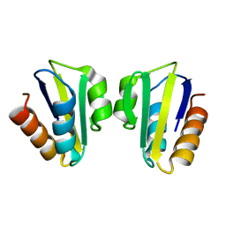
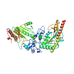
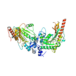
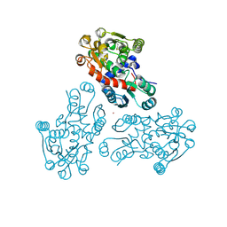
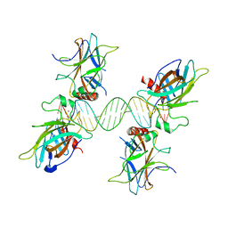
3US2
DownloadVisualize
BU of 3us2 by Molmil
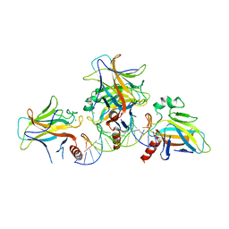
Structure of p63 DNA Binding Domain in Complex with a 19 Base Pair A/T Rich Response Element Containing Two Half Sites with a Single Base Pair Overlap
Descriptor: 5'-D(*AP*AP*AP*CP*AP*TP*GP*TP*TP*AP*AP*AP*CP*AP*TP*GP*TP*TP*T)-3', 5'-D(*AP*AP*AP*CP*AP*TP*GP*TP*TP*TP*AP*AP*CP*AP*TP*GP*TP*TP*T)-3', Tumor protein 63, ...
Authors:Chen, C, Herzberg, O.
Deposit date:2011-11-22
Release date:2012-02-01
Last modified:2024-02-28
Method:X-RAY DIFFRACTION (4.2 Å)
Cite:Pliable DNA Conformation of Response Elements Bound to Transcription Factor p63.
J.Biol.Chem., 287, 2012
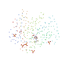
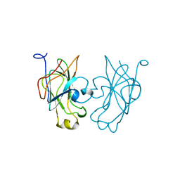
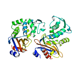
3US0
DownloadVisualize
BU of 3us0 by Molmil
Structure of p63 DNA Binding Domain in Complex with a 22 Base Pair A/T Rich Response Element Containing a Two Base Pair "AT" Spacer Between Half Sites
Descriptor: 5'-D(*AP*AP*AP*CP*AP*TP*GP*TP*TP*TP*AP*TP*AP*AP*AP*CP*AP*TP*GP*TP*TP*T)-3', Tumor protein 63, ZINC ION
Authors:Chen, C, Herzberg, O.
Deposit date:2011-11-22
Release date:2012-02-01
Last modified:2023-09-13
Method:X-RAY DIFFRACTION (2.5 Å)
Cite:Pliable DNA Conformation of Response Elements Bound to Transcription Factor p63.
J.Biol.Chem., 287, 2012
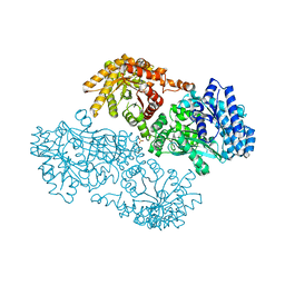
3US1
DownloadVisualize
BU of 3us1 by Molmil
Structure of p63 DNA Binding Domain in Complex with a 22 Base Pair Response Element Containing a Two Base Pair "GC" Spacer Between Half Sites
Descriptor: 5'-D(*AP*AP*AP*CP*AP*TP*GP*TP*TP*TP*GP*CP*AP*AP*AP*CP*AP*TP*GP*TP*TP*T)-3', Tumor protein 63, ZINC ION
Authors:Chen, C, Herzberg, O.
Deposit date:2011-11-22
Release date:2012-02-01
Last modified:2023-09-13
Method:X-RAY DIFFRACTION (2.8 Å)
Cite:Pliable DNA Conformation of Response Elements Bound to Transcription Factor p63.
J.Biol.Chem., 287, 2012
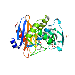
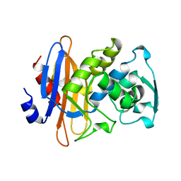
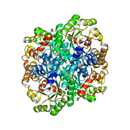
1GHM
DownloadVisualize
BU of 1ghm by Molmil
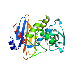
Structures of the acyl-enzyme complex of the staphylococcus aureus beta-lactamase mutant GLU166ASP:ASN170GLN with degraded cephaloridine
Descriptor: 5-METHYL-2-[2-OXO-1-(2-THIOPHEN-2-YL-ACETYLAMINO)-ETHYL]-3,6-DIHYDRO-2H-[1,3]THIAZINE-4-CARBOXYLIC ACID, BETA-LACTAMASE, CARBONATE ION, ...
Authors:Chen, C.C.H, Herzberg, O.
Deposit date:2000-12-19
Release date:2001-04-04
Last modified:2024-10-16
Method:X-RAY DIFFRACTION (1.86 Å)
Cite:Structures of the acyl-enzyme complexes of the Staphylococcus aureus beta-lactamase mutant Glu166Asp:Asn170Gln with benzylpenicillin and cephaloridine.
Biochemistry, 40, 2001
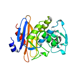
1GHP
DownloadVisualize
BU of 1ghp by Molmil
STRUCTURES OF THE ACYL-ENZYME COMPLEX OF THE STAPHYLOCOCCUS AUREUS BETA-LACTAMASE MUTANT GLU166ASP:ASN170GLN WITH DEGRADED BENZYLPENICILLIN
Descriptor: BETA-LACTAMASE, OPEN FORM - PENICILLIN G, SULFATE ION
Authors:Chen, C.C.H, Herzberg, O.
Deposit date:2000-12-21
Release date:2001-04-04
Last modified:2023-08-09
Method:X-RAY DIFFRACTION (1.76 Å)
Cite:Structures of the acyl-enzyme complexes of the Staphylococcus aureus beta-lactamase mutant Glu166Asp:Asn170Gln with benzylpenicillin and cephaloridine.
Biochemistry, 40, 2001
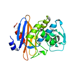
1DJB
DownloadVisualize
BU of 1djb by Molmil
STRUCTURE OF BETA-LACTAMASE PRECURSOR, S70A MUTANT, AT 298K
Descriptor: BETA-LACTAMASE
Authors:Chen, C.C.H, Herzberg, O.
Deposit date:1996-08-13
Release date:1997-03-12
Last modified:2024-02-07
Method:X-RAY DIFFRACTION (2.1 Å)
Cite:Structure and kinetics of the beta-lactamase mutants S70A and K73H from Staphylococcus aureus PC1.
Biochemistry, 35, 1996
1GHI
DownloadVisualize
BU of 1ghi by Molmil
STRUCTURE OF BETA-LACTAMASE GLU166ASP:ASN170GLN MUTANT
Descriptor: BETA-LACTAMASE, CARBONATE ION
Authors:Chen, C.C.H, Herzberg, O.
Deposit date:2000-12-18
Release date:2001-04-04
Last modified:2023-08-09
Method:X-RAY DIFFRACTION (2.3 Å)
Cite:Structures of the acyl-enzyme complexes of the Staphylococcus aureus beta-lactamase mutant Glu166Asp:Asn170Gln with benzylpenicillin and cephaloridine.
Biochemistry, 40, 2001
1GPR
DownloadVisualize
BU of 1gpr by Molmil
REFINED CRYSTAL STRUCTURE OF IIA DOMAIN OF THE GLUCOSE PERMEASE OF BACILLUS SUBTILIS AT 1.9 ANGSTROMS RESOLUTION
Descriptor: GLUCOSE PERMEASE
Authors:Liao, D.-I, Herzberg, O.
Deposit date:1991-09-25
Release date:1993-10-31
Last modified:2024-02-07
Method:X-RAY DIFFRACTION (1.9 Å)
Cite:An atomic model for protein-protein phosphoryl group transfer.
J.Biol.Chem., 267, 1992
1KGF
DownloadVisualize
BU of 1kgf by Molmil
STRUCTURE OF BETA-LACTAMASE ASN 170 GLN MUTANT
Descriptor: BETA-LACTAMASE
Authors:Chen, C.C.H, Zawadzke, L.E, Herzberg, O.
Deposit date:1996-10-17
Release date:1997-03-12
Last modified:2024-02-07
Method:X-RAY DIFFRACTION (2.2 Å)
Cite:Elimination of the hydrolytic water molecule in a class A beta-lactamase mutant: crystal structure and kinetics.
Biochemistry, 35, 1996
1KGE
DownloadVisualize
BU of 1kge by Molmil
STRUCTURE OF BETA-LACTAMASE ASN 170 MET MUTANT
Descriptor: BETA-LACTAMASE
Authors:Chen, C.C.H, Zawadzke, L.E, Herzberg, O.
Deposit date:1996-10-17
Release date:1997-04-01
Last modified:2024-02-07
Method:X-RAY DIFFRACTION (2 Å)
Cite:Elimination of the hydrolytic water molecule in a class A beta-lactamase mutant: crystal structure and kinetics.
Biochemistry, 35, 1996
3B8I
DownloadVisualize
BU of 3b8i by Molmil
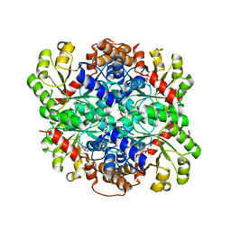
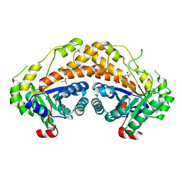
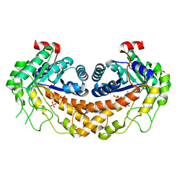
Crystal Structure of Oxaloacetate Decarboxylase from Pseudomonas Aeruginosa (PA4872) in complex with oxalate and Mg2+.
Descriptor: GLYCEROL, MAGNESIUM ION, OXALATE ION, ...
Authors:Narayanan, B.C, Herzberg, O.
Deposit date:2007-11-01
Release date:2008-01-01
Last modified:2024-02-21
Method:X-RAY DIFFRACTION (1.9 Å)
Cite:Structure and function of PA4872 from Pseudomonas aeruginosa, a novel class of oxaloacetate decarboxylase from the PEP mutase/isocitrate lyase superfamily.
Biochemistry, 47, 2008
5TIB
DownloadVisualize
BU of 5tib by Molmil
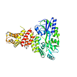
Gasdermin-B C-terminal domain containing the polymorphism residues Arg299:Ser306 fused to maltose binding protein
Descriptor: ACETATE ION, SODIUM ION, Sugar ABC transporter substrate-binding protein,Gasdermin-B, ...
Authors:Chao, K, Herzberg, O.
Deposit date:2016-10-01
Release date:2017-02-01
Last modified:2023-10-04
Method:X-RAY DIFFRACTION (2.6 Å)
Cite:Human Gasdermin-B and disease: Sulfatide Binding, Caspase cleavage, and Structural impact of Asthma- and IBS-Associated Polymorphism
Proc.Natl.Acad.Sci.Usa, 2017
5TJ4
DownloadVisualize
BU of 5tj4 by Molmil
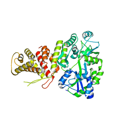
Gasdermin-B C-terminal domain containing the polymorphism residues Gly299:Pro306 fused to maltose binding protein
Descriptor: SODIUM ION, Sugar ABC transporter substrate-binding protein,Gasdermin-B fusion protein, alpha-D-glucopyranose-(1-4)-alpha-D-glucopyranose
Authors:Chao, L.K, Herzberg, O.
Deposit date:2016-10-03
Release date:2017-02-01
Last modified:2023-10-04
Method:X-RAY DIFFRACTION (3.5 Å)
Cite:Gene polymorphism linked to increased asthma and IBD risk alters gasdermin-B structure, a sulfatide and phosphoinositide binding protein.
Proc. Natl. Acad. Sci. U.S.A., 114, 2017
3GAY
DownloadVisualize
BU of 3gay by Molmil
Structure of Giardia fructose-1,6-biphosphate aldolase in complex with tagatose-1,6-biphosphate
Descriptor: 1,6-di-O-phosphono-D-tagatose, Fructose-bisphosphate aldolase, ZINC ION
Authors:Galkin, A, Herzberg, O.
Deposit date:2009-02-18
Release date:2009-03-31
Last modified:2023-09-06
Method:X-RAY DIFFRACTION (1.8 Å)
Cite:Structural insights into the substrate binding and stereoselectivity of giardia fructose-1,6-bisphosphate aldolase.
Biochemistry, 48, 2009
5TJ2
DownloadVisualize
BU of 5tj2 by Molmil
Gasdermin-B C-terminal domain containing the polymorphism residues Gly299:Ser306 fused to maltose binding protein
Descriptor: Sugar ABC transporter substrate-binding protein,Gasdermin-B fusion protein, alpha-D-glucopyranose-(1-4)-alpha-D-glucopyranose
Authors:Chao, L.K, Herzberg, O.
Deposit date:2016-10-03
Release date:2017-02-01
Last modified:2023-10-04
Method:X-RAY DIFFRACTION (2.8 Å)
Cite:Human Gasdermin-B and disease: Sulfatide Binding, Caspase cleavage, and Structural impact of Asthma- and IBS-Associated Polymorphism
Proc.Natl.Acad.Sci.Usa, 2017
1SPH
DownloadVisualize
BU of 1sph by Molmil
REFINED STRUCTURES OF THE ACTIVE S83C AND IMPAIRED S46D HPRS: EVIDENCE THAT PHOSPHORYLATION DOES NOT REQUIRE A BACKBONE CONFORMATIONAL TRANSITION
Descriptor: HISTIDINE-CONTAINING PHOSPHOCARRIER PROTEIN HPR
Authors:Liao, D.-I, Herzberg, O.
Deposit date:1994-11-03
Release date:1995-02-07
Last modified:2024-02-14
Method:X-RAY DIFFRACTION (2 Å)
Cite:Refined structures of the active Ser83-->Cys and impaired Ser46-->Asp histidine-containing phosphocarrier proteins.
Structure, 2, 1994
3L2D
DownloadVisualize
BU of 3l2d by Molmil
Glycocyamine kinase, beta-beta homodimer from marine worm Namalycastis sp.
Descriptor: Glycocyamine kinase beta chain
Authors:Lim, K, Pullalarevu, S, Herzberg, O.
Deposit date:2009-12-15
Release date:2010-03-02
Last modified:2023-09-06
Method:X-RAY DIFFRACTION (2.4 Å)
Cite:Structural basis for the mechanism and substrate specificity of glycocyamine kinase, a phosphagen kinase family member.
Biochemistry, 49, 2010
3L2E
DownloadVisualize
BU of 3l2e by Molmil
Glycocyamine kinase, alpha-beta heterodimer from marine worm Namalycastis sp.
Descriptor: Glycocyamine kinase alpha chain, Glycocyamine kinase beta chain
Authors:Lim, K, Pullalarevu, S, Herzberg, O.
Deposit date:2009-12-15
Release date:2010-03-02
Last modified:2023-09-06
Method:X-RAY DIFFRACTION (2.6 Å)
Cite:Structural basis for the mechanism and substrate specificity of glycocyamine kinase, a phosphagen kinase family member.
Biochemistry, 49, 2010
3GRF
DownloadVisualize
BU of 3grf by Molmil
X-ray Structure of Ornithine Transcarbamoylase from Giardia lamblia
Descriptor: NICKEL (II) ION, Ornithine carbamoyltransferase
Authors:Galkin, A, Herzberg, O.
Deposit date:2009-03-25
Release date:2009-06-30
Last modified:2024-10-16
Method:X-RAY DIFFRACTION (2 Å)
Cite:X-ray structure and kinetic properties of ornithine transcarbamoylase from the human parasite Giardia lamblia.
Proteins, 76, 2009
1OME
DownloadVisualize
BU of 1ome by Molmil
CRYSTAL STRUCTURE OF THE OMEGA LOOP DELETION MUTANT (RESIDUES 163-178 DELETED) OF BETA-LACTAMASE FROM STAPHYLOCOCCUS AUREUS PC1
Descriptor: BETA-LACTAMASE, CHLORIDE ION
Authors:Banerjee, S, Pieper, U, Herzberg, O.
Deposit date:1998-02-09
Release date:1998-05-27
Last modified:2024-05-22
Method:X-RAY DIFFRACTION (2.3 Å)
Cite:Role of the omega-loop in the activity, substrate specificity, and structure of class A beta-lactamase.
Biochemistry, 37, 1998
1KC7
DownloadVisualize
BU of 1kc7 by Molmil
Pyruvate Phosphate Dikinase with Bound Mg-phosphonopyruvate
Descriptor: MAGNESIUM ION, PHOSPHONOPYRUVATE, SULFATE ION, ...
Authors:Chen, C.C, Howard, A, Herzberg, O.
Deposit date:2001-11-07
Release date:2002-01-30
Last modified:2023-08-16
Method:X-RAY DIFFRACTION (2.2 Å)
Cite:Pyruvate site of pyruvate phosphate dikinase: crystal structure of the enzyme-phosphonopyruvate complex, and mutant analysis
Biochemistry, 41, 2002
3FA3
DownloadVisualize
BU of 3fa3 by Molmil
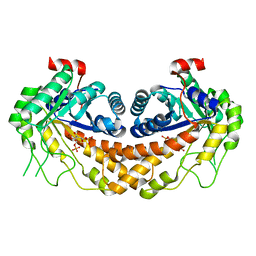
Crystal structure of 2,3-dimethylmalate lyase, a PEP mutase/isocitrate lyase superfamily member, trigonal crystal form
Descriptor: 2,2-difluoro-3,3-dihydroxybutanedioic acid, 2,3-dimethylmalate lyase, GLYCEROL, ...
Authors:Narayanan, B.C, Herzberg, O.
Deposit date:2008-11-14
Release date:2009-01-27
Last modified:2023-12-27
Method:X-RAY DIFFRACTION (2.6 Å)
Cite:Structure and function of 2,3-dimethylmalate lyase, a PEP mutase/isocitrate lyase superfamily member.
J.Mol.Biol., 386, 2009
3GAK
DownloadVisualize
BU of 3gak by Molmil
Structure of Giardia fructose-1,6-biphosphate aldolase
Descriptor: Fructose-bisphosphate aldolase, SULFATE ION, ZINC ION
Authors:Galkin, A, Herzberg, O.
Deposit date:2009-02-17
Release date:2009-03-31
Last modified:2023-09-06
Method:X-RAY DIFFRACTION (2.9 Å)
Cite:Structural insights into the substrate binding and stereoselectivity of giardia fructose-1,6-bisphosphate aldolase.
Biochemistry, 48, 2009
1KGG
DownloadVisualize
BU of 1kgg by Molmil
STRUCTURE OF BETA-LACTAMASE GLU166GLN:ASN170ASP MUTANT
Descriptor: PROTEIN (BETA-LACTAMASE), SULFATE ION
Authors:Chen, C.C.H, Herzberg, O.
Deposit date:1999-05-20
Release date:1999-05-28
Last modified:2023-08-16
Method:X-RAY DIFFRACTION (2.3 Å)
Cite:Relocation of the catalytic carboxylate group in class A beta-lactamase: the structure and function of the mutant enzyme Glu166-->Gln:Asn170-->Asp.
Protein Eng., 12, 1999
3GB6
DownloadVisualize
BU of 3gb6 by Molmil
Structure of Giardia fructose-1,6-biphosphate aldolase D83A mutant in complex with fructose-1,6-bisphosphate
Descriptor: 1,6-di-O-phosphono-D-fructose, Fructose-bisphosphate aldolase, ZINC ION
Authors:Galkin, A, Herzberg, O.
Deposit date:2009-02-18
Release date:2009-03-31
Last modified:2023-09-06
Method:X-RAY DIFFRACTION (2 Å)
Cite:Structural insights into the substrate binding and stereoselectivity of giardia fructose-1,6-bisphosphate aldolase.
Biochemistry, 48, 2009

226707

PDB entries from 2024-10-30

PDB statisticsPDBj update infoContact PDBjnumon