Loading
PDBj
メニューPDBj@FacebookPDBj@TwitterPDBj@YouTubewwPDB FoundationwwPDB
RCSB PDBPDBeBMRBAdv. SearchSearch help
PDB: 160 件

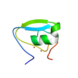
3NY1
DownloadVisualize
BU of 3ny1 by Molmil
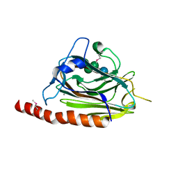
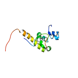
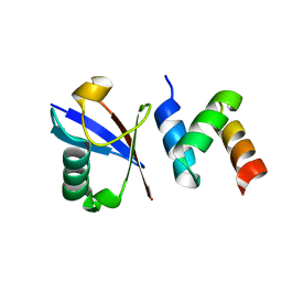
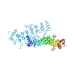
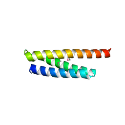
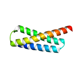
Structure of the ubr-box of the UBR1 ubiquitin ligase
分子名称: E3 ubiquitin-protein ligase UBR1, ZINC ION
著者Matta-Camacho, E, Kozlov, G, Li, F, Gehring, K.
登録日2010-07-14
公開日2010-08-11
最終更新日2024-02-21
実験手法X-RAY DIFFRACTION (2.085 Å)
主引用文献Structural basis of substrate recognition and specificity in the N-end rule pathway.
Nat.Struct.Mol.Biol., 17, 2010
3O0X
DownloadVisualize
BU of 3o0x by Molmil
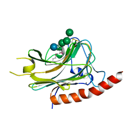
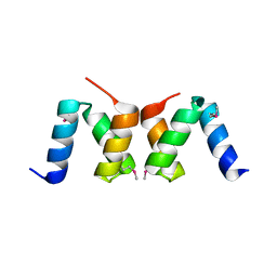
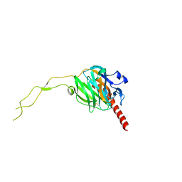
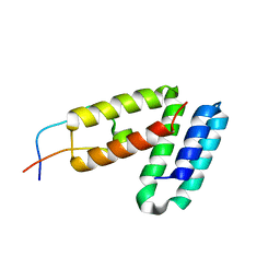
Structural basis of carbohydrate recognition by calreticulin
分子名称: CALCIUM ION, Calreticulin, alpha-D-glucopyranose-(1-3)-alpha-D-mannopyranose-(1-2)-alpha-D-mannopyranose-(1-2)-alpha-D-mannopyranose
著者Kozlov, G, Gehring, K.
登録日2010-07-20
公開日2010-09-29
最終更新日2023-12-06
実験手法X-RAY DIFFRACTION (2.01 Å)
主引用文献Structural basis of carbohydrate recognition by calreticulin.
J.Biol.Chem., 285, 2010
3NY2
DownloadVisualize
BU of 3ny2 by Molmil
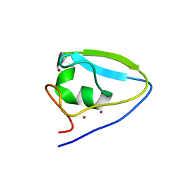
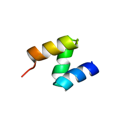
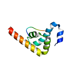
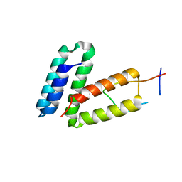
Structure of the ubr-box of UBR2 ubiquitin ligase
分子名称: E3 ubiquitin-protein ligase UBR2, ZINC ION
著者Matta-Camacho, E, Kozlov, G, Li, F, Gehring, K.
登録日2010-07-14
公開日2010-08-11
最終更新日2024-02-21
実験手法X-RAY DIFFRACTION (2.61 Å)
主引用文献Structural basis of substrate recognition and specificity in the N-end rule pathway.
Nat.Struct.Mol.Biol., 17, 2010
3O0W
DownloadVisualize
BU of 3o0w by Molmil
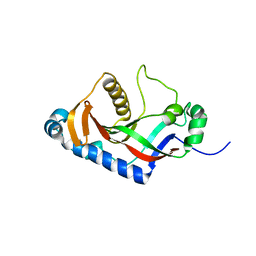
Structural basis of carbohydrate recognition by calreticulin
分子名称: CALCIUM ION, Calreticulin, alpha-D-glucopyranose-(1-3)-alpha-D-mannopyranose-(1-2)-alpha-D-mannopyranose-(1-2)-alpha-D-mannopyranose
著者Kozlov, G, Gehring, K.
登録日2010-07-20
公開日2010-09-29
最終更新日2020-07-29
実験手法X-RAY DIFFRACTION (1.95 Å)
主引用文献Structural basis of carbohydrate recognition by calreticulin.
J.Biol.Chem., 285, 2010
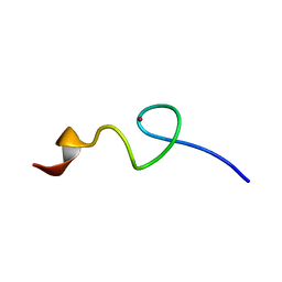
3NY3
DownloadVisualize
BU of 3ny3 by Molmil
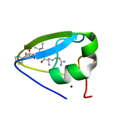
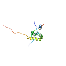
Structure of the ubr-box of UBR2 in complex with N-degron
分子名称: E3 ubiquitin-protein ligase UBR2, N-degron, ZINC ION
著者Matta-Camacho, E, Kozlov, G, Li, F, Gehring, K.
登録日2010-07-14
公開日2010-08-11
最終更新日2024-02-21
実験手法X-RAY DIFFRACTION (1.6 Å)
主引用文献Structural basis of substrate recognition and specificity in the N-end rule pathway.
Nat.Struct.Mol.Biol., 17, 2010
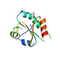
3UVT
DownloadVisualize
BU of 3uvt by Molmil
Crystal structure of the third catalytic domain of ERp46
分子名称: SULFATE ION, Thioredoxin domain-containing protein 5
著者Kozlov, G, Gulerez, I.E, Gehring, K.
登録日2011-11-30
公開日2012-04-11
最終更新日2023-09-13
実験手法X-RAY DIFFRACTION (2 Å)
主引用文献Structure of the third catalytic domain of the protein disulfide isomerase ERp46.
Acta Crystallogr.,Sect.F, 68, 2012
4Z2Z
DownloadVisualize
BU of 4z2z by Molmil
New crystal structure of yeast Ddi1 aspartyl protease reveals substrate engagement mode
分子名称: DNA damage-inducible protein 1
著者Trempe, J.-F, Feng, X, Gehring, K.
登録日2015-03-30
公開日2016-04-13
最終更新日2023-09-27
実験手法X-RAY DIFFRACTION (1.8 Å)
主引用文献Structural studies of the yeast DNA damage-inducible protein Ddi1 reveal domain architecture of this eukaryotic protein family.
Sci Rep, 6, 2016
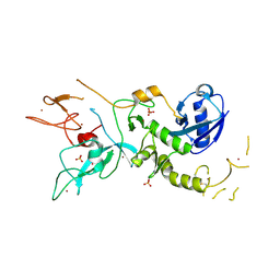
4ZYN
DownloadVisualize
BU of 4zyn by Molmil
Crystal Structure of Parkin E3 ubiquitin ligase (linker deletion; delta 86-130)
分子名称: E3 ubiquitin-protein ligase parkin, SULFATE ION, ZINC ION
著者Lilov, A, Sauve, V, Trempe, J.F, Rodionov, D, Wang, J, Gehring, K.
登録日2015-05-21
公開日2015-08-19
最終更新日2023-09-27
実験手法X-RAY DIFFRACTION (2.54 Å)
主引用文献A Ubl/ubiquitin switch in the activation of Parkin.
Embo J., 34, 2015
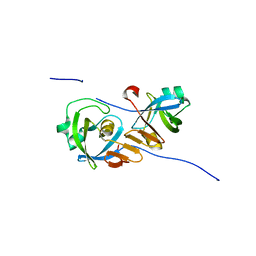
2MJC
DownloadVisualize
BU of 2mjc by Molmil
Zn-binding domain of eukaryotic translation initiation factor 3, subunit G
分子名称: Eukaryotic translation initiation factor 3 subunit G, ZINC ION
著者Al-Abdul-Wahid, M, Menade, M, Xie, J, Kozlov, G, Gehring, K.
登録日2014-01-03
公開日2015-01-07
最終更新日2024-05-15
実験手法SOLUTION NMR
主引用文献Solution NMR structure of the Zn-binding domain of eukaryotic translation initiation factor 3, subunit G
To be Published
1JGN
DownloadVisualize
BU of 1jgn by Molmil
Solution structure of the C-terminal PABC domain of human poly(A)-binding protein in complex with the peptide from Paip2
分子名称: polyadenylate-binding protein 1, polyadenylate-binding protein-interacting protein 2
著者Kozlov, G, Siddiqui, N, Coillet-Matillon, S, Ekiel, I, Gehring, K.
登録日2001-06-26
公開日2003-06-24
最終更新日2024-05-22
実験手法SOLUTION NMR
主引用文献Structural basis of ligand recognition by PABC, a highly specific peptide-binding domain found in poly(A)-binding protein and a HECT ubiquitin ligase
EMBO J., 23, 2004
2OO9
DownloadVisualize
BU of 2oo9 by Molmil
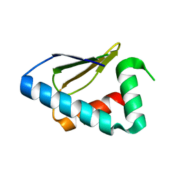
crystal structure of the UBA domain from human c-Cbl ubiquitin ligase
分子名称: E3 ubiquitin-protein ligase CBL
著者Kozlov, G, Gehring, K.
登録日2007-01-25
公開日2007-02-06
最終更新日2023-12-27
実験手法X-RAY DIFFRACTION (2.1 Å)
主引用文献Structural basis for UBA-mediated dimerization of c-Cbl ubiquitin ligase.
J.Biol.Chem., 282, 2007
2OOA
DownloadVisualize
BU of 2ooa by Molmil
crystal structure of the UBA domain from Cbl-b ubiquitin ligase
分子名称: E3 ubiquitin-protein ligase CBL-B
著者Kozlov, G, Gehring, K.
登録日2007-01-25
公開日2007-02-06
最終更新日2023-12-27
実験手法X-RAY DIFFRACTION (1.56 Å)
主引用文献Structural basis for ubiquitin-mediated dimerization and activation of the ubiquitin protein ligase Cbl-b.
Mol.Cell, 27, 2007
1JH4
DownloadVisualize
BU of 1jh4 by Molmil
Solution structure of the C-terminal PABC domain of human poly(A)-binding protein in complex with the peptide from Paip1
分子名称: polyadenylate-binding protein 1, polyadenylate-binding protein-interacting protein-1
著者Kozlov, G, Siddiqui, N, Coillet-Matillon, S, Ekiel, I, Gehring, K.
登録日2001-06-27
公開日2003-06-24
最終更新日2024-05-22
実験手法SOLUTION NMR
主引用文献Structural basis of ligand recognition by PABC, a highly specific peptide-binding domain found in poly(A)-binding protein and a HECT ubiquitin ligase
EMBO J., 23, 2004
2OOB
DownloadVisualize
BU of 2oob by Molmil
crystal structure of the UBA domain from Cbl-b ubiquitin ligase in complex with ubiquitin
分子名称: E3 ubiquitin-protein ligase CBL-B, Ubiquitin
著者Kozlov, G, Gehring, K.
登録日2007-01-25
公開日2007-02-06
最終更新日2023-08-30
実験手法X-RAY DIFFRACTION (1.9 Å)
主引用文献Structural basis for ubiquitin-mediated dimerization and activation of the ubiquitin protein ligase Cbl-b.
Mol.Cell, 27, 2007
3PDZ
DownloadVisualize
BU of 3pdz by Molmil
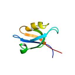
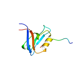
SOLUTION STRUCTURE OF THE PDZ2 DOMAIN FROM HUMAN PHOSPHATASE HPTP1E
分子名称: PROTEIN (TYROSINE PHOSPHATASE (PTP-BAS, TYPE 1))
著者Kozlov, G, Gehring, K, Ekiel, I.
登録日1999-05-10
公開日2000-03-17
最終更新日2023-12-27
実験手法SOLUTION NMR
主引用文献Solution structure of the PDZ2 domain from human phosphatase hPTP1E and its interactions with C-terminal peptides from the Fas receptor.
Biochemistry, 39, 2000
3PT3
DownloadVisualize
BU of 3pt3 by Molmil
Crystal structure of the C-terminal lobe of the human UBR5 HECT domain
分子名称: E3 ubiquitin-protein ligase UBR5
著者Matta-Camacho, E, Kozlov, G, Menade, M, Gehring, K.
登録日2010-12-02
公開日2012-01-25
最終更新日2023-09-06
実験手法X-RAY DIFFRACTION (1.97 Å)
主引用文献Structure of the HECT C-lobe of the UBR5 E3 ubiquitin ligase.
Acta Crystallogr.,Sect.F, 68, 2012
1D5G
DownloadVisualize
BU of 1d5g by Molmil
SOLUTION STRUCTURE OF THE PDZ2 DOMAIN FROM HUMAN PHOSPHATASE HPTP1E COMPLEXED WITH A PEPTIDE
分子名称: HUMAN PHOSPHATASE HPTP1E, PEPTIDE FADSEADENEQVSAV
著者Kozlov, G, Gehring, K, Ekiel, I.
登録日1999-10-07
公開日2002-07-24
最終更新日2024-05-22
実験手法SOLUTION NMR
主引用文献Solution Structure of the PDZ2 Domain from Cytosolic Human Phosphatase hPTP1E Complexed with a Peptide Reveals Contribution of the beta2-beta3 Loop to PDZ Domain-Ligand Interactions
J.Mol.Biol., 320, 2002
5VRQ
DownloadVisualize
BU of 5vrq by Molmil
Crystal structure of Legionella pneumophila effector AnkC
分子名称: Ankyrin repeat-containing protein
著者Kozlov, G, Wong, K, Wang, W, Skubak, P, Munoz-Escobar, J, Liu, Y, Pannu, N.S, Gehring, K, Montreal-Kingston Bacterial Structural Genomics Initiative (BSGI)
登録日2017-05-11
公開日2017-11-29
最終更新日2024-03-13
実験手法X-RAY DIFFRACTION (3.205 Å)
主引用文献Ankyrin repeats as a dimerization module.
Biochem. Biophys. Res. Commun., 495, 2018
3PKN
DownloadVisualize
BU of 3pkn by Molmil
Crystal structure of MLLE domain of poly(A) binding protein in complex with PAM2 motif of La-related protein 4 (LARP4)
分子名称: IODIDE ION, La-related protein 4, Polyadenylate-binding protein 1, ...
著者Xie, J, Kozlov, G, Gehring, K.
登録日2010-11-11
公開日2011-01-12
最終更新日2023-09-06
実験手法X-RAY DIFFRACTION (1.8 Å)
主引用文献La-Related Protein 4 Binds Poly(A), Interacts with the Poly(A)-Binding Protein MLLE Domain via a Variant PAM2w Motif, and Can Promote mRNA Stability.
Mol.Cell.Biol., 31, 2011
5WD8
DownloadVisualize
BU of 5wd8 by Molmil
Crystal structure of Legionella pneumophila effector lpg2328
分子名称: Lem22
著者Kozlov, G, Wong, K, Gehring, K, Montreal-Kingston Bacterial Structural Genomics Initiative (BSGI)
登録日2017-07-04
公開日2017-11-29
最終更新日2020-01-08
実験手法X-RAY DIFFRACTION (1.944 Å)
主引用文献Crystal structure of the Legionella effector Lem22.
Proteins, 86, 2018
3RG0
DownloadVisualize
BU of 3rg0 by Molmil
Structural and functional relationships between the lectin and arm domains of calreticulin
分子名称: CALCIUM ION, Calreticulin
著者Kozlov, G, Pocanschi, C.L, Brockmeier, U, Williams, D.B, Gehring, K.
登録日2011-04-07
公開日2011-06-01
最終更新日2023-09-13
実験手法X-RAY DIFFRACTION (2.57 Å)
主引用文献Structural and Functional Relationships between the Lectin and Arm Domains of Calreticulin.
J.Biol.Chem., 286, 2011
5WD9
DownloadVisualize
BU of 5wd9 by Molmil
Crystal structure of Legionella pneumophila effector lpg2328
分子名称: Lem22
著者Kozlov, G, Wong, K, Gehring, K, Montreal-Kingston Bacterial Structural Genomics Initiative (BSGI)
登録日2017-07-04
公開日2017-11-29
最終更新日2024-04-03
実験手法X-RAY DIFFRACTION (1.4 Å)
主引用文献Crystal structure of the Legionella effector Lem22.
Proteins, 86, 2018
5V8Z
DownloadVisualize
BU of 5v8z by Molmil
Crystal structure of ERp29 D-domain in complex with the P-domain of calmegin
分子名称: Calmegin, Endoplasmic reticulum resident protein 29
著者Kozlov, G, Munoz-Escobar, J, Gehring, K.
登録日2017-03-22
公開日2017-06-21
最終更新日2023-10-04
実験手法X-RAY DIFFRACTION (2.105 Å)
主引用文献Mapping the ER Interactome: The P Domains of Calnexin and Calreticulin as Plurivalent Adapters for Foldases and Chaperones.
Structure, 25, 2017
5V90
DownloadVisualize
BU of 5v90 by Molmil
Crystal structure of ERp29 D-domain in complex with the P-domain of calreticulin
分子名称: Calreticulin, Endoplasmic reticulum resident protein 29, GLYCEROL
著者Kozlov, G, Munoz-Escobar, J, Gehring, K.
登録日2017-03-22
公開日2017-06-21
最終更新日2023-10-04
実験手法X-RAY DIFFRACTION (3.255 Å)
主引用文献Mapping the ER Interactome: The P Domains of Calnexin and Calreticulin as Plurivalent Adapters for Foldases and Chaperones.
Structure, 25, 2017
2ILX
DownloadVisualize
BU of 2ilx by Molmil
Solution structure of catalytic domain of rat 2',3'-cyclic-nucleotide 3'-phosphodiesterase (CNP) protein
分子名称: 2',3'-cyclic-nucleotide 3'-phosphodiesterase
著者Denisov, A.Y, Kozlov, G, Gehring, K.
登録日2006-10-03
公開日2007-03-06
最終更新日2024-05-29
実験手法SOLUTION NMR
主引用文献Solution structure of the catalytic domain of RICH protein from goldfish.
Febs J., 274, 2007

221051

件を2024-06-12に公開中

PDB statisticsPDBj update infoContact PDBjnumon