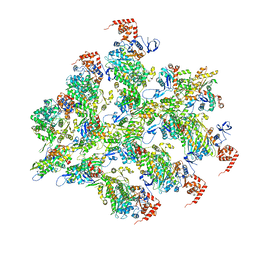
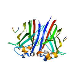
Loading
PDBj
メニューPDBj@FacebookPDBj@TwitterPDBj@YouTubewwPDB FoundationwwPDB
RCSB PDBPDBeBMRBAdv. SearchSearch help
PDB: 1718 件

8JA0
DownloadVisualize
BU of 8ja0 by Molmil
Cryo-EM structure of the NmeCas9-sgRNA-AcrIIC4 ternary complex
分子名称: CRISPR-associated endonuclease Cas9, RNA (117-MER), Uncharacterized protein
著者Yin, H, Li, Z, Yu, G.M, Li, X.Z.
登録日2023-05-05
公開日2023-08-09
最終更新日2024-07-03
実験手法ELECTRON MICROSCOPY (3.52 Å)
主引用文献Cryo-EM structure of the NmeCas9-sgRNA-AcrIIC4 ternary complex
To Be Published
7QRC
DownloadVisualize
BU of 7qrc by Molmil
X-ray structure of Trypanosoma cruzi PEX14 in complex with a PEX5-PEX14 PPI inhibitor
分子名称: GLYCEROL, Peroxin-14, ~{N}-(5-ethyl-6-oxidanylidene-benzo[b][1,4]benzothiazepin-2-yl)-2-(4-fluorophenyl)ethanamide
著者Napolitano, V, Popowicz, G.M, Dawidowski, M, Dubin, G.
登録日2022-01-10
公開日2022-11-23
最終更新日2024-01-31
実験手法X-RAY DIFFRACTION (2.18 Å)
主引用文献Structure-based design, synthesis and evaluation of a novel family of PEX5-PEX14 interaction inhibitors against Trypanosoma.
Eur.J.Med.Chem., 243, 2022
7REX
DownloadVisualize
BU of 7rex by Molmil
PreQ1-1 (type-1) riboswitch in complex with tandem stacked metabolites
分子名称: 7-DEAZA-7-AMINOMETHYL-GUANINE, MAGNESIUM ION, RNA (34-MER)
著者Jenkins, J.L, Schroeder, G.M, Wedekind, J.E.
登録日2021-07-13
公開日2021-12-29
最終更新日2023-10-18
実験手法X-RAY DIFFRACTION (2.6 Å)
主引用文献A small RNA that cooperatively senses two stacked metabolites in one pocket for gene control
Nat Commun, 13, 2022
1MDI
DownloadVisualize
BU of 1mdi by Molmil
HIGH RESOLUTION SOLUTION NMR STRUCTURE OF MIXED DISULFIDE INTERMEDIATE BETWEEN MUTANT HUMAN THIOREDOXIN AND A 13 RESIDUE PEPTIDE COMPRISING ITS TARGET SITE IN HUMAN NFKB
分子名称: TARGET SITE IN HUMAN NFKB, THIOREDOXIN
著者Clore, G.M, Qin, J, Gronenborn, A.M.
登録日1995-02-27
公開日1995-06-03
最終更新日2022-02-23
実験手法SOLUTION NMR
主引用文献Solution structure of human thioredoxin in a mixed disulfide intermediate complex with its target peptide from the transcription factor NF kappa B.
Structure, 3, 1995
1MDK
DownloadVisualize
BU of 1mdk by Molmil
HIGH RESOLUTION SOLUTION NMR STRUCTURE OF MIXED DISULFIDE INTERMEDIATE BETWEEN HUMAN THIOREDOXIN (C35A, C62A, C69A, C73A) MUTANT AND A 13 RESIDUE PEPTIDE COMPRISING ITS TARGET SITE IN HUMAN NFKB (RESIDUES 56-68 OF THE P50 SUBUNIT OF NFKB)
分子名称: TARGET SITE IN HUMAN NFKB, THIOREDOXIN
著者Clore, G.M, Qin, J, Gronenborn, A.M.
登録日1995-02-27
公開日1995-06-03
最終更新日2022-02-23
実験手法SOLUTION NMR
主引用文献Solution structure of human thioredoxin in a mixed disulfide intermediate complex with its target peptide from the transcription factor NF kappa B.
Structure, 3, 1995
1MDJ
DownloadVisualize
BU of 1mdj by Molmil
HIGH RESOLUTION SOLUTION NMR STRUCTURE OF MIXED DISULFIDE INTERMEDIATE BETWEEN HUMAN THIOREDOXIN (C35A, C62A, C69A, C73A) MUTANT AND A 13 RESIDUE PEPTIDE COMPRISING ITS TARGET SITE IN HUMAN NFKB (RESIDUES 56-68 OF THE P50 SUBUNIT OF NFKB)
分子名称: TARGET SITE IN HUMAN NFKB, THIOREDOXIN
著者Clore, G.M, Qin, J, Gronenborn, A.M.
登録日1995-02-27
公開日1995-06-03
最終更新日2022-02-23
実験手法SOLUTION NMR
主引用文献Solution structure of human thioredoxin in a mixed disulfide intermediate complex with its target peptide from the transcription factor NF kappa B.
Structure, 3, 1995
7AF2
DownloadVisualize
BU of 7af2 by Molmil
Salmonella typhimurium neuraminidase mutant (D62G)
分子名称: GLYCEROL, PHOSPHATE ION, Sialidase
著者Salinger, M.T, Kuhn, P, Laver, W.G, Pape, T, Schneider, T.R, Sheldrick, G.M, Vimr, E.R, Garman, E.F.
登録日2020-09-19
公開日2020-09-30
最終更新日2024-01-31
実験手法X-RAY DIFFRACTION (0.792 Å)
主引用文献Salmonella typhimurium neuraminidase mutant (D62G)
To Be Published
7AEY
DownloadVisualize
BU of 7aey by Molmil
Salmonella typhimurium neuraminidase in complex with isocarba-DANA.
分子名称: (3~{S},4~{S},5~{R})-4-acetamido-3-oxidanyl-5-[(1~{S},2~{R})-1,2,3-tris(oxidanyl)propyl]cyclohexane-1-carboxylic acid, GLYCEROL, PHOSPHATE ION, ...
著者Salinger, M.T, Kuhn, P, Laver, W.G, Pape, T, Schneider, T.R, Sheldrick, G.M, Vasella, A.T, Vimr, E.R, Vorwerk, S, Garman, E.F.
登録日2020-09-18
公開日2020-10-21
最終更新日2024-01-31
実験手法X-RAY DIFFRACTION (0.919 Å)
主引用文献Salmonella typhimurium neuraminidase in complex with isocarba-DANA.
To Be Published
3SAK
DownloadVisualize
BU of 3sak by Molmil
HIGH RESOLUTION SOLUTION NMR STRUCTURE OF THE OLIGOMERIZATION DOMAIN OF P53 BY MULTI-DIMENSIONAL NMR (SAC STRUCTURES)
分子名称: PROTEIN (TUMOR SUPPRESSOR P53)
著者Clore, G.M.
登録日1999-04-30
公開日1999-06-25
最終更新日2023-12-27
実験手法SOLUTION NMR
主引用文献Improving the Packing and Accuracy of NMR Structure with a Pseudopotential for the Radius of Gyration
J.Am.Chem.Soc., 121, 1999
6BNQ
DownloadVisualize
BU of 6bnq by Molmil
CryoEM structure of Myosin VI-Actin complex in the ADP state
分子名称: ADENOSINE-5'-DIPHOSPHATE, Actin, alpha skeletal muscle, ...
著者Gurel, P.G, Alushin, G.M.
登録日2017-11-17
公開日2018-01-10
最終更新日2024-03-13
実験手法ELECTRON MICROSCOPY (5.5 Å)
主引用文献Cryo-EM structures reveal specialization at the myosin VI-actin interface and a mechanism of force sensitivity.
Elife, 6, 2017
6BNW
DownloadVisualize
BU of 6bnw by Molmil
CryoEM structure of Myosin VI-Actin complex in the ADP state, backbone-averaged with side chains truncated to alanine
分子名称: Actin, alpha skeletal muscle, Unconventional myosin-VI
著者Gurel, P.G, Alushin, G.M.
登録日2017-11-17
公開日2018-01-10
最終更新日2024-03-13
実験手法ELECTRON MICROSCOPY (5.5 Å)
主引用文献Cryo-EM structures reveal specialization at the myosin VI-actin interface and a mechanism of force sensitivity.
Elife, 6, 2017
8QAO
DownloadVisualize
BU of 8qao by Molmil
Crystal structure of TP901-1 CI-NTD89 repressor N-terminal domain
分子名称: CI
著者Huang, Z, Hamad, G.M, Lo Leggio, L, Varming, A.K.
登録日2023-08-23
公開日2024-02-28
最終更新日2024-06-19
実験手法X-RAY DIFFRACTION (1.29 Å)
主引用文献CI:Mor interactions in the lysogeny switches of Lactococcus lactis TP901-1 and Staphylococcus aureus phi 13 bacteriophages.
Microbiome Res Rep, 3, 2024
8Q34
DownloadVisualize
BU of 8q34 by Molmil
Crystal structure of the first bromodomain of human BRD4 in complex with the ligand ZZ001229a
分子名称: 1,2-ETHANEDIOL, Bromodomain-containing protein 4, ~{N}-(1~{H}-imidazo[4,5-b]pyridin-2-ylmethyl)-3-(3-methyl-1,2-diazirin-3-yl)propanamide
著者MacLean, E.M, Gao, Q, Williams, E, Balcomb, B.H, von Delft, F, Bajusz, D, Keeley, A, Abranyi-Balogh, P, Koekemoer, L, Keseru, G.M.
登録日2023-08-03
公開日2024-02-07
実験手法X-RAY DIFFRACTION (1.48 Å)
主引用文献Mapping protein binding sites by photoreactive fragment pharmacophores
Res Sq, 2023
5WED
DownloadVisualize
BU of 5wed by Molmil
Structure of bacterial type II NADH dehydrogenase from Caldalkalibacillus thermarum at 2.15A resolution
分子名称: FAD-dependent pyridine nucleotide-disulfide oxidoreductase, FLAVIN-ADENINE DINUCLEOTIDE
著者Nakatani, Y, Aragao, D, Cook, G.M.
登録日2017-07-09
公開日2017-10-18
最終更新日2024-05-22
実験手法X-RAY DIFFRACTION (2.15 Å)
主引用文献Crystal structure of type II NADH:quinone oxidoreductase from Caldalkalibacillus thermarum with an improved resolution of 2.15 angstrom.
Acta Crystallogr F Struct Biol Commun, 73, 2017
6I41
DownloadVisualize
BU of 6i41 by Molmil
Co-crystal structure of human SPOP MATH domain (wild-type) and human BRD3 fragment
分子名称: Bromodomain-containing protein 3, Speckle-type POZ protein
著者Ostertag, M.S, Popowicz, G.M, Sattler, M.
登録日2018-11-08
公開日2019-05-08
最終更新日2024-01-24
実験手法X-RAY DIFFRACTION (1.9 Å)
主引用文献Structural Insights into BET Client Recognition of Endometrial and Prostate Cancer-Associated SPOP Mutants.
J.Mol.Biol., 431, 2019
6I5P
DownloadVisualize
BU of 6i5p by Molmil
Co-crystal structure of human SPOP MATH domain (E47K) and human BRD3 fragment
分子名称: Bromodomain-containing protein 3, Speckle-type POZ protein
著者Ostertag, M.S, Popowicz, G.M, Sattler, M.
登録日2018-11-14
公開日2019-05-08
最終更新日2024-01-24
実験手法X-RAY DIFFRACTION (1.81 Å)
主引用文献Structural Insights into BET Client Recognition of Endometrial and Prostate Cancer-Associated SPOP Mutants.
J.Mol.Biol., 431, 2019
6I68
DownloadVisualize
BU of 6i68 by Molmil
Co-crystal structure of human SPOP MATH domain (M117V) and human BRD3 fragment
分子名称: Bromodomain-containing protein 3, Speckle-type POZ protein
著者Ostertag, M.S, Popowicz, G.M, Sattler, M.
登録日2018-11-15
公開日2019-05-08
最終更新日2024-01-24
実験手法X-RAY DIFFRACTION (1.85 Å)
主引用文献Structural Insights into BET Client Recognition of Endometrial and Prostate Cancer-Associated SPOP Mutants.
J.Mol.Biol., 431, 2019
6GL1
DownloadVisualize
BU of 6gl1 by Molmil
HLA-E*01:03 in complex with the HIV epitope, RL9HIV
分子名称: ARG-MET-TYR-SER-PRO-THR-SER-ILE-LEU, Beta-2-microglobulin, MHC class I antigen, ...
著者Walters, L.C, Gillespie, G.M, McMichael, A.J, Rozbesky, D, Jones, E.Y, Harlos, K.
登録日2018-05-22
公開日2018-08-08
最終更新日2024-01-17
実験手法X-RAY DIFFRACTION (2.623 Å)
主引用文献Pathogen-derived HLA-E bound epitopes reveal broad primary anchor pocket tolerability and conformationally malleable peptide binding.
Nat Commun, 9, 2018
6I7A
DownloadVisualize
BU of 6i7a by Molmil
Co-crystal structure of human SPOP MATH domain (D140N) and human BRD3 fragment
分子名称: Bromodomain-containing protein 3, Speckle-type POZ protein
著者Ostertag, M.S, Popowicz, G.M, Sattler, M.
登録日2018-11-16
公開日2019-05-08
最終更新日2024-01-24
実験手法X-RAY DIFFRACTION (2.2 Å)
主引用文献Structural Insights into BET Client Recognition of Endometrial and Prostate Cancer-Associated SPOP Mutants.
J.Mol.Biol., 431, 2019
6GL8
DownloadVisualize
BU of 6gl8 by Molmil
Crystal structure of Bcl-2 in complex with the novel orally active inhibitor S55746
分子名称: Apoptosis regulator Bcl-2,Apoptosis regulator Bcl-2,Apoptosis regulator Bcl-2,Bcl-2-like protein 1,Apoptosis regulator Bcl-2,Apoptosis regulator Bcl-2,Apoptosis regulator Bcl-2, ~{N}-(4-hydroxyphenyl)-3-[6-[[(3~{S})-3-(morpholin-4-ylmethyl)-3,4-dihydro-1~{H}-isoquinolin-2-yl]carbonyl]-1,3-benzodioxol-5-yl]-~{N}-phenyl-5,6,7,8-tetrahydroindolizine-1-carboxamide
著者Casara, P, Davidson, J, Claperon, A, Le Toumelin-Braizat, G, Vogler, M, Bruno, A, Chanrion, M, Lysiak-Auvity, G, Le Diguarher, T, Starck, J.B, Chen, I, Whitehead, N, Graham, C, Matassova, N, Dokurno, P, Pedder, C, Wang, Y, Qiu, S, Girard, A.M, Schneider, E, Grave, F, Studeny, A, Guasconi, G, Rocchetti, F, Maiga, S, Henlin, J.M, Colland, F, Kraus-Berthier, L, Le Gouill, S, Dyer, M.J.S, Hubbard, R, Wood, M, Amiot, M, Cohen, G.M, Hickman, J.A, Morris, E, Murray, J, Geneste, O.
登録日2018-05-23
公開日2018-11-07
最終更新日2024-01-17
実験手法X-RAY DIFFRACTION (1.4 Å)
主引用文献S55746 is a novel orally active BCL-2 selective and potent inhibitor that impairs hematological tumor growth.
Oncotarget, 9, 2018
6HIR
DownloadVisualize
BU of 6hir by Molmil
SOLUTION STRUCTURE OF RECOMBINANT HIRUDIN AND THE LYS-47 (RIGHT ARROW) GLU MUTANT. A NUCLEAR MAGNETIC RESONANCE AND HYBRID DISTANCE GEOMETRY-DYNAMICAL SIMULATED ANNEALING STUDY
分子名称: HIRUDIN VARIANT-1
著者Clore, G.M, Gronenborn, A.M.
登録日1990-01-09
公開日1990-01-15
最終更新日2017-11-29
実験手法SOLUTION NMR
主引用文献Solution structure of recombinant hirudin and the Lys-47----Glu mutant: a nuclear magnetic resonance and hybrid distance geometry-dynamical simulated annealing study.
Biochemistry, 28, 1989
4TRX
DownloadVisualize
BU of 4trx by Molmil
HIGH-RESOLUTION THREE-DIMENSIONAL STRUCTURE OF REDUCED RECOMBINANT HUMAN THIOREDOXIN IN SOLUTION
分子名称: THIOREDOXIN
著者Forman-Kay, J.D, Clore, G.M, Gronenborn, A.M.
登録日1990-12-17
公開日1992-01-15
最終更新日2024-05-22
実験手法SOLUTION NMR
主引用文献High-resolution three-dimensional structure of reduced recombinant human thioredoxin in solution.
Biochemistry, 30, 1991
3ZNF
DownloadVisualize
BU of 3znf by Molmil
HIGH-RESOLUTION THREE-DIMENSIONAL STRUCTURE OF A SINGLE ZINC FINGER FROM A HUMAN ENHANCER BINDING PROTEIN IN SOLUTION
分子名称: ZINC FINGER, ZINC ION
著者Gronenborn, A.M, Clore, G.M, Omichinski, J.G.
登録日1990-07-09
公開日1992-01-15
最終更新日2024-05-22
実験手法SOLUTION NMR
主引用文献High-resolution three-dimensional structure of a single zinc finger from a human enhancer binding protein in solution.
Biochemistry, 29, 1990
3ZDT
DownloadVisualize
BU of 3zdt by Molmil
Crystal structure of basic patch mutant FAK FERM domain FAK31- 405 K216A, K218A, R221A, K222A
分子名称: FOCAL ADHESION KINASE 1
著者Goni, G.M, Epifano, C, Boskovic, J, Camacho-Artacho, M, Zhou, J, Martin, M.T, Eck, M.J, Kremer, L, Graeter, F, Gervasio, F.L, Perez-Moreno, M, Lietha, D.
登録日2012-11-30
公開日2012-12-12
最終更新日2023-12-20
実験手法X-RAY DIFFRACTION (3.15 Å)
主引用文献Phosphatidylinositol 4,5-Bisphosphate Triggers Activation of Focal Adhesion Kinase by Inducing Clustering and Conformational Changes.
Proc.Natl.Acad.Sci.USA, 111, 2014
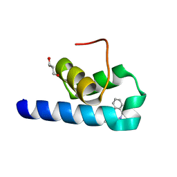
3V3B
DownloadVisualize
BU of 3v3b by Molmil
Structure of the Stapled p53 Peptide Bound to Mdm2
分子名称: CHLORIDE ION, E3 ubiquitin-protein ligase Mdm2, SAH-p53-8 stapled-peptide
著者Baek, S, Kutchukian, P.S, Verdine, G.L, Huber, R, Holak, T.A, Ki Won, L, Popowicz, G.M.
登録日2011-12-13
公開日2012-01-18
最終更新日2023-11-15
実験手法X-RAY DIFFRACTION (2 Å)
主引用文献Structure of the stapled p53 peptide bound to Mdm2.
J.Am.Chem.Soc., 134, 2012

222415

件を2024-07-10に公開中

PDB statisticsPDBj update infoContact PDBjnumon