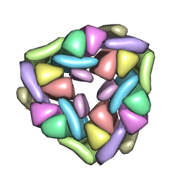
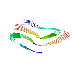
4UNU
 
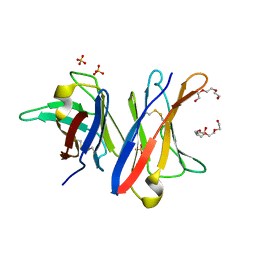 | | MCG - a dimer of lambda variable domains | | 分子名称: | IG LAMBDA CHAIN V-II REGION MGC, POLYETHYLENE GLYCOL (N=34), SULFATE ION | | 著者 | Brumshtein, B, Esswein, S.R, Landau, M, Ryan, C, Whitelegge, J, Casio, D, Sawaya, M.R, Eisenberg, D.S. | | 登録日 | 2014-05-30 | | 公開日 | 2014-08-27 | | 最終更新日 | 2024-01-10 | | 実験手法 | X-RAY DIFFRACTION (0.95 Å) | | 主引用文献 | Formation of Amyloid Fibers by Monomeric Light-Chain Variable Domains.
J.Biol.Chem., 289, 2014
|
|
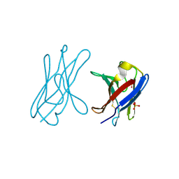
4UNT
 | | Induced monomer of the Mcg variable domain | | 分子名称: | IG LAMBDA CHAIN V-II REGION MGC, SULFATE ION | | 著者 | Brumshtein, B, Esswein, S.R, Landau, M, Ryan, C.M, Whitelegge, J.P, Phillips, M.L, Cascio, D, Sawaya, M.R, Eisenberg, D.S. | | 登録日 | 2014-05-30 | | 公開日 | 2014-08-27 | | 最終更新日 | 2024-01-10 | | 実験手法 | X-RAY DIFFRACTION (2.7 Å) | | 主引用文献 | Formation of Amyloid Fibers by Monomeric Light-Chain Variable Domains.
J.Biol.Chem., 289, 2014
|
|
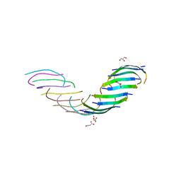
4UNV
 | | Covalent dimer of lambda variable domains | | 分子名称: | IG LAMBDA CHAIN V-II REGION MGC, SULFATE ION | | 著者 | Brumshtein, B, Esswein, S, Landau, M, Ryan, C, Whitelegge, J, Sawaya, M, Eisenberg, D.S. | | 登録日 | 2014-05-30 | | 公開日 | 2014-08-27 | | 最終更新日 | 2024-01-10 | | 実験手法 | X-RAY DIFFRACTION (1.6 Å) | | 主引用文献 | Formation of Amyloid Fibers by Monomeric Light-Chain Variable Domains.
J.Biol.Chem., 289, 2014
|
|
5VOS
 | | VGSNKGAIIGL from Amyloid Beta determined by MicroED | | 分子名称: | Amyloid beta A4 protein | | 著者 | Rodriguez, J.A, Sawaya, M.R, Cascio, D, Eisenberg, D.S, Griner, S.L, Gonen, T. | | 登録日 | 2017-05-03 | | 公開日 | 2018-01-03 | | 最終更新日 | 2024-03-13 | | 実験手法 | ELECTRON CRYSTALLOGRAPHY (1.42 Å) | | 主引用文献 | Common fibrillar spines of amyloid-beta and human islet amyloid polypeptide revealed by microelectron diffraction and structure-based inhibitors.
J. Biol. Chem., 293, 2018
|
|
5W50
 | | Crystal structure of the segment, LIIKGI, from the RRM2 of TDP-43, residues 248-253 | | 分子名称: | TAR DNA-binding protein 43 | | 著者 | Guenther, E.L, Trinh, H, Sawaya, M.R, Eisenberg, D.S. | | 登録日 | 2017-06-13 | | 公開日 | 2018-02-21 | | 最終更新日 | 2023-10-04 | | 実験手法 | X-RAY DIFFRACTION (1.4 Å) | | 主引用文献 | Atomic-level evidence for packing and positional amyloid polymorphism by segment from TDP-43 RRM2.
Nat. Struct. Mol. Biol., 25, 2018
|
|
5W52
 | | MicroED structure of the segment, DLIIKGISVHI, from the RRM2 of TDP-43, residues 247-257 | | 分子名称: | TAR DNA-binding protein 43 | | 著者 | Guenther, E.L, Sawaya, M.R, Cascio, D, Eisenberg, D.S. | | 登録日 | 2017-06-13 | | 公開日 | 2018-02-21 | | 最終更新日 | 2024-04-03 | | 実験手法 | ELECTRON CRYSTALLOGRAPHY (1.4 Å) | | 主引用文献 | Atomic-level evidence for packing and positional amyloid polymorphism by segment from TDP-43 RRM2.
Nat. Struct. Mol. Biol., 25, 2018
|
|
5WIA
 | | Crystal structure of the segment, GNNSYS, from the low complexity domain of TDP-43, residues 370-375 | | 分子名称: | TAR DNA-binding protein 43 | | 著者 | Guenther, E.L, Trinh, H, Sawaya, M.R, Eisenberg, D.S. | | 登録日 | 2017-07-18 | | 公開日 | 2018-04-25 | | 最終更新日 | 2024-04-03 | | 実験手法 | X-RAY DIFFRACTION (1.002 Å) | | 主引用文献 | Atomic structures of TDP-43 LCD segments and insights into reversible or pathogenic aggregation.
Nat. Struct. Mol. Biol., 25, 2018
|
|
5WKD
 | | Crystal structure of the segment, GNNQGSN, from the low complexity domain of TDP-43, residues 300-306 | | 分子名称: | TAR DNA-binding protein 43 | | 著者 | Guenther, E.L, Trinh, H, Sawaya, M.R, Cascio, D, Eisenberg, D.S. | | 登録日 | 2017-07-25 | | 公開日 | 2018-04-18 | | 最終更新日 | 2024-04-03 | | 実験手法 | X-RAY DIFFRACTION (1.8 Å) | | 主引用文献 | Atomic structures of TDP-43 LCD segments and insights into reversible or pathogenic aggregation.
Nat. Struct. Mol. Biol., 25, 2018
|
|
5WOR
 | |
5W7V
 | |
5WHN
 | | Crystal structure of the segment, NFGAFS, from the low complexity domain of TDP-43, residues 312-317 | | 分子名称: | Segment of TAR DNA-binding protein 43 | | 著者 | Guenther, E.L, Sawaya, M.R, Eisenberg, D.S. | | 登録日 | 2017-07-17 | | 公開日 | 2018-04-25 | | 最終更新日 | 2023-10-04 | | 実験手法 | X-RAY DIFFRACTION (1.1 Å) | | 主引用文献 | Atomic structures of TDP-43 LCD segments and insights into reversible or pathogenic aggregation.
Nat. Struct. Mol. Biol., 25, 2018
|
|
5WHP
 | | Crystal structure of the segment, NFGTFS, from the A315T familial variant of the low complexity domain of TDP-43, residues 312-317 | | 分子名称: | Segment of TAR DNA-binding protein 43 | | 著者 | Guenther, E.L, Sawaya, M.R, Eisenberg, D.S. | | 登録日 | 2017-07-17 | | 公開日 | 2018-05-23 | | 最終更新日 | 2024-03-13 | | 実験手法 | X-RAY DIFFRACTION (1 Å) | | 主引用文献 | Atomic structures of TDP-43 LCD segments and insights into reversible or pathogenic aggregation.
Nat. Struct. Mol. Biol., 25, 2018
|
|
5WIQ
 | | Crystal structure of the segment, GFNGGFG, from the low complexity domain of TDP-43, residues 396-402 | | 分子名称: | TAR DNA-binding protein 43 | | 著者 | Guenther, E.L, Sawaya, M.R, Eisenberg, D.S. | | 登録日 | 2017-07-19 | | 公開日 | 2018-04-18 | | 最終更新日 | 2023-10-04 | | 実験手法 | X-RAY DIFFRACTION (1.25 Å) | | 主引用文献 | Atomic structures of TDP-43 LCD segments and insights into reversible or pathogenic aggregation.
Nat. Struct. Mol. Biol., 25, 2018
|
|
5WKB
 | | MicroED structure of the segment, NFGEFS, from the A315E familial variant of the low complexity domain of TDP-43, residues 312-317 | | 分子名称: | TAR DNA-binding protein 43 | | 著者 | Guenther, E.L, Sawaya, M.R, Cascio, D, Eisenberg, D.S. | | 登録日 | 2017-07-24 | | 公開日 | 2018-05-23 | | 最終更新日 | 2024-03-13 | | 実験手法 | ELECTRON CRYSTALLOGRAPHY (1 Å) | | 主引用文献 | Atomic structures of TDP-43 LCD segments and insights into reversible or pathogenic aggregation.
Nat. Struct. Mol. Biol., 25, 2018
|
|
6N3A
 | | SegA-long, conformation of TDP-43 low complexity domain segment A long | | 分子名称: | TAR DNA-binding protein 43, segA long small | | 著者 | Cao, Q, Boyer, D.R, Sawaya, M.R, Eisenberg, D.S. | | 登録日 | 2018-11-14 | | 公開日 | 2019-06-26 | | 最終更新日 | 2024-03-20 | | 実験手法 | ELECTRON MICROSCOPY (3.3 Å) | | 主引用文献 | Cryo-EM structures of four polymorphic TDP-43 amyloid cores.
Nat.Struct.Mol.Biol., 26, 2019
|
|
6N37
 | | SegA-sym, conformation of TDP-43 low complexity domain segment A sym | | 分子名称: | TAR DNA-binding protein 43 | | 著者 | Cao, Q, Boyer, D.R, Sawaya, M.R, Eisenberg, D.S. | | 登録日 | 2018-11-14 | | 公開日 | 2019-06-26 | | 最終更新日 | 2024-03-20 | | 実験手法 | ELECTRON MICROSCOPY (3.8 Å) | | 主引用文献 | Cryo-EM structures of four polymorphic TDP-43 amyloid cores.
Nat.Struct.Mol.Biol., 26, 2019
|
|
6N3C
 | | SegB, conformation of TDP-43 low complexity domain segment A | | 分子名称: | TAR DNA-binding protein 43 | | 著者 | Cao, Q, Boyer, D.R, Sawaya, M.R, Eisenberg, D.S. | | 登録日 | 2018-11-14 | | 公開日 | 2019-06-26 | | 最終更新日 | 2024-03-20 | | 実験手法 | ELECTRON MICROSCOPY (3.3 Å) | | 主引用文献 | Cryo-EM structures of four polymorphic TDP-43 amyloid cores.
Nat.Struct.Mol.Biol., 26, 2019
|
|
6N3B
 | | SegA-asym, conformation of TDP-43 low complexity domain segment A asym | | 分子名称: | TAR DNA-binding protein 43 | | 著者 | Cao, Q, Boyer, D.R, Sawaya, M.R, Eisenberg, D.S. | | 登録日 | 2018-11-14 | | 公開日 | 2019-06-26 | | 最終更新日 | 2024-03-20 | | 実験手法 | ELECTRON MICROSCOPY (3.8 Å) | | 主引用文献 | Cryo-EM structures of four polymorphic TDP-43 amyloid cores.
Nat.Struct.Mol.Biol., 26, 2019
|
|
7N8R
 | | FGTGFG segment from the Nucleoporin p54, residues 63-68 | | 分子名称: | FGTGFG segment from the Nucleoporin p54, residues 63-68, trifluoroacetic acid | | 著者 | Hughes, M.P, Sawaya, M.R, Eisenberg, D.S. | | 登録日 | 2021-06-15 | | 公開日 | 2022-02-16 | | 最終更新日 | 2024-05-22 | | 実験手法 | X-RAY DIFFRACTION (1.2 Å) | | 主引用文献 | Extended beta-Strands Contribute to Reversible Amyloid Formation.
Acs Nano, 16, 2022
|
|
5FOY
 | | De novo structure of the binary mosquito larvicide BinAB at pH 7 | | 分子名称: | 41.9 KDA INSECTICIDAL TOXIN, LARVICIDAL TOXIN 51 KDA PROTEIN | | 著者 | Colletier, J.P, Sawaya, M.R, Gingery, M, Rodriguez, J.A, Cascio, D, Brewster, A.S, Michels-Clark, T, Boutet, S, Williams, G.J, Messerschmidt, M, DePonte, D.P, Sierra, R.G, Laksmono, H, Koglin, J.E, Hunter, M.S, W Park, H, Uervirojnangkoorn, M, Bideshi, D.L, Brunger, A.T, Federici, B.A, Sauter, N.K, Eisenberg, D.S. | | 登録日 | 2015-11-26 | | 公開日 | 2016-10-05 | | 最終更新日 | 2019-08-28 | | 実験手法 | X-RAY DIFFRACTION (2.25 Å) | | 主引用文献 | De Novo Phasing with X-Ray Laser Reveals Mosquito Larvicide Binab Structure.
Nature, 539, 2016
|
|
5FOZ
 | | De novo structure of the binary mosquito larvicide BinAB at pH 10 | | 分子名称: | LARVICIDAL TOXIN PROTEIN, SODIUM ION, TOXIN | | 著者 | Colletier, J.P, Sawaya, M.R, Gingery, M, Rodriguez, J.A, Cascio, D, Brewster, A.S, Michels-Clark, T, Boutet, S, Williams, G.J, Messerschmidt, M, DePonte, D.P, Sierra, R.G, Laksmono, H, Koglin, J.E, Hunter, M.S, W Park, H, Uervirojnangkoorn, M, Bideshi, D.L, Brunger, A.T, Federici, B.A, Sauter, N.K, Eisenberg, D.S. | | 登録日 | 2015-11-26 | | 公開日 | 2016-10-05 | | 最終更新日 | 2024-01-10 | | 実験手法 | X-RAY DIFFRACTION (2.4 Å) | | 主引用文献 | De Novo Phasing with X-Ray Laser Reveals Mosquito Larvicide Binab Structure.
Nature, 539, 2016
|
|
5G37
 | | MR structure of the binary mosquito larvicide BinAB at pH 5 | | 分子名称: | 41.9 KDA INSECTICIDAL TOXIN, LARVICIDAL TOXIN 51 KDA PROTEIN | | 著者 | Colletier, J.P, Sawaya, M.R, Gingery, M, Rodriguez, J.A, Cascio, D, Brewster, A.S, Michels-Clark, T, Boutet, S, Williams, G.J, Messerschmidt, M, DePonte, D.P, Sierra, R.G, Laksmono, H, Koglin, J.E, Hunter, M.S, W Park, H, Uervirojnangkoorn, M, Bideshi, D.L, Brunger, A.T, Federici, B.A, Sauter, N.K, Eisenberg, D.S. | | 登録日 | 2016-04-24 | | 公開日 | 2016-10-05 | | 最終更新日 | 2024-01-10 | | 実験手法 | X-RAY DIFFRACTION (2.5 Å) | | 主引用文献 | De Novo Phasing with X-Ray Laser Reveals Mosquito Larvicide Binab Structure.
Nature, 539, 2016
|
|
5HGE
 | | Filamentous Assembly of Green Fluorescent Protein Supported by a C-terminal fusion of 18-residues, viewed in space group P212121 | | 分子名称: | (4S)-2-METHYL-2,4-PENTANEDIOL, Green fluorescent protein | | 著者 | Sawaya, M.R, Heller, D.M, McPartland, L, Hochschild, A, Eisenberg, D.S. | | 登録日 | 2016-01-08 | | 公開日 | 2017-01-11 | | 最終更新日 | 2023-11-15 | | 実験手法 | X-RAY DIFFRACTION (1.863 Å) | | 主引用文献 | Green Fluorescent Protein Fusion that Self Assembles as Polar Filaments
to be published
|
|
5HW9
 | | Filamentous Assembly of Green Fluorescent Protein Supported by a C-terminal fusion of 18-residues, viewed in space group P21 | | 分子名称: | Green fluorescent protein | | 著者 | Sawaya, M.R, Heller, D.M, McPartland, L, Hochschild, A, Eisenberg, D.S. | | 登録日 | 2016-01-29 | | 公開日 | 2017-02-01 | | 最終更新日 | 2023-11-15 | | 実験手法 | X-RAY DIFFRACTION (3 Å) | | 主引用文献 | Green Fluorescent Protein Fusion that Self Assembles as Polar Filaments
to be published
|
|
5HBD
 | | Filamentous Assembly of Green Fluorescent Protein Supported by a C-terminal fusion of 18-residues, viewed in space group C2 | | 分子名称: | Green fluorescent protein | | 著者 | Sawaya, M.R, Hochschild, A, Heller, D.M, McPartland, L, Eisenberg, D.S. | | 登録日 | 2015-12-31 | | 公開日 | 2017-01-04 | | 最終更新日 | 2024-03-06 | | 実験手法 | X-RAY DIFFRACTION (1.65 Å) | | 主引用文献 | Green Fluorescent Protein Fusion that Self Assembles as Polar Filaments
to be published
|
|


