3MAS
 
 | |
4ZA1
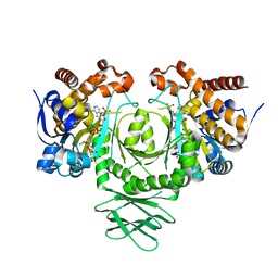
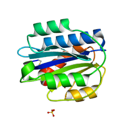
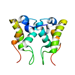
 | | Crystal Structure of NosA Involved in Nosiheptide Biosynthesis | | 分子名称: | 2,3-DIHYDROXY-1,4-DITHIOBUTANE, NosA | | 著者 | Liu, S, Guo, H, Zhang, T, Han, L, Yao, P, Zhang, Y, Rong, N, Yu, Y, Lan, W, Wang, C, Ding, J, Wang, R, Liu, W, Cao, C. | | 登録日 | 2015-04-13 | | 公開日 | 2015-08-19 | | 最終更新日 | 2024-03-20 | | 実験手法 | X-RAY DIFFRACTION (2.5 Å) | | 主引用文献 | Structure-based Mechanistic Insights into Terminal Amide Synthase in Nosiheptide-Represented Thiopeptides Biosynthesis
Sci Rep, 5, 2015
|
|
3MAR
 | |
5C3O
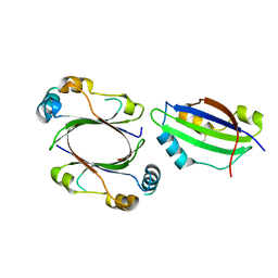
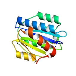
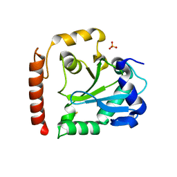
 | | Crystal structure of the C-terminal truncated Neurospora crassa T7H (NcT7HdeltaC) in apo form | | 分子名称: | 1,2-ETHANEDIOL, CALCIUM ION, Thymine dioxygenase | | 著者 | Li, W, Zhang, T, Ding, J. | | 登録日 | 2015-06-17 | | 公開日 | 2015-10-21 | | 最終更新日 | 2015-12-02 | | 実験手法 | X-RAY DIFFRACTION (2.3 Å) | | 主引用文献 | Molecular basis for the substrate specificity and catalytic mechanism of thymine-7-hydroxylase in fungi
Nucleic Acids Res., 43, 2015
|
|
3DOE
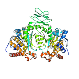
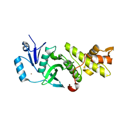
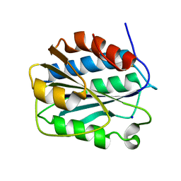
 | | Complex of ARL2 and BART, Crystal Form 1 | | 分子名称: | ADP-ribosylation factor-like protein 2, ADP-ribosylation factor-like protein 2-binding protein, GUANOSINE-5'-TRIPHOSPHATE, ... | | 著者 | Zhang, T, Li, S, Ding, J. | | 登録日 | 2008-07-04 | | 公開日 | 2009-03-03 | | 最終更新日 | 2023-11-01 | | 実験手法 | X-RAY DIFFRACTION (2.25 Å) | | 主引用文献 | Crystal structure of the ARL2-GTP-BART complex reveals a novel recognition and binding mode of small GTPase with effector
Structure, 17, 2009
|
|
4N3X
 | |
4N3Z
 | |
1R0A
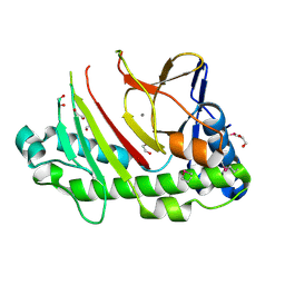
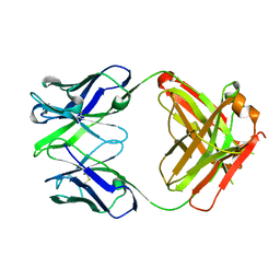
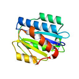
 | | Crystal structure of HIV-1 reverse transcriptase covalently tethered to DNA template-primer solved to 2.8 angstroms | | 分子名称: | 5'-D(*A*TP*GP*CP*AP*TP*CP*GP*GP*CP*GP*CP*TP*CP*GP*AP*AP*CP*AP*GP*GP*GP*AP*CP*GP*GP*T)-3', 5'-D(*C*CP*GP*TP*CP*CP*CP*TP*GP*TP*TP*CP*GP*AP*GP*CP*GP*CP*CP*GP*(2DA))-3', GLYCEROL, ... | | 著者 | Tuske, S, Ding, J, Arnold, E. | | 登録日 | 2003-09-19 | | 公開日 | 2004-08-03 | | 最終更新日 | 2021-10-27 | | 実験手法 | X-RAY DIFFRACTION (2.8 Å) | | 主引用文献 | Nonnucleoside inhibitor binding affects the interactions of the fingers subdomain of human immunodeficiency virus type 1 reverse transcriptase with DNA.
J.Virol., 78, 2004
|
|
1T03
 | | HIV-1 reverse transcriptase crosslinked to tenofovir terminated template-primer (complex P) | | 分子名称: | MAGNESIUM ION, POL polyprotein, Synthetic oligonucleotide primer, ... | | 著者 | Tuske, S, Sarafianos, S.G, Ding, J, Arnold, E. | | 登録日 | 2004-04-07 | | 公開日 | 2004-05-11 | | 最終更新日 | 2023-08-23 | | 実験手法 | X-RAY DIFFRACTION (3.1 Å) | | 主引用文献 | Structure of HIV-1 RT-DNA complexes before and after incorporation of the anti-AIDS drug tenofovir
Nat.Struct.Mol.Biol., 11, 2004
|
|
3PPV
 | | Crystal structure of an engineered VWF A2 domain (N1493C and C1670S) | | 分子名称: | CALCIUM ION, SULFATE ION, von Willebrand factor | | 著者 | Zhou, M, Dong, X, Zhong, C, Ding, J. | | 登録日 | 2010-11-25 | | 公開日 | 2011-05-04 | | 最終更新日 | 2023-11-01 | | 実験手法 | X-RAY DIFFRACTION (1.9 Å) | | 主引用文献 | A novel calcium-binding site of von Willebrand factor A2 domain regulates its cleavage by ADAMTS13
Blood, 117, 2011
|
|
3PPX
 | | Crystal structure of the N1602A mutant of an engineered VWF A2 domain (N1493C and C1670S) | | 分子名称: | SODIUM ION, von Willebrand factor | | 著者 | Zhou, M, Dong, X, Zhong, C, Ding, J. | | 登録日 | 2010-11-25 | | 公開日 | 2011-05-04 | | 最終更新日 | 2023-11-01 | | 実験手法 | X-RAY DIFFRACTION (1.91 Å) | | 主引用文献 | A novel calcium-binding site of von Willebrand factor A2 domain regulates its cleavage by ADAMTS13
Blood, 117, 2011
|
|
3QAH
 | | Crystal structure of apo-form human MOF catalytic domain | | 分子名称: | Probable histone acetyltransferase MYST1, ZINC ION | | 著者 | Sun, B, Tang, Q, Zhong, C, Ding, J. | | 登録日 | 2011-01-11 | | 公開日 | 2011-07-06 | | 最終更新日 | 2023-12-06 | | 実験手法 | X-RAY DIFFRACTION (2.1 Å) | | 主引用文献 | Regulation of the histone acetyltransferase activity of hMOF via autoacetylation of Lys274
Cell Res., 21, 2011
|
|
3EO9
 | | Crystal structure the Fab fragment of Efalizumab | | 分子名称: | Efalizumab Fab fragment, heavy chain, light chain | | 著者 | Li, S, Ding, J. | | 登録日 | 2008-09-26 | | 公開日 | 2009-04-14 | | 最終更新日 | 2023-11-01 | | 実験手法 | X-RAY DIFFRACTION (1.8 Å) | | 主引用文献 | Efalizumab binding to the LFA-1 alphaL I domain blocks ICAM-1 binding via steric hindrance.
Proc.Natl.Acad.Sci.USA, 106, 2009
|
|
2MYJ
 | |
3SP4
 | | Crystal structure of aprataxin ortholog Hnt3 from Schizosaccharomyces pombe | | 分子名称: | Aprataxin-like protein, SULFATE ION, ZINC ION | | 著者 | Gong, Y, Zhu, D, Ding, J, Dou, C, Ren, X, Jiang, T, Wang, D. | | 登録日 | 2011-07-01 | | 公開日 | 2011-10-12 | | 最終更新日 | 2013-07-03 | | 実験手法 | X-RAY DIFFRACTION (1.8 Å) | | 主引用文献 | Crystal structures of aprataxin ortholog Hnt3 reveal the mechanism for reversal of 5'-adenylated DNA
Nat.Struct.Mol.Biol., 18, 2011
|
|
3PPY
 | | Crystal structure of the D1596A/N1602A double mutant of an engineered VWF A2 domain (N1493C and C1670S) | | 分子名称: | SODIUM ION, von Willebrand factor | | 著者 | Zhou, M, Dong, X, Zhong, C, Ding, J. | | 登録日 | 2010-11-25 | | 公開日 | 2011-05-04 | | 最終更新日 | 2023-11-01 | | 実験手法 | X-RAY DIFFRACTION (2 Å) | | 主引用文献 | A novel calcium-binding site of von Willebrand factor A2 domain regulates its cleavage by ADAMTS13
Blood, 117, 2011
|
|
3PPW
 | | Crystal structure of the D1596A mutant of an engineered VWF A2 domain (N1493C and C1670S) | | 分子名称: | SODIUM ION, von Willebrand factor | | 著者 | Zhou, M, Dong, X, Zhong, C, Ding, J. | | 登録日 | 2010-11-25 | | 公開日 | 2011-05-04 | | 最終更新日 | 2023-11-01 | | 実験手法 | X-RAY DIFFRACTION (1.9 Å) | | 主引用文献 | A novel calcium-binding site of von Willebrand factor A2 domain regulates its cleavage by ADAMTS13
Blood, 117, 2011
|
|
5Y38
 | | Crystal structure of C7orf59-HBXIP complex | | 分子名称: | Ragulator complex protein LAMTOR4, Ragulator complex protein LAMTOR5, SULFATE ION | | 著者 | Zhang, T, Ding, J. | | 登録日 | 2017-07-28 | | 公開日 | 2017-12-06 | | 最終更新日 | 2023-11-22 | | 実験手法 | X-RAY DIFFRACTION (2.8 Å) | | 主引用文献 | Structural basis for Ragulator functioning as a scaffold in membrane-anchoring of Rag GTPases and mTORC1
Nat Commun, 8, 2017
|
|
3SR9
 | | Crystal structure of mouse PTPsigma | | 分子名称: | Receptor-type tyrosine-protein phosphatase S | | 著者 | Wang, J, Hou, L, Li, J, Ding, J. | | 登録日 | 2011-07-07 | | 公開日 | 2012-05-30 | | 最終更新日 | 2023-11-01 | | 実験手法 | X-RAY DIFFRACTION (2.4 Å) | | 主引用文献 | Structural insights into the homology and differences between mouse protein tyrosine phosphatase-sigma and human protein tyrosine phosphatase-sigma
Acta Biochim.Biophys.Sin., 43, 2011
|
|
4M6W
 | | Crystal structure of the C-terminal segment of FANCM in complex with FAAP24 | | 分子名称: | Fanconi anemia group M protein, Fanconi anemia-associated protein of 24 kDa, SULFATE ION | | 著者 | Yang, H, Zhang, T, Tong, L, Ding, J. | | 登録日 | 2013-08-11 | | 公開日 | 2013-10-02 | | 最終更新日 | 2024-03-20 | | 実験手法 | X-RAY DIFFRACTION (2.9 Å) | | 主引用文献 | Structural insights into the functions of the FANCM-FAAP24 complex in DNA repair.
Nucleic Acids Res., 41, 2013
|
|
7F00
 | | Crystal structure of SPD_0310 | | 分子名称: | SULFATE ION, UPF0371 protein SPRM200_0309 | | 著者 | Cao, K, Zhang, T, Li, N, Yang, X, Ding, J, He, Q, Sun, X. | | 登録日 | 2021-06-03 | | 公開日 | 2022-04-27 | | 最終更新日 | 2023-11-29 | | 実験手法 | X-RAY DIFFRACTION (2.7 Å) | | 主引用文献 | Identification and Tetramer Structure of Hemin-Binding Protein SPD_0310 Linked to Iron Homeostasis and Virulence of Streptococcus pneumoniae.
Msystems, 7, 2022
|
|


