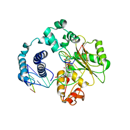
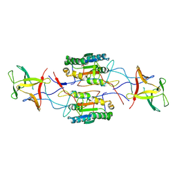
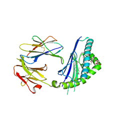
5F9T
 
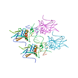
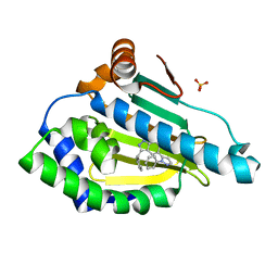
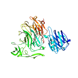 | | Crystal Structure of Streptococcus pneumoniae NanC, covalent complex with a fluorinated Neu5Ac derivative | | 分子名称: | (2R,3R,4R,5R,6R)-5-acetamido-2,3-difluoro-4-hydroxy-6-[(1R,2R)-1,2,3-trihydroxypropyl]tetrahydro-2H-pyran-2-carboxylic acid, 5-acetamido-3,5-dideoxy-3-fluoro-D-erythro-alpha-L-manno-non-2-ulopyranosonic acid, GLYCEROL, ... | | 著者 | Owen, C.D, Lukacik, P, Potter, J.A, Walsh, M, Taylor, G.L. | | 登録日 | 2015-12-10 | | 公開日 | 2015-12-23 | | 最終更新日 | 2024-01-10 | | 実験手法 | X-RAY DIFFRACTION (2.05 Å) | | 主引用文献 | Streptococcus pneumoniae NanC: STRUCTURAL INSIGHTS INTO THE SPECIFICITY AND MECHANISM OF A SIALIDASE THAT PRODUCES A SIALIDASE INHIBITOR.
J.Biol.Chem., 290, 2015
|
|
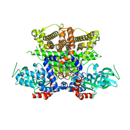
6V4A
 | |
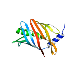
7KCU
 | | Joint neutron/X-ray structure of Oxyferrous Dehaloperoxidase B | | 分子名称: | Dehaloperoxidase B, OXYGEN MOLECULE, PROTOPORPHYRIN IX CONTAINING FE, ... | | 著者 | Carey, L.M, Ghiladi, R.A, Meilleur, F, Myles, D. | | 登録日 | 2020-10-07 | | 公開日 | 2021-10-13 | | 最終更新日 | 2023-10-25 | | 実験手法 | NEUTRON DIFFRACTION (2.2 Å), X-RAY DIFFRACTION | | 主引用文献 | Complementarity of neutron, XFEL and synchrotron crystallography for defining the structures of metalloenzymes at room temperature.
Iucrj, 9, 2022
|
|
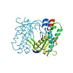
7K4M
 | | Crystal structure of MetAP2 Modified Hemoglobin S | | 分子名称: | CARBON MONOXIDE, Hemoglobin subunit alpha, Hemoglobin subunit beta, ... | | 著者 | Musayev, F.N, Safo, M.K, Light, D.R. | | 登録日 | 2020-09-15 | | 公開日 | 2021-10-13 | | 最終更新日 | 2023-10-18 | | 実験手法 | X-RAY DIFFRACTION (2.5 Å) | | 主引用文献 | MetAP2 inhibition modifies hemoglobin S to delay polymerization and improves blood flow in sickle cell disease.
Blood Adv, 5, 2021
|
|
3HX0
 | | ternary complex of L277A, H511A, R514 mutant pol lambda bound to a 2 nucleotide gapped DNA substrate with a scrunched dA | | 分子名称: | 2',3'-DIDEOXY-THYMIDINE-5'-TRIPHOSPHATE, 5'-D(*CP*AP*GP*TP*AP*T)-3', 5'-D(*CP*GP*GP*CP*AP*AP*AP*TP*AP*CP*TP*G)-3', ... | | 著者 | Garcia-Diaz, M, Bebenek, K, Larrea, A.A, Havener, J.M, Perera, L, Krahn, J.M, Pedersen, L.C, Ramsden, D.A, Kunkel, T.A. | | 登録日 | 2009-06-19 | | 公開日 | 2009-08-25 | | 最終更新日 | 2023-09-06 | | 実験手法 | X-RAY DIFFRACTION (3 Å) | | 主引用文献 | Scrunching During DNA Repair Synthesis
To be Published
|
|
7JXA
 | | CRYSTAL STRUCTURE OF NATIVE BOVINE ARRESTIN 1 IN COMPLEX WITH INOSITOL 1,4,5-TRIPHOSPHATE | | 分子名称: | 2-ETHOXYETHANOL, D-MYO-INOSITOL-1,4,5-TRIPHOSPHATE, S-arrestin, ... | | 著者 | Sander, C.L, Palczewski, K, Kiser, P.D. | | 登録日 | 2020-08-26 | | 公開日 | 2021-10-13 | | 最終更新日 | 2024-05-22 | | 実験手法 | X-RAY DIFFRACTION (2.4 Å) | | 主引用文献 | Structural evidence for visual arrestin priming via complexation of phosphoinositols.
Structure, 30, 2022
|
|
5K3H
 | | Crystals structure of Acyl-CoA oxidase-1 in Caenorhabditis elegans, Apo form-II | | 分子名称: | Acyl-coenzyme A oxidase | | 著者 | Zhang, X, Li, K, Jones, R.A, Bruner, S.D, Butcher, R.A. | | 登録日 | 2016-05-19 | | 公開日 | 2016-08-24 | | 最終更新日 | 2023-09-27 | | 実験手法 | X-RAY DIFFRACTION (2.48 Å) | | 主引用文献 | Structural characterization of acyl-CoA oxidases reveals a direct link between pheromone biosynthesis and metabolic state in Caenorhabditis elegans.
Proc.Natl.Acad.Sci.USA, 113, 2016
|
|
8CM1
 | |
5MBG
 | | Structure of a bacterial light-regulated adenylyl cyclase | | 分子名称: | Beta subunit of photoactivated adenylyl cyclase, IODIDE ION | | 著者 | Lindner, R, Hartmann, E, Tarnawski, M, Winkler, A, Frey, D, Reinstein, J, Meinhart, A, Schlichting, I. | | 登録日 | 2016-11-08 | | 公開日 | 2017-04-05 | | 最終更新日 | 2024-01-17 | | 実験手法 | X-RAY DIFFRACTION (2.3 Å) | | 主引用文献 | Photoactivation Mechanism of a Bacterial Light-Regulated Adenylyl Cyclase.
J. Mol. Biol., 429, 2017
|
|
8SV1
 | |
5C14
 | | Crystal structure of PECAM-1 D1D2 domain | | 分子名称: | 2-AMINO-2-HYDROXYMETHYL-PROPANE-1,3-DIOL, 2-acetamido-2-deoxy-beta-D-glucopyranose, COPPER (II) ION, ... | | 著者 | Zhou, D, Paddock, C, Newman, P, Zhu, J. | | 登録日 | 2015-06-12 | | 公開日 | 2016-01-06 | | 最終更新日 | 2020-07-29 | | 実験手法 | X-RAY DIFFRACTION (2.8 Å) | | 主引用文献 | Structural basis for PECAM-1 homophilic binding.
Blood, 127, 2016
|
|
7NL3
 | |
7RYT
 | |
5MF7
 | | New Insights into the Role of DNA Shape on Its Recognition by p53 Proteins (complex p53DBD-GADD45) | | 分子名称: | Cellular tumor antigen p53, DI(HYDROXYETHYL)ETHER, DNA, ... | | 著者 | Rozenberg, H, Diskin-Posner, Y, Golovenko, D, Shakked, Z. | | 登録日 | 2016-11-17 | | 公開日 | 2018-05-30 | | 最終更新日 | 2024-01-17 | | 実験手法 | X-RAY DIFFRACTION (1.59 Å) | | 主引用文献 | New Insights into the Role of DNA Shape on Its Recognition by p53 Proteins.
Structure, 26, 2018
|
|
6S1C
 | | P3221 crystal form of the Ctf18-1-8/Pol2(1-528) complex | | 分子名称: | Chromosome transmission fidelity protein 18, Chromosome transmission fidelity protein 8, DNA polymerase epsilon catalytic subunit A, ... | | 著者 | Grabarczyk, D.B. | | 登録日 | 2019-06-18 | | 公開日 | 2020-07-08 | | 最終更新日 | 2024-01-24 | | 実験手法 | X-RAY DIFFRACTION (6.1 Å) | | 主引用文献 | Ctf18-RFC and DNA Pol ε form a stable leading strand polymerase/clamp loader complex required for normal and perturbed DNA replication.
Nucleic Acids Res., 48, 2020
|
|
5MUS
 | | Structure of the C-terminal domain of a reptarenavirus L protein | | 分子名称: | CHLORIDE ION, GLYCEROL, L protein | | 著者 | Rosenthal, M, Gogrefe, N, Reguera, J, Vogel, D, Rauschenberger, B, Cusack, S, Gunther, S, Reindl, S. | | 登録日 | 2017-01-14 | | 公開日 | 2017-05-17 | | 最終更新日 | 2017-05-24 | | 実験手法 | X-RAY DIFFRACTION (2.009 Å) | | 主引用文献 | Structural insights into reptarenavirus cap-snatching machinery.
PLoS Pathog., 13, 2017
|
|
6HF4
 | | The structure of BoMan26B, a GH26 beta-mannanase from Bacteroides ovatus, complexed with G1M4 | | 分子名称: | CALCIUM ION, CHLORIDE ION, Glycosyl hydrolase family 26, ... | | 著者 | Bagenholm, V, Logan, D.T, Stalbrand, H. | | 登録日 | 2018-08-21 | | 公開日 | 2019-04-24 | | 最終更新日 | 2024-05-15 | | 実験手法 | X-RAY DIFFRACTION (1.781 Å) | | 主引用文献 | A surface-exposed GH26 beta-mannanase fromBacteroides ovatus: Structure, role, and phylogenetic analysis ofBoMan26B.
J.Biol.Chem., 294, 2019
|
|
5TW2
 | | Structure of mouse CD1d with bound alpha-galactosylsphingamide JG168 | | 分子名称: | (5R,6S,7S)-5,6-dihydroxy-7-(octanoylamino)-N-(6-phenylhexyl)-8-{[(2S,3R,4S,5R,6R)-3,4,5-trihydroxy-6-(hydroxymethyl)tetrahydro-2H-pyran-2-yl]oxy}octanamide, 2-acetamido-2-deoxy-beta-D-glucopyranose, 2-acetamido-2-deoxy-beta-D-glucopyranose-(1-4)-[alpha-L-fucopyranose-(1-6)]2-acetamido-2-deoxy-beta-D-glucopyranose, ... | | 著者 | Zajonc, D.M, Wang, J. | | 登録日 | 2016-11-11 | | 公開日 | 2017-09-20 | | 最終更新日 | 2023-10-04 | | 実験手法 | X-RAY DIFFRACTION (1.75 Å) | | 主引用文献 | Galactosylsphingamides: new alpha-GalCer analogues to probe the F'-pocket of CD1d.
Sci Rep, 7, 2017
|
|
7PJC
 | |
6EX1
 | | Crystal structure of human carbonic anhydrase I in complex with the 4-[(3S)-3 benzyl-4-(4-sulfamoylbenzoyl)piperazine -1-carbonyl]benzene-1-sulfonamide inhibitor | | 分子名称: | 4-[(3~{R})-3-(phenylmethyl)piperazin-1-yl]carbonylbenzenesulfonamide, ACETATE ION, Carbonic anhydrase 1, ... | | 著者 | Ferraroni, M, Supuran, C.T, Chiapponi, D, Chiaramonte, N. | | 登録日 | 2017-11-07 | | 公開日 | 2018-10-10 | | 最終更新日 | 2024-01-17 | | 実験手法 | X-RAY DIFFRACTION (1.6 Å) | | 主引用文献 | 2-Benzylpiperazine: A new scaffold for potent human carbonic anhydrase inhibitors. Synthesis, enzyme inhibition, enantioselectivity, computational and crystallographic studies and in vivo activity for a new class of intraocular pressure lowering agents.
Eur J Med Chem, 151, 2018
|
|
5C5G
 | |
6EYB
 | |
5JR1
 | |
6V37
 | |
7Q6D
 | | E. coli FtsA 1-405 ATP 3 Ni | | 分子名称: | ADENOSINE-5'-TRIPHOSPHATE, Cell division protein FtsA, MAGNESIUM ION, ... | | 著者 | Nierhaus, T, Kureisaite-Ciziene, D, Lowe, J. | | 登録日 | 2021-11-06 | | 公開日 | 2022-09-21 | | 最終更新日 | 2024-01-31 | | 実験手法 | X-RAY DIFFRACTION (2.8 Å) | | 主引用文献 | Bacterial divisome protein FtsA forms curved antiparallel double filaments when binding to FtsN.
Nat Microbiol, 7, 2022
|
|


