Loading
PDBj
MenuPDBj@FacebookPDBj@TwitterPDBj@YouTubewwPDB FoundationwwPDB
RCSB PDBPDBeBMRBAdv. SearchSearch help
PDB: 837 results

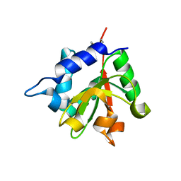
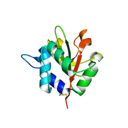
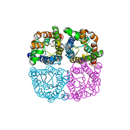
3RMR
DownloadVisualize
BU of 3rmr by Molmil
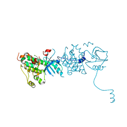
Crystal structure of Hyaloperonospora arabidopsidis ATR1 effector domain
Descriptor: Avirulence protein
Authors:Chou, S, Krasileva, K.V, Holton, J.M, Staskawicz, B.J, Alber, T.
Deposit date:2011-04-21
Release date:2011-07-20
Last modified:2019-07-17
Method:X-RAY DIFFRACTION (2.3 Å)
Cite:The Hyaloperonospora arabidopsidis ATR1 effector has distributed recognition surfaces and a structural subdomain conserved across oomycete species
To be Published
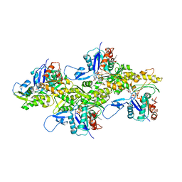
7K20
DownloadVisualize
BU of 7k20 by Molmil
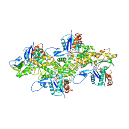
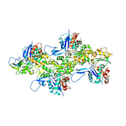
Cryo-EM structure of pyrene-labeled ADP-actin filaments
Descriptor: ADENOSINE-5'-DIPHOSPHATE, Actin, alpha skeletal muscle, ...
Authors:Chou, S.Z, Pollard, T.D.
Deposit date:2020-09-08
Release date:2020-11-04
Last modified:2020-12-02
Method:ELECTRON MICROSCOPY (3.2 Å)
Cite:Cryo-electron microscopy structures of pyrene-labeled ADP-P i - and ADP-actin filaments.
Nat Commun, 11, 2020
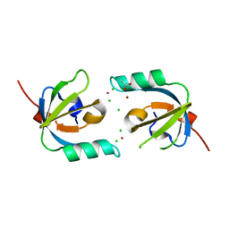
3MHX
DownloadVisualize
BU of 3mhx by Molmil
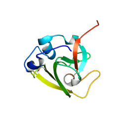
Crystal Structure of Stenotrophomonas maltophilia FeoA complexed with Zinc: A Unique Procaryotic SH3 Domain Protein Possibly Acting as a Bacterial Ferrous Iron Transport Activating Factor
Descriptor: ACETATE ION, CHLORIDE ION, Putative ferrous iron transport protein A, ...
Authors:Chou, S.-H, Su, Y.-C, Chin, K.-H, Hung, H.-C, Shen, G.-H, Wang, A.H.-J.
Deposit date:2010-04-09
Release date:2010-06-23
Last modified:2024-03-20
Method:X-RAY DIFFRACTION (1.7 Å)
Cite:Structure of Stenotrophomonas maltophilia FeoA complexed with zinc: a unique prokaryotic SH3-domain protein that possibly acts as a bacterial ferrous iron-transport activating factor
Acta Crystallogr.,Sect.F, 66, 2010
7K21
DownloadVisualize
BU of 7k21 by Molmil
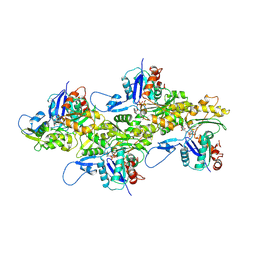
Cryo-EM structure of pyrene-labeled ADP-Pi-actin filaments
Descriptor: ADENOSINE-5'-DIPHOSPHATE, Actin, alpha skeletal muscle, ...
Authors:Chou, S.Z, Pollard, T.D.
Deposit date:2020-09-08
Release date:2020-11-04
Last modified:2020-12-02
Method:ELECTRON MICROSCOPY (3 Å)
Cite:Cryo-electron microscopy structures of pyrene-labeled ADP-P i - and ADP-actin filaments.
Nat Commun, 11, 2020
4EOB
DownloadVisualize
BU of 4eob by Molmil
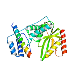
Structure of the type VI peptidoglycan amidase effector Tse1 from Pseudomonas aeruginosa
Descriptor: type VI amidase effector Tse1
Authors:Chou, S, Mougous, J.D.
Deposit date:2012-04-13
Release date:2012-05-30
Last modified:2024-10-16
Method:X-RAY DIFFRACTION (2.611 Å)
Cite:Structure of a peptidoglycan amidase effector targeted to Gram-negative bacteria by the type VI secretion system.
Cell Rep, 1, 2012
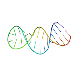
2GIO
DownloadVisualize
BU of 2gio by Molmil
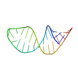
Solution Structure of a portion of the 5'UTR of HspA mRNA of Bradyrhizobium japonicum
Descriptor: 29-MER
Authors:Chowdhury, S, Maris, C, Allain, F.H, Narberhaus, F.
Deposit date:2006-03-29
Release date:2006-06-20
Last modified:2024-05-29
Method:SOLUTION NMR
Cite:Molecular basis for temperature sensing by an RNA thermometer.
Embo J., 25, 2006
3GOP
DownloadVisualize
BU of 3gop by Molmil
Crystal structure of the EGF receptor juxtamembrane and kinase domains
Descriptor: Epidermal growth factor receptor
Authors:Choi, S.H, Alvarado, D, Moravcevic, K, Lemmon, M.A.
Deposit date:2009-03-19
Release date:2009-07-07
Last modified:2023-09-06
Method:X-RAY DIFFRACTION (2.8 Å)
Cite:The juxtamembrane region of the EGF receptor functions as an activation domain.
Mol.Cell, 34, 2009
3M4U
DownloadVisualize
BU of 3m4u by Molmil
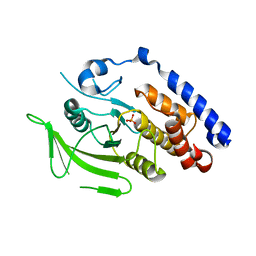
Crystal Structure of Trypanosoma brucei Protein Tyrosine Phosphatase TbPTP1
Descriptor: PHOSPHATE ION, Tyrosine specific protein phosphatase, putative
Authors:Chou, S, Alber, T, Grundner, C.
Deposit date:2010-03-12
Release date:2010-05-05
Last modified:2024-02-21
Method:X-RAY DIFFRACTION (2.392 Å)
Cite:The Trypanosoma brucei life cycle switch TbPTP1 is structurally conserved and dephosphorylates the nucleolar protein NOPP44/46.
J.Biol.Chem., 285, 2010
6WIN
DownloadVisualize
BU of 6win by Molmil
Type 6 secretion amidase effector 2 (Tae2)
Descriptor: Type 6 secretion amidase effector 2
Authors:Chou, S, Radkov, A.D.
Deposit date:2020-04-10
Release date:2020-04-22
Last modified:2024-10-23
Method:X-RAY DIFFRACTION (2.05 Å)
Cite:Ticks Resist Skin Commensals with Immune Factor of Bacterial Origin.
Cell, 183, 2020
3J41
DownloadVisualize
BU of 3j41 by Molmil
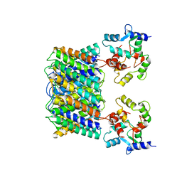
Pseudo-atomic model of the Aquaporin-0/Calmodulin complex derived from electron microscopy
Descriptor: CALCIUM ION, Calmodulin, Lens fiber major intrinsic protein
Authors:Reichow, S.L, Clemens, D.M, Freites, J.A, Nemeth-Cahalan, K.L, Heyden, M, Tobias, D.J, Hall, J.E, Gonen, T.
Deposit date:2013-05-31
Release date:2013-07-31
Last modified:2024-02-21
Method:ELECTRON MICROSCOPY (25 Å)
Cite:Allosteric mechanism of water-channel gating by Ca(2+)-calmodulin.
Nat.Struct.Mol.Biol., 20, 2013
2GIP
DownloadVisualize
BU of 2gip by Molmil
Solution structure of a portion of the 5'UTR of HspA mRNA from Bradyrhizobium janponicum having deleted G83
Descriptor: 28-MER
Authors:Chowdhury, S, Maris, C, Allain, F.H, Narberhaus, F.
Deposit date:2006-03-29
Release date:2006-06-20
Last modified:2024-05-29
Method:SOLUTION NMR
Cite:Molecular basis for temperature sensing by an RNA thermometer.
Embo J., 25, 2006
3J4Q
DownloadVisualize
BU of 3j4q by Molmil
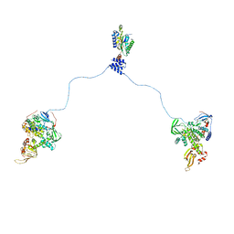
Pseudo-atomic model of the AKAP18-PKA complex in a bent conformation derived from electron microscopy
Descriptor: A-kinase anchor protein 18, cAMP-dependent protein kinase catalytic subunit alpha, cAMP-dependent protein kinase type II-alpha regulatory subunit
Authors:Reichow, S.L, Gonen, T.
Deposit date:2013-09-25
Release date:2013-11-13
Last modified:2024-02-21
Method:ELECTRON MICROSCOPY (35 Å)
Cite:Intrinsic disorder within an AKAP-protein kinase A complex guides local substrate phosphorylation.
Elife, 2, 2013
7E42
DownloadVisualize
BU of 7e42 by Molmil
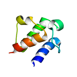
Solution strucutre of holo acyl carrier protein from Acinetobacter baumannii
Descriptor: Acyl carrier protein
Authors:Choi, S, Park, J, Kim, Y.
Deposit date:2021-02-10
Release date:2022-02-16
Last modified:2024-05-15
Method:SOLUTION NMR
Cite:solution structure of holo acyl carrier protein in acinetobacter baumannii
To Be Published
3J4R
DownloadVisualize
BU of 3j4r by Molmil
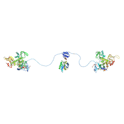
Pseudo-atomic model of the AKAP18-PKA Complex in a linear conformation derived from electron microscopy
Descriptor: A-kinase anchor protein 18, cAMP-dependent protein kinase catalytic subunit alpha, cAMP-dependent protein kinase type II-alpha regulatory subunit
Authors:Reichow, S.L, Gonen, T.
Deposit date:2013-09-25
Release date:2013-11-13
Last modified:2024-02-21
Method:ELECTRON MICROSCOPY (35 Å)
Cite:Intrinsic disorder within an AKAP-protein kinase A complex guides local substrate phosphorylation.
Elife, 2, 2013
4F4M
DownloadVisualize
BU of 4f4m by Molmil
Structure of the type VI peptidoglycan amidase effector Tse1 (C30A) from Pseudomonas aeruginosa
Descriptor: papain peptidoglycan amidase effector Tse1
Authors:Chou, S, Mougous, J.D.
Deposit date:2012-05-10
Release date:2012-05-30
Last modified:2024-10-30
Method:X-RAY DIFFRACTION (2.677 Å)
Cite:Structure of a peptidoglycan amidase effector targeted to Gram-negative bacteria by the type VI secretion system.
Cell Rep, 1, 2012
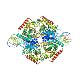
5HTO
DownloadVisualize
BU of 5hto by Molmil
Crystal structure of Plasmodium Vivax LDH in complex with a DNA aptamer called pL1 (tetrameric LDH in an asymmetric unit)
Descriptor: DNA (30-MER), DNA (34-MER), L-lactate dehydrogenase, ...
Authors:Choi, S.J, Ban, C.
Deposit date:2016-01-27
Release date:2016-10-19
Last modified:2023-11-08
Method:X-RAY DIFFRACTION (1.9 Å)
Cite:Crystal structure of a DNA aptamer bound to PvLDH elucidates novel single-stranded DNA structural elements for folding and recognition
Sci Rep, 6, 2016
5HS4
DownloadVisualize
BU of 5hs4 by Molmil
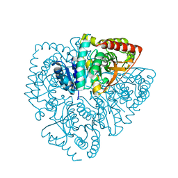
Plasmdoium Vivax Lactate dehydrogenase
Descriptor: L-lactate dehydrogenase
Authors:Choi, S.J, Ban, C.
Deposit date:2016-01-25
Release date:2016-10-19
Last modified:2023-11-08
Method:X-RAY DIFFRACTION (1.339 Å)
Cite:Crystal structure of a DNA aptamer bound to PvLDH elucidates novel single-stranded DNA structural elements for folding and recognition
Sci Rep, 6, 2016
5HRU
DownloadVisualize
BU of 5hru by Molmil
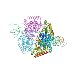
Crystal structure of Plasmodium vivax LDH in complex with a DNA aptamer called pL1
Descriptor: DNA (32-MER), L-lactate dehydrogenase, MAGNESIUM ION
Authors:Choi, S.J, Ban, C.
Deposit date:2016-01-24
Release date:2016-10-19
Last modified:2023-11-08
Method:X-RAY DIFFRACTION (1.71 Å)
Cite:Crystal structure of a DNA aptamer bound to PvLDH elucidates novel single-stranded DNA structural elements for folding and recognition
Sci Rep, 6, 2016
6QIM
DownloadVisualize
BU of 6qim by Molmil
Structure of AtPIP2;4
Descriptor: Probable aquaporin PIP2-4
Authors:Schoebel, S, Wang, H.
Deposit date:2019-01-21
Release date:2019-11-20
Last modified:2024-01-24
Method:X-RAY DIFFRACTION (3.7 Å)
Cite:Characterization of aquaporin-driven hydrogen peroxide transport.
Biochim Biophys Acta Biomembr, 1862, 2020
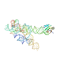
3BO3
DownloadVisualize
BU of 3bo3 by Molmil
A relaxed active site following exon ligation by a group I intron
Descriptor: Group I intron P9, MAGNESIUM ION, POTASSIUM ION, ...
Authors:Lipchock, S.V, Strobel, S.A.
Deposit date:2007-12-17
Release date:2008-04-01
Last modified:2024-02-21
Method:X-RAY DIFFRACTION (3.4 Å)
Cite:A relaxed active site after exon ligation by the group I intron
Proc.Natl.Acad.Sci.Usa, 105, 2008
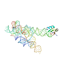
3BO4
DownloadVisualize
BU of 3bo4 by Molmil
A relaxed active site following exon ligation by a group I intron
Descriptor: DNA/RNA (5'-R(*AP*AP*GP*CP*CP*AP*CP*AP*CP*AP*AP*AP*CP*CP*A)-D(P*DG)-3'), DNA/RNA (5'-R(*CP*A)-D(P*DU)-R(P*AP*CP*GP*GP*CP*C)-3'), Group I intron P9, ...
Authors:Lipchock, S.V, Strobel, S.A.
Deposit date:2007-12-17
Release date:2008-04-01
Last modified:2024-02-21
Method:X-RAY DIFFRACTION (3.33 Å)
Cite:A relaxed active site after exon ligation by the group I intron
Proc.Natl.Acad.Sci.Usa, 105, 2008
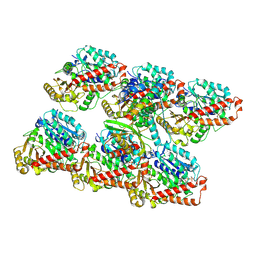
7ZCW
DownloadVisualize
BU of 7zcw by Molmil
Cryo-EM structure of GMPCPP-microtubules in complex with VASH2-SVBP
Descriptor: GUANOSINE-5'-TRIPHOSPHATE, MAGNESIUM ION, PHOSPHOMETHYLPHOSPHONIC ACID GUANYLATE ESTER, ...
Authors:Choi, S.R, Blum, T, Steinmetz, M.O.
Deposit date:2022-03-29
Release date:2022-12-14
Last modified:2024-07-24
Method:ELECTRON MICROSCOPY (3.6 Å)
Cite:VASH1-SVBP and VASH2-SVBP generate different detyrosination profiles on microtubules.
J.Cell Biol., 222, 2023
6DJO
DownloadVisualize
BU of 6djo by Molmil
Cryo-EM structure of ADP-actin filaments
Descriptor: ADENOSINE-5'-DIPHOSPHATE, Actin, alpha skeletal muscle, ...
Authors:Chou, S.Z, Pollard, T.D.
Deposit date:2018-05-25
Release date:2019-02-27
Last modified:2024-04-10
Method:ELECTRON MICROSCOPY (3.6 Å)
Cite:Mechanism of actin polymerization revealed by cryo-EM structures of actin filaments with three different bound nucleotides.
Proc.Natl.Acad.Sci.USA, 116, 2019
6DJM
DownloadVisualize
BU of 6djm by Molmil
Cryo-EM structure of AMPPNP-actin filaments
Descriptor: Actin, alpha skeletal muscle, MAGNESIUM ION, ...
Authors:Chou, S.Z, Pollard, T.D.
Deposit date:2018-05-25
Release date:2019-02-27
Last modified:2020-01-08
Method:ELECTRON MICROSCOPY (3.1 Å)
Cite:Mechanism of actin polymerization revealed by cryo-EM structures of actin filaments with three different bound nucleotides.
Proc.Natl.Acad.Sci.USA, 116, 2019
6QBY
DownloadVisualize
BU of 6qby by Molmil
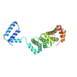
Crystal structure of VASH 2 in complex with SVBP
Descriptor: Small vasohibin-binding protein, Tubulinyl-Tyr carboxypeptidase 2
Authors:Choi, S.R, Olieric, V, Steinmetz, M.O, Olieric, N.
Deposit date:2018-12-24
Release date:2019-04-24
Last modified:2024-05-15
Method:X-RAY DIFFRACTION (2.09 Å)
Cite:Structural basis of tubulin detyrosination by the vasohibin-SVBP enzyme complex.
Nat.Struct.Mol.Biol., 26, 2019

226707

PDB entries from 2024-10-30

PDB statisticsPDBj update infoContact PDBjnumon