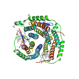
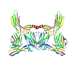
6BEV
 
 | |
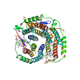
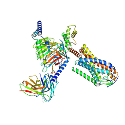
6VK7
 | | Crystal Structure of reduced Methylosinus trichosporium OB3b Soluble Methane Monooxygenase Hydroxylase | | 分子名称: | FE (III) ION, Methane monooxygenase, Methane monooxygenase component A alpha chain | | 著者 | Jones, J.C, Banerjee, R, Shi, K, Aihara, H, Lipscomb, J.D. | | 登録日 | 2020-01-18 | | 公開日 | 2020-08-05 | | 最終更新日 | 2023-10-11 | | 実験手法 | X-RAY DIFFRACTION (2.12 Å) | | 主引用文献 | Structural Studies of theMethylosinus trichosporiumOB3b Soluble Methane Monooxygenase Hydroxylase and Regulatory Component Complex Reveal a Transient Substrate Tunnel.
Biochemistry, 59, 2020
|
|
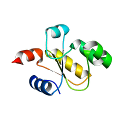
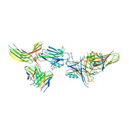
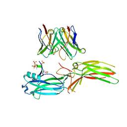
4K12
 | | Structural Basis for Host Specificity of Factor H Binding by Streptococcus pneumoniae | | 分子名称: | Choline binding protein A, Complement factor H | | 著者 | Liu, A, Achila, D, Banerjee, R, Martinez-Hackert, E, Li, Y, Yan, H. | | 登録日 | 2013-04-04 | | 公開日 | 2014-04-09 | | 最終更新日 | 2017-11-15 | | 実験手法 | X-RAY DIFFRACTION (1.079 Å) | | 主引用文献 | Structural determinants of host specificity of complement Factor H recruitment by Streptococcus pneumoniae.
Biochem.J., 465, 2015
|
|
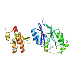
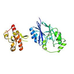
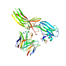
4HI2
 | |
8JA3
 | | Structure of beta-arrestin1 in complex with C3aRpp | | 分子名称: | Beta-arrestin-1, C3a anaphylatoxin chemotactic receptor, Fab30 heavy chain, ... | | 著者 | Maharana, J, Sarma, P, Yadav, M.K, Chami, M, Banerjee, R, Shukla, A.K. | | 登録日 | 2023-05-05 | | 公開日 | 2023-12-27 | | 最終更新日 | 2024-01-17 | | 実験手法 | ELECTRON MICROSCOPY (3.94 Å) | | 主引用文献 | Molecular insights into atypical modes of beta-arrestin interaction with seven transmembrane receptors.
Science, 383, 2024
|
|
8IYW
 | | Structure of GSK256073-GPR109A-G-protein complex | | 分子名称: | 8-chloranyl-3-pentyl-7H-purine-2,6-dione, Guanine nucleotide-binding protein G(I)/G(S)/G(O) subunit gamma-2, Guanine nucleotide-binding protein G(I)/G(S)/G(T) subunit beta-1, ... | | 著者 | Yadav, M.K, Sarma, P, Chami, M, Banerjee, R, Shukla, A.K. | | 登録日 | 2023-04-06 | | 公開日 | 2024-03-06 | | 最終更新日 | 2024-03-20 | | 実験手法 | ELECTRON MICROSCOPY (3.45 Å) | | 主引用文献 | Structure-guided engineering of biased-agonism in the human niacin receptor via single amino acid substitution.
Nat Commun, 15, 2024
|
|
8IY9
 | | Structure of Niacin-GPR109A-G protein complex | | 分子名称: | Guanine nucleotide-binding protein G(I)/G(S)/G(O) subunit gamma-2, Guanine nucleotide-binding protein G(I)/G(S)/G(T) subunit beta-1, Guanine nucleotide-binding protein G(o) subunit alpha, ... | | 著者 | Yadav, M.K, Sarma, P, Chami, M, Banerjee, R, Shukla, A.K. | | 登録日 | 2023-04-04 | | 公開日 | 2024-03-06 | | 最終更新日 | 2024-03-20 | | 実験手法 | ELECTRON MICROSCOPY (3.37 Å) | | 主引用文献 | Structure-guided engineering of biased-agonism in the human niacin receptor via single amino acid substitution.
Nat Commun, 15, 2024
|
|
8JER
 | | Structure of Acipimox-GPR109A-G protein complex | | 分子名称: | 5-methyl-4-oxidanyl-pyrazin-4-ium-2-carboxylic acid, Guanine nucleotide-binding protein G(I)/G(S)/G(O) subunit gamma-2, Guanine nucleotide-binding protein G(I)/G(S)/G(T) subunit beta-1, ... | | 著者 | Yadav, M.K, Sarma, P, Chami, M, Banerjee, R, Shukla, A.K. | | 登録日 | 2023-05-16 | | 公開日 | 2024-03-06 | | 最終更新日 | 2024-03-20 | | 実験手法 | ELECTRON MICROSCOPY (3.45 Å) | | 主引用文献 | Structure-guided engineering of biased-agonism in the human niacin receptor via single amino acid substitution.
Nat Commun, 15, 2024
|
|
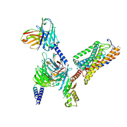
6D5X
 | | Structure of Human ATP:Cobalamin Adenosyltransferase bound to ATP, Adenosylcobalamin, and Triphosphate | | 分子名称: | 5'-DEOXYADENOSINE, ADENOSINE-5'-TRIPHOSPHATE, COBALAMIN, ... | | 著者 | Dodge, G.J, Campanello, G, Smith, J.L, Banerjee, R. | | 登録日 | 2018-04-19 | | 公開日 | 2018-10-10 | | 最終更新日 | 2024-03-13 | | 実験手法 | X-RAY DIFFRACTION (2.4 Å) | | 主引用文献 | Sacrificial Cobalt-Carbon Bond Homolysis in Coenzyme B12as a Cofactor Conservation Strategy.
J. Am. Chem. Soc., 140, 2018
|
|
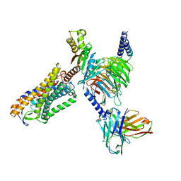
6D5K
 | | Structure of Human ATP:Cobalamin Adenosyltransferase bound to ATP, and Adenosylcobalamin | | 分子名称: | 4-(2-HYDROXYETHYL)-1-PIPERAZINE ETHANESULFONIC ACID, 5'-DEOXYADENOSINE, ADENOSINE-5'-TRIPHOSPHATE, ... | | 著者 | Dodge, G.J, Campanello, G, Smith, J.L, Banerjee, R. | | 登録日 | 2018-04-19 | | 公開日 | 2018-10-10 | | 最終更新日 | 2023-10-04 | | 実験手法 | X-RAY DIFFRACTION (2.85 Å) | | 主引用文献 | Sacrificial Cobalt-Carbon Bond Homolysis in Coenzyme B12as a Cofactor Conservation Strategy.
J. Am. Chem. Soc., 140, 2018
|
|
5VE5
 | | Crystal structure of persulfide dioxygenase rhodanese fusion protein with rhodanese domain inactivating mutation (C314S) from Burkholderia phytofirmans in complex with glutathione | | 分子名称: | BpPRF, CHLORIDE ION, FE (III) ION, ... | | 著者 | Motl, N, Skiba, M.A, Smith, J.L, Banerjee, R. | | 登録日 | 2017-04-03 | | 公開日 | 2017-07-19 | | 最終更新日 | 2023-10-04 | | 実験手法 | X-RAY DIFFRACTION (2.35 Å) | | 主引用文献 | Structural and biochemical analyses indicate that a bacterial persulfide dioxygenase-rhodanese fusion protein functions in sulfur assimilation.
J. Biol. Chem., 292, 2017
|
|
3SC0
 | | Crystal Structure of MMACHC (1-238), a human B12 processing enzyme, complexed with MethylCobalamin | | 分子名称: | CO-METHYLCOBALAMIN, Methylmalonic aciduria and homocystinuria type C protein | | 著者 | Koutmos, M, Gherasim, C, Smith, J.L, Banerjee, R. | | 登録日 | 2011-06-06 | | 公開日 | 2011-06-22 | | 最終更新日 | 2023-09-13 | | 実験手法 | X-RAY DIFFRACTION (1.95 Å) | | 主引用文献 | Structural basis of multifunctionality in a vitamin B12-processing enzyme.
J.Biol.Chem., 286, 2011
|
|
3SBZ
 | | Crystal Structure of Apo-MMACHC (1-244), a human B12 processing enzyme | | 分子名称: | GLYCEROL, MALONATE ION, Methylmalonic aciduria and homocystinuria type C protein | | 著者 | Koutmos, M, Gherasim, C, Smith, J.L, Banerjee, R. | | 登録日 | 2011-06-06 | | 公開日 | 2011-06-22 | | 最終更新日 | 2023-09-13 | | 実験手法 | X-RAY DIFFRACTION (2 Å) | | 主引用文献 | Structural basis of multifunctionality in a vitamin B12-processing enzyme.
J.Biol.Chem., 286, 2011
|
|
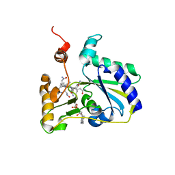
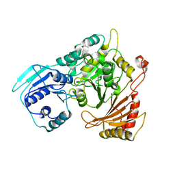
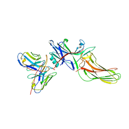
4HI1
 | |
5UCU
 | | STRUCTURAL AND MECHANISTIC INSIGHTS INTO HEMOGLOBIN-CATALYZED HYDROGEN SULFIDE OXIDATION AND THE FATE OF POLYSULFIDE PRODUCTS | | 分子名称: | HYDROSULFURIC ACID, Hemoglobin subunit alpha, Hemoglobin subunit beta, ... | | 著者 | Vitvitsky, V, Yadav, P.K, An, S, Seravalli, J, Cho, U.-S, Banerjee, R. | | 登録日 | 2016-12-22 | | 公開日 | 2017-03-01 | | 最終更新日 | 2023-10-04 | | 実験手法 | X-RAY DIFFRACTION (1.797 Å) | | 主引用文献 | Structural and Mechanistic Insights into Hemoglobin-catalyzed Hydrogen Sulfide Oxidation and the Fate of Polysulfide Products.
J. Biol. Chem., 292, 2017
|
|
5VE4
 | | Crystal structure of persulfide dioxygenase-rhodanese fusion protein with rhodanese domain inactivating mutation (C314S) from Burkholderia phytofirmans | | 分子名称: | BpPRF, CHLORIDE ION, DI(HYDROXYETHYL)ETHER, ... | | 著者 | Motl, N, Skiba, M.A, Smith, J.L, Banerjee, R. | | 登録日 | 2017-04-03 | | 公開日 | 2017-07-19 | | 最終更新日 | 2023-10-04 | | 実験手法 | X-RAY DIFFRACTION (2.65 Å) | | 主引用文献 | Structural and biochemical analyses indicate that a bacterial persulfide dioxygenase-rhodanese fusion protein functions in sulfur assimilation.
J. Biol. Chem., 292, 2017
|
|
5VE3
 | | Crystal structure of wild-type persulfide dioxygenase-rhodanese fusion protein from Burkholderia phytofirmans | | 分子名称: | BpPRF, FE (III) ION | | 著者 | Motl, N, Skiba, M.A, Smith, J.L, Banerjee, R. | | 登録日 | 2017-04-03 | | 公開日 | 2017-07-19 | | 最終更新日 | 2023-11-15 | | 実験手法 | X-RAY DIFFRACTION (1.793 Å) | | 主引用文献 | Structural and biochemical analyses indicate that a bacterial persulfide dioxygenase-rhodanese fusion protein functions in sulfur assimilation.
J. Biol. Chem., 292, 2017
|
|
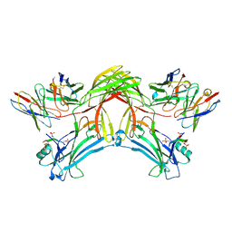
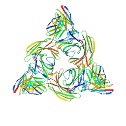
4QG5
 | |
8J8Z
 | | Structure of beta-arrestin1 in complex with D6Rpp | | 分子名称: | Atypical chemokine receptor 2, Beta-arrestin-1, Fab30 Heavy Chain, ... | | 著者 | Maharana, J, Sarma, P, Yadav, M.K, Chami, M, Banerjee, R, Shukla, A.K. | | 登録日 | 2023-05-02 | | 公開日 | 2023-12-27 | | 最終更新日 | 2024-01-17 | | 実験手法 | ELECTRON MICROSCOPY (3.4 Å) | | 主引用文献 | Molecular insights into atypical modes of beta-arrestin interaction with seven transmembrane receptors.
Science, 383, 2024
|
|
8J8V
 | | Structure of beta-arrestin2 in complex with D6Rpp (Local Refine) | | 分子名称: | Atypical chemokine receptor 2, Beta-arrestin-2, Fab30 Heavy Chain, ... | | 著者 | Maharana, J, Sarma, P, Yadav, M.K, Chami, M, Banerjee, R, Shukla, A.K. | | 登録日 | 2023-05-02 | | 公開日 | 2023-12-27 | | 最終更新日 | 2024-01-17 | | 実験手法 | ELECTRON MICROSCOPY (3.22 Å) | | 主引用文献 | Molecular insights into atypical modes of beta-arrestin interaction with seven transmembrane receptors.
Science, 383, 2024
|
|
8J97
 | | Structure of Muscarinic receptor (M2R) in complex with beta-arrestin1 (Local refine, cross-linked) | | 分子名称: | Beta-arrestin-1, Fab30 Heavy Chain, Fab30 Light Chain, ... | | 著者 | Maharana, J, Sano, F.K, Shihoya, W, Banerjee, R, Nureki, O, Shukla, A.K. | | 登録日 | 2023-05-02 | | 公開日 | 2023-12-27 | | 最終更新日 | 2024-01-17 | | 実験手法 | ELECTRON MICROSCOPY (3.2 Å) | | 主引用文献 | Molecular insights into atypical modes of beta-arrestin interaction with seven transmembrane receptors.
Science, 383, 2024
|
|
8JAF
 | | Structure of Muscarinic receptor (M2R) in complex with beta-arrestin1 (Local Refine, non-cross linked) | | 分子名称: | Beta-arrestin-1, Fab30 heavy chain, Fab30 light chain, ... | | 著者 | Maharana, J, Sano, F.K, Shihoya, W, Banerjee, R, Nureki, O, Shukla, A.K. | | 登録日 | 2023-05-05 | | 公開日 | 2023-12-27 | | 最終更新日 | 2024-01-17 | | 実験手法 | ELECTRON MICROSCOPY (3.1 Å) | | 主引用文献 | Molecular insights into atypical modes of beta-arrestin interaction with seven transmembrane receptors.
Science, 383, 2024
|
|
8J9K
 | | Structure of basal beta-arrestin2 | | 分子名称: | Beta-arrestin-2, Fab6 heavy chain, Fab6 light chain | | 著者 | Maharana, J, Sarma, P, Yadav, M.K, Chami, M, Banerjee, R, Shukla, A.K. | | 登録日 | 2023-05-03 | | 公開日 | 2023-12-27 | | 最終更新日 | 2024-01-17 | | 実験手法 | ELECTRON MICROSCOPY (3.5 Å) | | 主引用文献 | Molecular insights into atypical modes of beta-arrestin interaction with seven transmembrane receptors.
Science, 383, 2024
|
|
8J8R
 | | Structure of beta-arrestin2 in complex with M2Rpp | | 分子名称: | Beta-arrestin-2, Fab30 Heavy Chain, Fab30 Light Chain, ... | | 著者 | Maharana, J, Sano, F.K, Shihoya, W, Banerjee, R, Nureki, O, Shukla, A.K. | | 登録日 | 2023-05-02 | | 公開日 | 2023-12-27 | | 最終更新日 | 2024-01-17 | | 実験手法 | ELECTRON MICROSCOPY (2.9 Å) | | 主引用文献 | Molecular insights into atypical modes of beta-arrestin interaction with seven transmembrane receptors.
Science, 383, 2024
|
|
8JHN
 | | Structure of MMF-GPR109A-G protein complex | | 分子名称: | (E)-4-methoxy-4-oxidanylidene-but-2-enoic acid, G protein subunit alpha o1,Guanine nucleotide-binding protein G(o) subunit alpha, Guanine nucleotide-binding protein G(I)/G(S)/G(O) subunit gamma-2, ... | | 著者 | Yadav, M.K, Sarma, P, Chami, M, Banerjee, R, Shukla, A.K. | | 登録日 | 2023-05-24 | | 公開日 | 2024-03-06 | | 最終更新日 | 2024-03-20 | | 実験手法 | ELECTRON MICROSCOPY (3.75 Å) | | 主引用文献 | Structure-guided engineering of biased-agonism in the human niacin receptor via single amino acid substitution.
Nat Commun, 15, 2024
|
|


