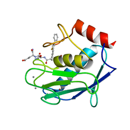
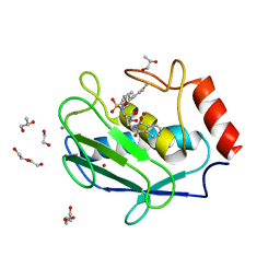
2C8G
 
 | | Structure of the PN loop Q182A mutant C3bot1 Exoenzyme (Free state, crystal form I) | | 分子名称: | MONO-ADP-RIBOSYLTRANSFERASE C3, SULFATE ION | | 著者 | Stura, E.A, Menetrey, J, Flatau, G, Boquet, P, Menez, A. | | 登録日 | 2005-12-03 | | 公開日 | 2007-02-27 | | 最終更新日 | 2023-12-13 | | 実験手法 | X-RAY DIFFRACTION (2 Å) | | 主引用文献 | Structural Properties of Wild-Type and Two Artt Motif Mutants Clostridium Botulinum C3 Exoenzyme Isoform 1 in Different Substrate Complexed States and Crystal Forms.
To be Published
|
|
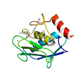
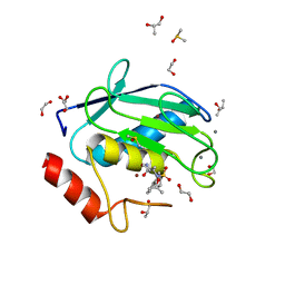
2C8H
 | | Structure of the PN loop Q182A mutant C3bot1 Exoenzyme (NAD-bound state, crystal form I) | | 分子名称: | MONO-ADP-RIBOSYLTRANSFERASE C3, NICOTINAMIDE-ADENINE-DINUCLEOTIDE, SULFATE ION | | 著者 | Stura, E.A, Menetrey, J, Flatau, G, Boquet, P, Menez, A. | | 登録日 | 2005-12-03 | | 公開日 | 2007-02-27 | | 最終更新日 | 2023-12-13 | | 実験手法 | X-RAY DIFFRACTION (1.65 Å) | | 主引用文献 | Structural Properties of Wild-Type and Two Artt Motif Mutants Clostridium Botulinum C3 Exoenzyme Isoform 1 in Different Substrate Complexed States and Crystal Forms.
To be Published
|
|
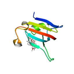
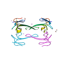
3QUM
 | | Crystal structure of human prostate specific antigen (PSA) in Fab sandwich with a high affinity and a PCa selective antibody | | 分子名称: | Fab 5D3D11 Heavy Chain, Fab 5D3D11 Light Chain, Fab 5D5A5 Heavy Chain, ... | | 著者 | Stura, E.A, Muller, B.H, Michel, S, Ducancel, F. | | 登録日 | 2011-02-24 | | 公開日 | 2011-11-09 | | 最終更新日 | 2023-09-13 | | 実験手法 | X-RAY DIFFRACTION (3.2 Å) | | 主引用文献 | Crystal structure of human prostate-specific antigen in a sandwich antibody complex.
J.Mol.Biol., 414, 2011
|
|
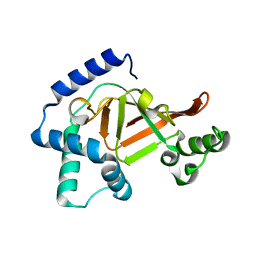
4ELC
 | | Crystal structure of the catalytic domain of botulinum neurotoxin BoNT/A C134 mutant with MTSEA modified Cys-165 | | 分子名称: | (2S)-2-hydroxybutanedioic acid, (4S)-2-METHYL-2,4-PENTANEDIOL, Botulinum neurotoxin A light chain, ... | | 著者 | Stura, E.A, Vera, L, Ptchelkine, D, Bakirci, H, Garcia, S, Dive, V. | | 登録日 | 2012-04-10 | | 公開日 | 2012-08-15 | | 最終更新日 | 2023-09-20 | | 実験手法 | X-RAY DIFFRACTION (1.8 Å) | | 主引用文献 | Structural Framework for Covalent Inhibition of Clostridium botulinum Neurotoxin A by Targeting Cys165.
J.Biol.Chem., 287, 2012
|
|
4EJ5
 | | Crystal structure of the catalytic domain of botulinum neurotoxin BoNT/A wild-type | | 分子名称: | 1,2-ETHANEDIOL, Botulinum neurotoxin A light chain, CARBONATE ION, ... | | 著者 | Stura, E.A, Vera, L, Dive, V. | | 登録日 | 2012-04-06 | | 公開日 | 2012-08-15 | | 最終更新日 | 2023-09-20 | | 実験手法 | X-RAY DIFFRACTION (1.87 Å) | | 主引用文献 | Structural Framework for Covalent Inhibition of Clostridium botulinum Neurotoxin A by Targeting Cys165.
J.Biol.Chem., 287, 2012
|
|
4EL4
 | | Crystal structure of the catalytic domain of botulinum neurotoxin BoNT/A C134S/C165S double mutant | | 分子名称: | 1,2-ETHANEDIOL, Botulinum neurotoxin A light chain, GLYCEROL, ... | | 著者 | Stura, E.A, Vera, L, Ptchelkine, D, Dive, V. | | 登録日 | 2012-04-10 | | 公開日 | 2012-08-15 | | 最終更新日 | 2023-09-20 | | 実験手法 | X-RAY DIFFRACTION (1.2 Å) | | 主引用文献 | Structural Framework for Covalent Inhibition of Clostridium botulinum Neurotoxin A by Targeting Cys165.
J.Biol.Chem., 287, 2012
|
|
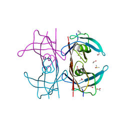
4FU4
 | | Human collagenase 3 (MMP-13) with peptide from pro-domain | | 分子名称: | 1,2-ETHANEDIOL, CALCIUM ION, CHLORIDE ION, ... | | 著者 | Stura, E.A, Vera, L, Visse, R, Nagase, H, Dive, V. | | 登録日 | 2012-06-28 | | 公開日 | 2013-08-21 | | 最終更新日 | 2023-09-13 | | 実験手法 | X-RAY DIFFRACTION (2.849 Å) | | 主引用文献 | Crystal structure of full-length human collagenase 3 (MMP-13) with peptides in the active site defines exosites in the catalytic domain.
Faseb J., 27, 2013
|
|
4FVL
 | | Human collagenase 3 (MMP-13) full form with peptides from pro-domain | | 分子名称: | CALCIUM ION, CHLORIDE ION, Collagenase 3, ... | | 著者 | Stura, E.A, Vera, L, Visse, R, Nagase, H, Dive, V. | | 登録日 | 2012-06-29 | | 公開日 | 2013-08-21 | | 最終更新日 | 2023-09-13 | | 実験手法 | X-RAY DIFFRACTION (2.436 Å) | | 主引用文献 | Crystal structure of full-length human collagenase 3 (MMP-13) with peptides in the active site defines exosites in the catalytic domain.
Faseb J., 27, 2013
|
|
4G0D
 | | Human collagenase 3 (MMP-13) full form with peptides from pro-domain | | 分子名称: | CALCIUM ION, CHLORIDE ION, Collagenase 3, ... | | 著者 | Stura, E.A, Vera, L, Visse, R, Nagase, H, Dive, V. | | 登録日 | 2012-07-09 | | 公開日 | 2013-08-21 | | 最終更新日 | 2023-09-13 | | 実験手法 | X-RAY DIFFRACTION (2.54 Å) | | 主引用文献 | Crystal structure of full-length human collagenase 3 (MMP-13) with peptides in the active site defines exosites in the catalytic domain.
Faseb J., 27, 2013
|
|
3KZ7
 | |
3TS4
 | | Human MMP12 in complex with L-glutamate motif inhibitor | | 分子名称: | CALCIUM ION, GLYCEROL, IMIDAZOLE, ... | | 著者 | Stura, E.A, Dive, V, Devel, L, Czarny, B, Beau, F, Vera, L. | | 登録日 | 2011-09-12 | | 公開日 | 2012-06-20 | | 最終更新日 | 2023-09-13 | | 実験手法 | X-RAY DIFFRACTION (1.587 Å) | | 主引用文献 | Simple pseudo-dipeptides with a P2' glutamate: a novel inhibitor family of matrix metalloproteases and other metzincins.
J.Biol.Chem., 287, 2012
|
|
3TSK
 | | Human MMP12 in complex with L-glutamate motif inhibitor | | 分子名称: | CALCIUM ION, Macrophage metalloelastase, N~2~-{3-[4-(4-phenylthiophen-2-yl)phenyl]propanoyl}-L-glutaminyl-L-alpha-glutamine, ... | | 著者 | Stura, E.A, Dive, V, Devel, L, Czarny, B, Beau, F, Vera, L. | | 登録日 | 2011-09-13 | | 公開日 | 2012-06-20 | | 最終更新日 | 2023-09-13 | | 実験手法 | X-RAY DIFFRACTION (2 Å) | | 主引用文献 | Simple pseudo-dipeptides with a P2' glutamate: a novel inhibitor family of matrix metalloproteases and other metzincins.
J.Biol.Chem., 287, 2012
|
|
5K1J
 | | Human TTR altered by a rhenium tris-carbonyl Pyta-C8 derivative | | 分子名称: | 1,2-ETHANEDIOL, ACETATE ION, DIMETHYL SULFOXIDE, ... | | 著者 | Stura, E.A, Ciccone, L, Shepard, W. | | 登録日 | 2016-05-18 | | 公開日 | 2016-07-20 | | 最終更新日 | 2024-01-10 | | 実験手法 | X-RAY DIFFRACTION (1.69 Å) | | 主引用文献 | Human TTR conformation altered by rhenium tris-carbonyl derivatives.
J.Struct.Biol., 195, 2016
|
|
4WZV
 | | Crystal structure of a hydroxamate based inhibitor EN140 in complex with the MMP-9 catalytic domain | | 分子名称: | (2R)-4-(1,3-dioxo-1,3-dihydro-2H-isoindol-2-yl)-N-hydroxy-2-{[(4'-methoxybiphenyl-4-yl)sulfonyl](propan-2-yloxy)amino}butanamide, 1,2-ETHANEDIOL, AZIDE ION, ... | | 著者 | Stura, E.A, Vera, L, Cassar-Lajeunesse, E, Nuti, E, Dive, V, Rossello, A. | | 登録日 | 2014-11-20 | | 公開日 | 2015-08-26 | | 最終更新日 | 2024-01-10 | | 実験手法 | X-RAY DIFFRACTION (1.65 Å) | | 主引用文献 | N-O-Isopropyl Sulfonamido-Based Hydroxamates as Matrix Metalloproteinase Inhibitors: Hit Selection and in Vivo Antiangiogenic Activity.
J.Med.Chem., 58, 2015
|
|
4XCT
 | | Crystal structure of a hydroxamate based inhibitor ARP101 (EN73) in complex with the MMP-9 catalytic domain. | | 分子名称: | (2S,3S)-butane-2,3-diol, (2~{R})-3-methyl-~{N}-oxidanylidene-2-[(4-phenylphenyl)sulfonyl-propan-2-yloxy-amino]butanamide, 1,2-ETHANEDIOL, ... | | 著者 | Stura, E.A, Tepshi, L, Nuti, E, Dive, V, Cassar-Lajeunesse, E, Vera, L, Rossello, A. | | 登録日 | 2014-12-18 | | 公開日 | 2015-04-22 | | 最終更新日 | 2024-01-10 | | 実験手法 | X-RAY DIFFRACTION (1.3 Å) | | 主引用文献 | N-O-Isopropyl Sulfonamido-Based Hydroxamates as Matrix Metalloproteinase Inhibitors: Hit Selection and in Vivo Antiangiogenic Activity.
J.Med.Chem., 58, 2015
|
|
5DO6
 | | Crystal structure of Dendroaspis polylepis venom mambalgin-1 T23A mutant | | 分子名称: | 1,2-ETHANEDIOL, IODIDE ION, Mambalgin-1, ... | | 著者 | Stura, E.A, Tepshi, L, Kessler, P, Gilles, M, Servent, D. | | 登録日 | 2015-09-10 | | 公開日 | 2015-12-30 | | 最終更新日 | 2017-01-25 | | 実験手法 | X-RAY DIFFRACTION (1.697 Å) | | 主引用文献 | Mambalgin-1 Pain-relieving Peptide, Stepwise Solid-phase Synthesis, Crystal Structure, and Functional Domain for Acid-sensing Ion Channel 1a Inhibition.
J.Biol.Chem., 291, 2016
|
|
2C89
 | | Structure of the wild-type C3bot1 Exoenzyme (Free state, crystal form I) | | 分子名称: | MONO-ADP-RIBOSYLTRANSFERASE C3, SULFATE ION | | 著者 | Stura, E.A, Menetrey, J, Flatau, G, Boquet, P, Menez, A. | | 登録日 | 2005-12-03 | | 公開日 | 2007-02-27 | | 最終更新日 | 2023-12-13 | | 実験手法 | X-RAY DIFFRACTION (1.85 Å) | | 主引用文献 | Structural Basis for the Nad-Hydrolysis Mechanism and the Artt-Loop Plasticity of C3 Exoenzymes.
Protein Sci., 17, 2008
|
|
2C8F
 | | Structure of the ARTT motif E214N mutant C3bot1 Exoenzyme (NAD-bound state, crystal form III) | | 分子名称: | MONO-ADP-RIBOSYLTRANSFERASE C3, NICOTINAMIDE-ADENINE-DINUCLEOTIDE | | 著者 | Stura, E.A, Menetrey, J, Flatau, G, Boquet, P, Menez, A. | | 登録日 | 2005-12-03 | | 公開日 | 2007-02-27 | | 最終更新日 | 2023-12-13 | | 実験手法 | X-RAY DIFFRACTION (2.5 Å) | | 主引用文献 | Structural Basis for the Nad-Hydrolysis Mechanism and the Artt-Loop Plasticity of C3 Exoenzymes.
Protein Sci., 17, 2008
|
|
2C8A
 | | Structure of the wild-type C3bot1 Exoenzyme (Nicotinamide-bound state, crystal form I) | | 分子名称: | MONO-ADP-RIBOSYLTRANSFERASE C3, NICOTINAMIDE, SULFATE ION | | 著者 | Stura, E.A, Menetrey, J, Flatau, G, Boquet, P, Menez, A. | | 登録日 | 2005-12-03 | | 公開日 | 2007-02-27 | | 最終更新日 | 2023-12-13 | | 実験手法 | X-RAY DIFFRACTION (1.7 Å) | | 主引用文献 | Structural Basis for the Nad-Hydrolysis Mechanism and the Artt-Loop Plasticity of C3 Exoenzymes.
Protein Sci., 17, 2008
|
|
2C8C
 | | Structure of the ARTT motif Q212A mutant C3bot1 Exoenzyme (NAD-bound state, crystal form I) | | 分子名称: | ADENOSINE-5'-DIPHOSPHATE, MONO-ADP-RIBOSYLTRANSFERASE C3, NICOTINAMIDE-ADENINE-DINUCLEOTIDE | | 著者 | Stura, E.A, Menetrey, J, Flatau, G, Boquet, P, Menez, A. | | 登録日 | 2005-12-03 | | 公開日 | 2007-02-27 | | 最終更新日 | 2023-12-13 | | 実験手法 | X-RAY DIFFRACTION (2.7 Å) | | 主引用文献 | Structural Basis for the Nad-Hydrolysis Mechanism and the Artt-Loop Plasticity of C3 Exoenzymes.
Protein Sci., 17, 2008
|
|
2C8D
 | | Structure of the ARTT motif Q212A mutant C3bot1 Exoenzyme (Free state, crystal form I) | | 分子名称: | MONO-ADP-RIBOSYLTRANSFERASE C3, SULFATE ION | | 著者 | Stura, E.A, Menetrey, J, Flatau, G, Boquet, P, Menez, A. | | 登録日 | 2005-12-03 | | 公開日 | 2007-02-27 | | 最終更新日 | 2023-12-13 | | 実験手法 | X-RAY DIFFRACTION (2.2 Å) | | 主引用文献 | Structural Basis for the Nad-Hydrolysis Mechanism and the Artt-Loop Plasticity of C3 Exoenzymes.
Protein Sci., 17, 2008
|
|
2C8E
 | | Structure of the ARTT motif E214N mutant C3bot1 Exoenzyme (Free state, crystal form III) | | 分子名称: | MONO-ADP-RIBOSYLTRANSFERASE C3, SULFATE ION | | 著者 | Stura, E.A, Menetrey, J, Flatau, G, Boquet, P, Menez, A. | | 登録日 | 2005-12-03 | | 公開日 | 2007-02-27 | | 最終更新日 | 2023-12-13 | | 実験手法 | X-RAY DIFFRACTION (1.6 Å) | | 主引用文献 | Structural Basis for the Nad-Hydrolysis Mechanism and the Artt-Loop Plasticity of C3 Exoenzymes.
Protein Sci., 17, 2008
|
|
2C8B
 | | Structure of the ARTT motif Q212A mutant C3bot1 Exoenzyme (Free state, crystal form II) | | 分子名称: | MONO-ADP-RIBOSYLTRANSFERASE C3, SULFATE ION | | 著者 | Stura, E.A, Menetrey, J, Flatau, G, Boquet, P, Menez, A. | | 登録日 | 2005-12-03 | | 公開日 | 2007-02-27 | | 最終更新日 | 2023-12-13 | | 実験手法 | X-RAY DIFFRACTION (1.7 Å) | | 主引用文献 | Structural Basis for the Nad-Hydrolysis Mechanism and the Artt-Loop Plasticity of C3 Exoenzymes.
Protein Sci., 17, 2008
|
|
3F5V
 | | C2 Crystal form of mite allergen DER P 1 | | 分子名称: | CALCIUM ION, Der p 1 allergen, HEXAETHYLENE GLYCOL | | 著者 | Stura, E.A, Minor, W, Chruszcz, M, Saint Remy, J.M. | | 登録日 | 2008-11-04 | | 公開日 | 2009-02-10 | | 最終更新日 | 2023-09-06 | | 実験手法 | X-RAY DIFFRACTION (1.36 Å) | | 主引用文献 | Crystal structures of mite allergens Der f 1 and Der p 1 reveal differences in surface-exposed residues that may influence antibody binding.
J.Mol.Biol., 386, 2009
|
|
3FEV
 | | Crystal structure of the chimeric muscarinic toxin MT7 with loop 1 from MT1. | | 分子名称: | Fusion of Muscarinic toxin 1, Muscarinic m1-toxin1, SULFATE ION | | 著者 | Stura, E.A, Menez, R, Mourier, G, Fruchart-Gaillard, C, Menez, A, Servant, D. | | 登録日 | 2008-12-01 | | 公開日 | 2009-12-22 | | 最終更新日 | 2023-11-01 | | 実験手法 | X-RAY DIFFRACTION (1.3 Å) | | 主引用文献 | Engineering of three-finger fold toxins creates ligands with original pharmacological profiles for muscarinic and adrenergic receptors.
Plos One, 7, 2012
|
|


