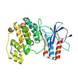
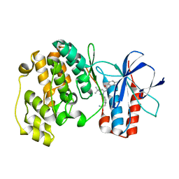
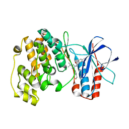
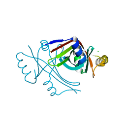
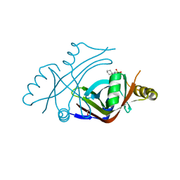
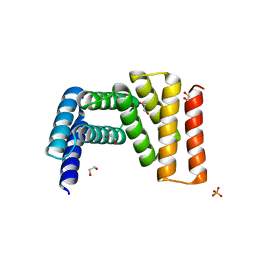
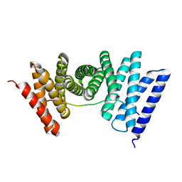
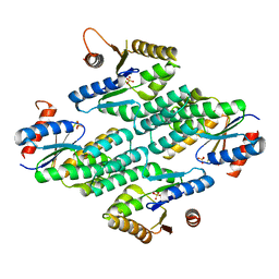
3GMT
 
 | | Crystal structure of adenylate kinase from burkholderia pseudomallei | | 分子名称: | Adenylate kinase, SULFATE ION | | 著者 | Abendroth, J, Staker, B.L, Robinson, H, Buchko, G.W, Hewitt, S.N, Napuli, A.J, Van Voorhis, W, Stacy, R, Myler, P.J, Stewart, L, Seattle Structural Genomics Center for Infectious Disease (SSGCID) | | 登録日 | 2009-03-15 | | 公開日 | 2009-06-02 | | 最終更新日 | 2013-10-30 | | 実験手法 | X-RAY DIFFRACTION (2.1 Å) | | 主引用文献 | Structural characterization of Burkholderia pseudomallei adenylate kinase (Adk): profound asymmetry in the crystal structure of the 'open' state.
Biochem.Biophys.Res.Commun., 394, 2010
|
|
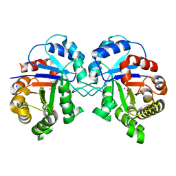
3NNU
 | | Crystal structure of P38 alpha in complex with DP1376 | | 分子名称: | 2-{3-[(5E)-5-{[(2,3-dichlorophenyl)carbamoyl]imino}-3-thiophen-2-yl-2,5-dihydro-1H-pyrazol-1-yl]phenyl}acetamide, Mitogen-activated protein kinase 14 | | 著者 | Abendroth, J. | | 登録日 | 2010-06-24 | | 公開日 | 2010-09-15 | | 最終更新日 | 2023-12-27 | | 実験手法 | X-RAY DIFFRACTION (2.4 Å) | | 主引用文献 | Switch control pocket inhibitors of p38-MAP kinase. Durable type II inhibitors that do not require binding into the canonical ATP hinge region
Bioorg.Med.Chem.Lett., 20, 2010
|
|
3NNX
 | | Crystal structure of phosphorylated P38 alpha in complex with DP802 | | 分子名称: | 2-[3-(3-tert-butyl-5-{[(2,3-dichlorophenyl)carbamoyl]imino}-2,5-dihydro-1H-pyrazol-1-yl)phenyl]acetamide, Mitogen-activated protein kinase 14 | | 著者 | Abendroth, J. | | 登録日 | 2010-06-24 | | 公開日 | 2010-09-15 | | 最終更新日 | 2023-12-27 | | 実験手法 | X-RAY DIFFRACTION (2.28 Å) | | 主引用文献 | Switch control pocket inhibitors of p38-MAP kinase. Durable type II inhibitors that do not require binding into the canonical ATP hinge region
Bioorg.Med.Chem.Lett., 20, 2010
|
|
3NNW
 | | Crystal structure of P38 alpha in complex with DP802 | | 分子名称: | 2-[3-(3-tert-butyl-5-{[(2,3-dichlorophenyl)carbamoyl]imino}-2,5-dihydro-1H-pyrazol-1-yl)phenyl]acetamide, Mitogen-activated protein kinase 14 | | 著者 | Abendroth, J. | | 登録日 | 2010-06-24 | | 公開日 | 2010-09-15 | | 最終更新日 | 2023-12-27 | | 実験手法 | X-RAY DIFFRACTION (1.89 Å) | | 主引用文献 | Switch control pocket inhibitors of p38-MAP kinase. Durable type II inhibitors that do not require binding into the canonical ATP hinge region
Bioorg.Med.Chem.Lett., 20, 2010
|
|
3NNV
 | | Crystal structure of P38 alpha in complex with DP437 | | 分子名称: | 1-{3-tert-butyl-1-[4-(hydroxymethyl)phenyl]-1H-pyrazol-5-yl}-3-naphthalen-1-ylurea, Mitogen-activated protein kinase 14 | | 著者 | Abendroth, J. | | 登録日 | 2010-06-24 | | 公開日 | 2010-09-15 | | 最終更新日 | 2023-12-27 | | 実験手法 | X-RAY DIFFRACTION (2.1 Å) | | 主引用文献 | Switch control pocket inhibitors of p38-MAP kinase. Durable type II inhibitors that do not require binding into the canonical ATP hinge region
Bioorg.Med.Chem.Lett., 20, 2010
|
|
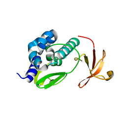
8T5E
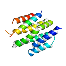 | | De novo design of high-affinity protein binders to bioactive helical peptides | | 分子名称: | Bcl-2-like protein 11, Bim_fulldiff | | 著者 | Torres, S.V, Leung, P.J.Y, Bera, A.K, Baker, D, Kang, A. | | 登録日 | 2023-06-13 | | 公開日 | 2024-01-10 | | 最終更新日 | 2024-02-14 | | 実験手法 | X-RAY DIFFRACTION (3 Å) | | 主引用文献 | De novo design of high-affinity binders of bioactive helical peptides.
Nature, 626, 2024
|
|
8T5F
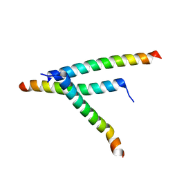 | | De novo design of high-affinity protein binders to bioactive helical peptides | | 分子名称: | Parathyroid hormone | | 著者 | Torres, S.V, Leung, P.J.Y, Bera, A.K, Baker, D, Kang, A. | | 登録日 | 2023-06-13 | | 公開日 | 2024-01-10 | | 最終更新日 | 2024-02-14 | | 実験手法 | X-RAY DIFFRACTION (1.99 Å) | | 主引用文献 | De novo design of high-affinity binders of bioactive helical peptides.
Nature, 626, 2024
|
|
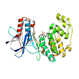
3UAM
 | |
3TSM
 | |
3U5W
 | |
3U0I
 | |
3UJH
 | |
7UDK
 | | Crystal structure of designed helical repeat protein RPB_LRP2_R4 bound to LRPx4 peptide | | 分子名称: | 4xLRP, Designed helical repeat protein (DHR) RPB_LRP2_R4 | | 著者 | Chang, Y, Redler, R.L, Bhabha, G, Ekiert, D.C. | | 登録日 | 2022-03-20 | | 公開日 | 2023-03-22 | | 最終更新日 | 2024-04-03 | | 実験手法 | X-RAY DIFFRACTION (3.18 Å) | | 主引用文献 | De novo design of modular peptide-binding proteins by superhelical matching.
Nature, 616, 2023
|
|
7UDO
 | | Crystal structure of designed helical repeat protein RPB_LRP2_R4 (proteolysis fragment?), forming pseudopolymeric filaments | | 分子名称: | 1,2-ETHANEDIOL, Designed helical repeat protein (DHR) RPB_LRP2_R4, PHOSPHATE ION | | 著者 | Redler, R.L, Chang, Y, Bhabha, G, Ekiert, D.C. | | 登録日 | 2022-03-20 | | 公開日 | 2023-03-22 | | 最終更新日 | 2024-04-03 | | 実験手法 | X-RAY DIFFRACTION (2.5 Å) | | 主引用文献 | De novo design of modular peptide-binding proteins by superhelical matching.
Nature, 616, 2023
|
|
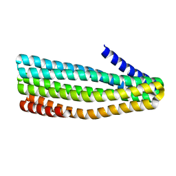
7UDJ
 | | Crystal structure of designed helical repeat protein RPB_PEW3_R4 bound to PAWx4 peptide | | 分子名称: | 4xPAW peptide, De novo designed helical repeat protein RPB_PEW3_R4 | | 著者 | Redler, R.L, Chang, Y, Bhabha, G, Ekiert, D. | | 登録日 | 2022-03-20 | | 公開日 | 2023-03-22 | | 最終更新日 | 2024-04-03 | | 実験手法 | X-RAY DIFFRACTION (2.7 Å) | | 主引用文献 | De novo design of modular peptide-binding proteins by superhelical matching.
Nature, 616, 2023
|
|
7UDL
 | | Crystal structure of designed helical repeat protein RPB_PLP1_R6 bound to PLPx6 peptide | | 分子名称: | 1,2-ETHANEDIOL, 6xPLP Peptide, Designed helical repeat protein (DHR) RPB_PLP1_R6 | | 著者 | Chang, Y, Redler, R.L, Bhabha, G, Ekiert, D.C. | | 登録日 | 2022-03-20 | | 公開日 | 2023-03-22 | | 最終更新日 | 2024-04-03 | | 実験手法 | X-RAY DIFFRACTION (2.15 Å) | | 主引用文献 | De novo design of modular peptide-binding proteins by superhelical matching.
Nature, 616, 2023
|
|
7UDN
 | |
7UE2
 | |
7UDM
 | | Crystal structure of designed helical repeat protein RPB_PLP1_R6 in alternative conformation 1 (with peptide) | | 分子名称: | 6xPLP, Designed helical repeat protein (DHR) RPB_PLP1_R6 | | 著者 | Chang, Y, Redler, R.L, Bhabha, G, Ekiert, D.C. | | 登録日 | 2022-03-20 | | 公開日 | 2023-03-22 | | 最終更新日 | 2024-04-03 | | 実験手法 | X-RAY DIFFRACTION (2.65 Å) | | 主引用文献 | De novo design of modular peptide-binding proteins by superhelical matching.
Nature, 616, 2023
|
|
3L56
 | | Crystal structure of the large c-terminal domain of polymerase basic protein 2 from influenza virus a/viet nam/1203/2004 (h5n1) | | 分子名称: | Polymerase PB2 | | 著者 | Staker, B.L, Edwards, T, Eric, S, Raymond, A, Stewart, L, Seattle Structural Genomics Center for Infectious Disease (SSGCID) | | 登録日 | 2009-12-21 | | 公開日 | 2010-03-09 | | 最終更新日 | 2023-09-06 | | 実験手法 | X-RAY DIFFRACTION (2.3 Å) | | 主引用文献 | Biological and structural characterization of a host-adapting amino acid in influenza virus.
Plos Pathog., 6, 2010
|
|
3KRS
 | |
3LD9
 | |
3LR3
 | |
3LR0
 | |
3LR5
 | |


